09.09.2021 Nail and skin fungus is a common problem. It is enough to swim in a pool, take a shared shower or visit a bathhouse to become infected with this disease. Diseased nails become yellow-gray and brittle, and noticeably thicken. The disease not only spoils the appearance, but also causes severe physical discomfort: itching and burning of the affected areas of the skin.
If the fungus is not treated, then soon its pathogen will spread throughout the body. The spores will enter the bloodstream, leading to allergic reactions. It is recommended to start treatment in the early stages and not stop therapy until all symptoms have passed.
Modern pharmacology offers many options for anti-fungal drugs in various dosage forms. The choice of remedy depends on the type of fungus, the degree of the disease and the individual reactions of the body.
Classification of fungi
The following types of fungus are distinguished, depending on the distribution of the process:
- Keratomycosis. The process of spore dissemination occurs in the epidermis;
- Dermatomycosis. Affects skin, hair and the top layer of nail plates;
- Candidiasis. Another name for the fungus is “thrush”. It usually affects the mucous membranes.
With systemic mycoses, not only the skin is infected, but also internal organs.
By origin, fungi are:
- yeast;
- moldy;
- dimorphic.
Only yeast are a natural component of the human microflora. The rest are classified as pathogenic agents.
Causes of candidiasis
Skin candidiasis begins to actively develop when it becomes pathogenic. This can be caused by several reasons:
- metabolic disease;
- avitaminosis;
- pathologies of the endocrine system;
- long-term use of antibiotics;
- postoperative therapy;
- immunodeficiency;
- gastrointestinal diseases.
As a rule, it is internal factors that provoke the development of this disease. But occasionally it can be triggered by external factors, such as:
- microtraumas;
- wearing synthetic items or too warm clothes that increase sweating;
- poor hygiene;
- exposure to harmful substances on the skin.
People whose professions involve chemical production and have a lot of contact with water should especially carefully monitor the condition of their skin.
Types of drugs for fungus
Before choosing a suitable remedy, you need to pay attention to the main types of drugs for mycoses:
- Products for topical use. This category includes varnishes, ointments, gels and solutions. They help well in the initial stages of the development of the disease;
- Tablets and capsules. They are used internally, according to the purpose and instructions. They are necessary if the disease is advanced and local remedies are no longer suitable.
To achieve an optimal result, it is necessary to undergo diagnostics and identify the pathogen. In this case, the therapy will bring the desired effect.
Recommendations for effective treatment
In the process of undergoing medical therapy, it is necessary to give up bad habits and limit yourself to excessive consumption of foods containing large amounts of carbohydrates.
Treatment is prescribed only by a doctor; self-medication can lead to serious, irreversible consequences.
If you are faced with a problem such as skin candidiasis , immediately contact a dermatovenerologist. Only proper treatment will relieve you of the problem.
List of drugs
When choosing drugs for fungus, it is necessary to focus on effectiveness, the presence of side effects, symptoms and course of the disease. It is important to take into account individual intolerance to the components.
Antifungal antibiotics
If the fungal infection is systemic in nature, an integrated approach is required. Deep tissue damage, separation of the nail plates and severe itching of the skin against the background of deteriorating general health suggest the use of not only local remedies, but also tablets.
Among external remedies, it is worth highlighting medications that contain naftifine. This component has an antibacterial and anti-inflammatory effect. When taken regularly and in combination with other therapeutic agents, recovery occurs quickly.
Preparations with naftifine:
| Exostat solution 1% 15ml |
| Mycoderil cream 1% 15g |
| Exoderil cream 1% 15g |
External preparations for dermatomycosis and keratomycosis
Yeast, mold and dimorphic fungi can be cured with Terbinafine-based products. Popular drugs with this active ingredient:
| Lamicide drops for nails 15ml |
| Lamicide spray for legs 15ml |
| Lamisil cream 1% 15g |
| Fungoterbin 1% 15g |
| Exifin gel 1% 15g |
| Terbizil cream 15g |
| Terbix cream 1% 10g |
| Exiter cream 1% 15g |
Medicines containing ketoconazole effectively fight fungal infections of the head and groin area. List of drugs:
| Shampoo Sulsen forte 250ml ketoconazole |
| Shampoo Sulsen mite for dandruff 1% 250ml |
| Sulsen forte paste 2% 75g ketoconazole |
| Sulsen mite paste 1% 75g |
| Shampoo Sulsen mite for dandruff 1% 150ml |
| Shampoo Sulsen forte from perch 150ml |
| Nizoral cream 2% 15g |
| Mycozoral ointment 2% 15g |
| Sebozol shampoo 100ml |
Products based on miconazole, a synthetic substance with an antifungal effect, are effective against dermatomycetes and yeast, as well as the causative agent of lichen versicolor. Preparations containing miconazole:
| Mykozon cream 2% 15g |
| Ginocaps vag caps x 10 |
Antifungal agents for systemic candidiasis
The Candida fungus spreads inside the body: on the respiratory system, in the digestive system, on the genitals. Sometimes it infects the nervous and cardiovascular systems.
Often, with systemic candidiasis, the fungus also affects external tissues - nail plates, skin on the legs and arms. To get rid of the disease, it is important to start therapy in a timely manner. To treat candidiasis, drugs containing clotrimazole are used.
Popular remedies for the treatment of mycotic diseases caused by the Candida fungus:
| Flucorem 0.5% gel 15g |
| Kanizon plus cream 15g |
| Kanizon cream 1% 20g |
| Candide cream 15g |
| Clotrimazole-Akrikhin ointment 1% 20g |
| Clotrimazole-Akos ointment 1% 20g |
| Candide B cream 15g |
| Candiderm cream 15g |
In recent years, there has been a steady increase in the incidence of diseases caused by opportunistic fungi, among which candidiasis is the most frequently recorded. The increase in the incidence of candidiasis is associated, first of all, with the use of modern therapeutic agents, as well as with environmental changes, in particular, with an increase in background radiation and other factors that weaken the immune system.
Etiology
Candidiasis is caused by yeast-like fungi of the genus Candida
, widespread in nature and classified as opportunistic microorganisms.
of Candida
are registered as causative agents of candidiasis , which are considered pathogenic for humans;
these include C. albicans, C. tropicalis, C. krusei
, etc.
Epidemiology
The causative agents of candidiasis are isolated from the intestines, genitals and bronchial secretions on average in every third person. Primary colonization of the body occurs in the birth canal, and after birth - through contact and nutrition. Infection of a child can occur due to candidiasis of the mother's nipples, from service personnel, through household items, etc. There have been outbreaks of candidiasis in newborns in maternity hospitals. Sexual transmission of candidiasis cannot be ruled out.
Pathogenesis
The main factor in the development of candidiasis is considered to be a background condition or disease of the body, in which opportunistic pathogens acquire pathogenic properties. In recent years, many researchers have come to the conclusion that the main predisposing factor for the occurrence of superficial forms of candidiasis, incl. in HIV infection, is mainly a violation of cellular immunity. A certain role in the development of candidiasis is played by the frequent and not always justified prescription of broad-spectrum antibiotics, incl. for prophylactic purposes, as well as the widespread use of drugs that have an immunosuppressive effect - glucocorticoid hormones and cytostatics. According to Zh.V. Stepanova and L.L. Smolyakova, in children staying in somatic departments of hospitals and receiving antibacterial therapy, damage to the oral mucosa caused by yeast-like fungi is observed in 6.6% of cases, of the oral mucosa and skin - in 15%, of the intestines - in 2.5% , candidiasis in the intestines – in 9.2%.
Candidal paronychia and onychia on the fingers, as a rule, develops in women who have frequent contact with water, while separation of the nail skin (eponychion) from the nail plate is observed, creating favorable conditions for fungal infection in the matrix area. The disease can occur in people suffering from diabetes mellitus, HIV-infected people, who have been receiving antibacterial drugs, corticosteroids, immunosuppressants, etc. for a long time. In recent years, women have begun to use false nails, and therefore another risk factor has emerged for the development of fungal and bacterial infections.
Yeast-like fungi of the genus Candida
can cause damage to the mucous membranes, skin and its appendages, and internal organs. The most common forms of mycosis are superficial.
Clinical picture: Candidiasis of smooth skin
The main localization of the disease is large (inguinal-femoral, intergluteal, under the mammary glands, axillary cavities) and small (interdigital) folds, but rashes can occur on the smooth skin of the trunk and limbs, incl. palms and soles. Outside the folds, foci of candidiasis develop mainly in infants, as well as in adults suffering from diabetes mellitus or severe general illness. In large folds small bubbles, the size of millet grains, and sometimes pustules appear, which open to form erosions. Due to peripheral growth, erosions quickly increase in size and merge, forming large areas of damage. The lesions are dark red in color with a burgundy tint, shiny, with a moist surface, irregular in shape, with a strip of exfoliating epidermis along the periphery. Fresh small erosions (screenings) form around large foci. In children, especially weakened ones, from the folds the lesion spreads to the skin of the thighs, buttocks, abdomen, and sometimes to the entire skin. There may be painful cracks deep in the folds. Candidiasis of smooth skin outside the folds (chest, abdomen, shoulders, forearms, legs, etc.) in children has a clinical picture of seborrheic dermatitis; in adults, the disease can manifest itself in the form of erythematous spots with peeling in the center and small blisters along the periphery. Candidiasis of smooth skin of small folds (interdigital erosion) most often occurs between the 3rd and 4th, 4th and 5th fingers of the hands, less often of the feet, and is characterized by the formation of eroded foci of a rich red color with a smooth, shiny, as if varnished surface, clear boundaries, with flaking of the horny layer of the epidermis along the periphery. The disease can begin with the appearance of small blisters on the lateral touching hyperemic surfaces of the skin, then spreads to the area of the interdigital fold, swelling, maceration, and peeling appear. Interdigital candidal erosion is observed mainly in individuals who have prolonged contact with water, which contributes to the development of skin maceration; in addition, favorable conditions are created for the development of candidal infection. In addition to the third and fourth folds, others may be affected, not only on one, but also on both hands. Patients are concerned about itching, burning, and if there are cracks, pain. The course of the disease is chronic, with frequent relapses. Candidiasis of the smooth skin of the nipples in nursing women deserves attention. Its clinical manifestations can be different: slight hyperemia in the area of the isola; lesion near the nipple with maceration, clear boundaries; fissure with maceration along the periphery and small bubbles between the nipple and the isola.
Candidiasis of the palms and soles is rare. On the palms, the disease can occur as dry lamellar dyshidrosis (superficial lamellar, ring-shaped or garland-shaped peeling), vesicular-pustular form (vesicles and pustules against the background of hyperemic and edematous skin) and hyperkeratotic eczema (against the background of diffuse hyperkeratosis or individual areas of keratinized skin are observed sharply limited wide skin grooves with a dirty brown color). Candidiasis of the skin of the soles occurs mainly in children and is characterized by the presence of small blisters and pustules, hyperemic spots with peeling and exfoliating macerated epidermis along the periphery.
Candidiasis of the nail folds and nails (candidal paronychia and onychia)
The disease begins from the posterior ridge, most often in the area of its transition to the lateral ridge, with the appearance of hyperemia, swelling and swelling of the skin. Then the inflammatory phenomena spread to the entire roller, which becomes thicker and seems to hang over the nail, and peeling is observed along the edge of the roller. The skin of the roller becomes thin, shiny, and the nail skin (eponychion) disappears. When the roller is pressed, ichor, a lump of white crumbly mass, or a drop of pus may be released (due to the addition of a secondary infection). Later, the nail plate changes: it becomes dull, in the area of the lunula it is separated from the bed, destroyed as onycholysis, or transverse grooves and elevations appear on it. These changes are associated with impaired blood supply in the matrix area, i.e. they are trophic in nature and are caused by inflammation in the area of the cushion. When the fungus penetrates the nail plate, and this happens from the lateral edges, the nails become thinner, separate from the bed, acquire a yellow-brown color and look as if they are trimmed on the sides. In young children, inflammatory phenomena in the area of the cushion are more pronounced, and the nail plate changes from the distal edge. There is candidiasis of the nails without inflammation of the ridge. In this case, the change in the plate begins from the distal edge and develops according to the type of onycholysis; the plate becomes thinner, does not adhere to the bed, and there may be multiple lesions of the nails.
Diagnosis and differential diagnosis
In superficial forms of candidiasis, the diagnosis is based on the presence of a characteristic clinical picture in the patient and identification of the fungus in pathological material (skin flakes, scrapings from nails) during microscopic examination. The diagnosis can be considered reliable if pseudomycelium or true mycelium and budding cells are detected. Sowing on a nutrient medium is carried out to identify the type of yeast-like fungus of the genus Candida
. Isolating only a culture of the fungus has no diagnostic value, since it can be obtained by culturing scrapings from the skin and nails of healthy people.
With candidiasis of smooth skin of large folds and outside the folds, the disease should be differentiated from seborrheic eczema, psoriasis, and other mycoses - inguinal athlete's foot, superficial trichophytosis, pseudomycosis erythrasma (complicated form); with interdigital candidal erosion on the hands - from dyshidrotic eczema, on the feet - from mycosis caused by Trichophyton interdigitale
and
Trichophyton rubrum
; when nails and ridges are affected - from onychia and paronychia of bacterial etiology, eczema and psoriasis.
Treatment
Considering that candidiasis is an opportunistic mycosis, first of all, it is necessary to identify and, if possible, eliminate the pathogenetic factors of the disease (study of the immune and endocrine status, gastrointestinal tract and corrective therapy). Limited and sometimes widespread forms of smooth skin lesions, especially those that developed during treatment with antibacterial drugs, as a rule, are easily treated with local antifungal agents and can resolve without treatment after discontinuation of antibiotics. Local and systemic antimycotics are prescribed as etiotropic therapy. In recent years, azole drugs with a broad spectrum of action, as well as polyene antibiotics, have been widely used in the treatment of candidiasis.
For candidiasis of large folds of smooth skin with acute inflammatory phenomena, treatment should begin with the use of an aqueous solution of brilliant green (1-2%) in combination with an indifferent powder and continue for 2-3 days, then antimycotic drugs should be prescribed until the clinical manifestations resolve.
For candidiasis of large, small folds and other areas of smooth skin, antifungal agents are used in the form of cream, ointment and solution: ketoconazole, clotrimazole, oxyconazole, bifonazole, natamycin. The cream or ointment is applied in a thin layer to the lesions and rubbed in 1-2 times a day, the duration of treatment is until the clinical manifestations resolve, then it is recommended to continue using the cream for another 7-10 days to prevent relapse. In case of widespread processes on the skin and the ineffectiveness of local therapy, systemic antimycotics are prescribed: fluconazole - for adults at a dose of 100-200 mg, for children at a dose of 5 mg/kg body weight; itraconazole - adults at a dose of 100-200 mg; nizoral - adults at a dose of 200 mg 1 time per day daily, as well as the polyene antibiotic natamycin (adults 100 mg 4 times a day, children 50 mg 2-4 times a day). The duration of therapy is 2-4 weeks.
For candidiasis of the nail folds and nails, anti-inflammatory treatment of the fold is first carried out using applications of pure ichthyol, which are done once a day until the inflammatory phenomena subside. Then apply antimycotic agents (ketoconazole, oxiconazole, isoconazole, clotrinazole, etc.) for topical use, rubbing them under and around the roller. The procedures are carried out 2 times a day, in the evening the drugs can be used under an occlusive dressing. It is possible to combine an ointment (cream) with a solution, alternating them. If the nail plate is involved in the process, you can not remove it or clean off the infected areas after softening with keratolytic agents (bifonazole in the nail treatment kit), and then treat the nail bed with antifungals for external use. Therapy is carried out until clinical manifestations resolve and a healthy nail plate grows. If it is ineffective, systemic antimycotics are prescribed: fluconazole according to an intermittent regimen (adults 150 mg once a week, children 5-7 mg/kg body weight); itraconazole (adults) using the pulse therapy method (200 mg 2 times a day for 7 days, then a 3-week break) for 2-3 months; nizoral (adults) 200 mg per day daily for 2-4 months.
Prevention
- To prevent the development of candida infection in children who are in somatic departments of hospitals and receiving antibacterial drugs, they need to be prescribed fluconazole at a rate of 3-5 mg/kg body weight once a day. The daily dose depends on the degree of risk, treatment is carried out during the main therapy.
- Patients with candidiasis in the intestines are prescribed nystatin 2-4 ml IU per day or natamycin 50 mg for children and 100 mg for adults 2 times a day for 15 days.
- Prevention of the development of candidiasis in patients with severe somatic and endocrine diseases, as well as with immunodeficiency (multiple mycological studies).
- Prevention of dysbiosis.
- Prevention of the development of candidiasis in newborns.
How to choose drugs for fungus
To combat mild fungal infections, topical preparations—varnishes, ointments, sprays, shampoos, and special solutions—are sufficient. For advanced diseases, both local and systemic agents are used. Anti-fungal tablets serve as a supplement, ridding the subungual skin and other tissues of the pathogen. In the “Antifungal Drugs” section on our website you will find the medications listed in this article, as well as their analogues.
Any drugs for fungus should be selected with the help of a specialist who can determine the severity of the disease and take into account the individual characteristics of the body.
The offer is not an offer, the drugs presented are medicines, specialist consultation is required.
Diagnosis and treatment
It is not enough to see only the clinical manifestations and make a diagnosis; it is necessary to make a scraping with removal of separated particles from the affected area and undergo a series of other tests. Only after laboratory testing and a thorough examination can a doctor make an accurate diagnosis.
The disease requires a course of treatment, the basis of which is antifungal drugs. To achieve maximum therapeutic results, an integrated approach is required:
- carry out therapy to combat the pathogen;
- destroy the endogenous source of the pathogen;
- cure concomitant diseases that provoke skin candidiasis;
- carry out restorative therapy.
Candidiasis of skin folds in adults
The incidence of cutaneous candidiasis in adults is low compared to candidiasis of the mucous membranes. The incidence is higher in the elderly and inpatients; risk groups include patients with obesity and diabetes mellitus.
In adults, the inguinal-femoral, intergluteal folds, scrotum, armpits, area under the mammary glands, as well as abdominal folds in obese people are affected.
Skin candidiasis in women
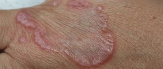
In these places, redness appears in the depths of the folds, crimson-colored erosions and a damp surface with a varnish sheen.
Skin candidiasis, photo
They increase in size and, merging, form extensive foci on the contacting surfaces of the folds.
Candidiasis of skin folds
The clinical picture of candidiasis at this stage is very typical. The lesions have polycyclic outlines, clearly delimited from the surrounding healthy skin along their periphery by a narrow fringe of exfoliating white epidermis.
Candidiasis
Around lesions on healthy skin, there are almost always screenings in the form of small pustules, erosions, and red spots.
Candidiasis of the intergluteal and inguinal-femoral folds is often combined with lesions of the genitals in women and the scrotum in men and is accompanied by painful itching.
Skin lesions due to candidiasis
Candidiasis of interdigital skin folds
Candidiasis of the interdigital folds is often an occupational disease and occurs in people employed in production using Candida spp. for industrial purposes, or those where there is a high probability of contamination by them (confectionery, canning, processing of vegetables and fruits, etc.), as well as for women who do a lot of housework without protective gloves, nurses.
Hand skin candidiasis
More often, the folds between the third and fourth fingers of the hands and the lateral surfaces of these fingers are affected, where the skin is macerated, whitish in color, and is easily torn off, exposing an eroded shiny surface.