An ovarian cyst is a cavity filled with contents. This benign disease can be congenital or acquired. Cystic formations are detected in women of different age groups, and even girls who are not sexually active.
Often the disease is asymptomatic, without causing discomfort, and is detected during an ultrasound examination.
What is a cyst
The content of the article
More than a third of women of all ages develop neoplasms in the ovaries, namely cysts. An ovarian cyst is a neoplasm located on the wall of an organ and filled with fluid. In most cases, a woman does not even suspect that any disturbances have occurred in her body.
Cysts can be dangerous and harmless. Safe (functional - small, without inclusions of pathological cells) disappear over time and have virtually no effect on a woman’s life. Other types of tumors are dangerous to health and must be removed surgically. If you delay treatment for an ovarian cyst, remember about possible complications: the development of a malignant tumor, suppuration and rupture of the cyst, followed by peritonitis.
The main types of cystic formations
Ovarian
This is a fluid-filled bladder that causes the ovary to enlarge, leading to pain and sometimes infertility. The main reason for its appearance is hormonal imbalance. The disease often occurs without symptoms; when the formation is large, the cycle is disrupted, pain is felt in the lower abdomen, a false urge to urinate occurs, and bleeding occurs outside of menstruation. Methods of surgical treatment: oophorectomy (complete removal of the ovary), laparoscopic cystectomy (excision of the cyst), adenxectomy (surgery to remove the uterine appendages). In most cases, the choice is not classical surgery, but gentle endoscopic removal of the cyst while preserving the patient’s reproductive function.
Read more about ovarian cyst removal
Coccygeal
Most often occurs in men aged 15-30 years. It is an opening in the area of the gluteal fold, approximately 10 cm from the anus. Externally it looks like a fistula. It can be congenital or acquired - due to too much hair in this area. It manifests itself as pain when walking, sitting, redness of the tailbone, a feeling of discomfort and the presence of a foreign body in the area where it is located. At a later stage, pus is released from the hole.
Read more about the treatment of coccygeal cyst
Bartholin gland
Appears due to blockage of the gland duct due to infection or chronic inflammation. The formation has a capsule and is filled with secretion, which gradually gels. If it is large, it interferes with walking and sitting, makes intimate intimacy inaccessible, and can become infected and lead to an abscess. Usually reaches 2 cm, but there are formations up to 9 cm. The main causes are chronic bartholinitis, candidiasis, bacterial vaginosis, decreased local immunity.
Where and how to remove a Bartholin gland cyst
Eye
A hollow formation filled with non-inflammatory fluid - products of the activity of the cornea or conjunctiva. May originate from the cornea, iris, conjunctiva and other eye membranes. Causes: inflammation and trauma, congenital anomalies. The disease is manifested by pain, a feeling of fullness, blurred vision, and the presence of translucent dots in the field of vision.
Read more about surgical treatment of cysts in ophthalmology
Maxillary sinus
A cavity containing fluid and having a membrane attached to the wall (usually the lower) of the maxillary sinus. A cystic formation can be true (its walls consist of mucous membrane) or pseudocyst (the mucous membrane is split and fluid accumulates in it). It manifests itself as headache, difficulty breathing through the nose, a feeling of fullness and heaviness in the eye and cheek area, mucus discharge from the nose and its flow down the wall of the pharynx, discharge of yellow transparent fluid from the nose, frequent sinusitis with suppuration. It is formed due to the peculiarities of the anatomical structure of the nose, blockage of the excretory duct of the glands of the maxillary sinus, inflammation of the teeth, spreading to the roots.
Read more about the treatment of abscesses and cysts of the ENT organs
Mammary gland
A cavity bounded by a capsule of connective tissue and filled with fluid is formed in the ducts and can be single or multiple. It is formed due to an increase in the duct of the mammary gland, the accumulation of secretions in it. The lesion may be round, oval, or irregular in shape. The disease is asymptomatic for a long time; over time, pain and burning appear in the mammary gland, and may be accompanied by suppuration and inflammation. One of the varieties is a fatty cystic formation that occurs when the sebaceous gland of the skin is blocked and filled with secretions. The main provoking factors are mastitis, thyroid disease, inflammation of the genital organs, and ovarian dysfunction.
Learn more about symptoms and treatments
Uterus and cervix
These are dilated and clogged glands, inside of which secretions (mucus) accumulate. The disease occurs against the background of endocervitis, cervitis. Provoking factors: abortion, childbirth, infections, menopause, hormonal imbalance, use of an intrauterine device, infections. Nabothian cystic formations are localized in the vaginal area of the uterus and are not removed until they reach a certain size. Retention occurs due to an excessive amount of secretion in the gland duct. The disease is often asymptomatic; its indirect signs are frequent inflammation.
Read more about surgical treatment of uterine and cervical cysts
Brain
A cystic formation is an accumulation of fluid in the substance or membranes of the brain. When large in size, it entails intracranial hypertension and puts pressure on the surrounding brain structures. Can form at any age. Depending on the location, cerebral (intracerebral) and arachnoid formations are distinguished. The first are found in the internal structures of the brain, in areas of necrosis. The second ones are in the meninges. Provoking factors: inflammatory diseases, trauma, including birth, cerebrovascular accident, parasites, complications after surgery. They manifest themselves as nausea, a feeling of pressure on the eyes, decreased performance, sleep disturbances, pulsation or noise in the head, and visual impairment.
Thyroid gland
Small formations (up to 5 mm) may not have pronounced symptoms. If the thyroid cyst has dense inclusions and a complicated structure, then special studies (for example, ultrasound), tests and biopsy are needed, because such a condition may be a sign of malignant degeneration.
Read more about surgical treatment of thyroid cysts
Larynx
Cysts of the larynx can be located in any part of the larynx. They do not grow into the mucous membrane, but grow towards the lumen of the larynx and thereby narrow it. The cause of retention cysts is blockage of the excretory ducts of the laryngeal glands. There are cysts of the vocal cords that arise due to constant irritation.
More information about surgical treatment of laryngeal cyst (laryngocele)
You can find out the prices for cyst removal endoscopically or by another method, as well as the cost of preoperative diagnostics on our website or by calling honey. +7 (812) 435 55 55.
Causes
The main reason for the formation of cysts is hormonal changes in the body associated with endocrine diseases, ovarian dysfunction and other changes, such as age-related ones.
It is noted that cysts often occur after abortions, stress and other traumatic factors. All the causes of cysts have not yet been precisely studied, but it is known that they are provoked by:
- inflammatory processes in the ovaries;
- sexual infections;
- Ovarian endometriosis is a disease in which cells from the uterus (endometrium) enter the ovarian tissue;
- congenital abnormalities of the reproductive system;
- termination of pregnancy, especially those carried out by surgical curettage;
- excess weight;
- metabolic diseases;
- imbalance of sex hormones.
Acquired ovarian cysts
- Follicular
- occurs if the follicle does not rupture and the egg does not come out. During ultrasound, they can be observed on both ovaries. Sometimes there are several such formations. - Endometrioid cysts
are manifestations of ovarian endometriosis. They are formed from endometrial uterine cells that enter the ovary. These cellular structures react to hormonal changes caused by the menstrual cycle - they increase in size before the onset of menstruation, and then begin to bleed. There is nowhere for the released blood to flow out, so it forms cysts, which are called “chocolate” due to the coagulated dark bloody contents. - Corpus luteum cysts
occur due to hormonal imbalances. After the follicle emerges, the corpus luteum produces progesterone for some time, which is necessary for pregnancy. But, if conception does not occur, this formation should resolve. Sometimes this does not happen and a cystic formation forms in the ovary. Sometimes the formation reaches a large size and begins to bleed. - Cysts in polycystic disease
can be multiple. Polycystic ovarian syndrome is associated with metabolism, so women experience infertility, miscarriages, cardiovascular disease, diabetes and even mental illness.
Congenital ovarian cysts
- Dermoid
, formed from embryonic glandular tissue. These formations may contain hair, fat, teeth, cartilage, and bone tissue. Dermoid cysts can reach a size of 25 cm. Although malignant degeneration of the cyst occurs only in 1-2 percent of cases, a large tumor puts pressure on neighboring organs, causing pain and dysfunction; - Paraovarian cysts
, arising from embryonic tissue, are located next to the ovaries. The reason for their appearance is a violation of the intrauterine formation of the girl’s internal genital organs. Paraovarian cysts grow very slowly, but can reach enormous sizes (up to 30 cm).
Symptoms
In some cases, cysts (especially functional ones) do not show any symptoms. Therefore, it is important to regularly make preventive visits to the gynecologist (1-2 times a year).
Specific symptoms depend on the type of cyst and how quickly it grows. Painful sensations are observed on the side on which the tumor is located (right, left, or both). The disease can manifest itself with signs such as:
- nagging or aching pain (or a feeling of fullness in the lower abdomen), intensifying during sexual intercourse, physical activity, urination or menstruation;
- menstrual irregularities (no periods or their period is prolonged);
- infertility;
- abdominal enlargement (if the cyst is large);
- bleeding from the vagina;
- increased frequency of urination, constipation (with cyst growth and, as a consequence, compression of internal organs);
- a lump (that can be felt) in the abdominal cavity.
Additional symptoms (which may occur in addition to those listed above) include:
- increased volume of menstrual flow;
- constant strong thirst;
- sudden weight loss or gain without objective reasons;
- blood pressure disorders;
- facial hair growth;
- increased body temperature (from 38 degrees and above);
- nausea, vomiting.
Diagnosis of cysts
In order for the gynecologist to determine whether the patient has a cyst, he first of all conducts an examination and sends her for an ultrasound examination of the pelvic organs. If the ultrasound does not give accurate results, then the following procedures are performed:
- Ultrasound of the ovaries;
- blood tests for biochemistry and tumor markers;
- test to determine the presence of pregnancy, including ectopic.
Additional studies performed to diagnose complications associated with the formation of an ovarian cyst:
- a urine test is carried out to find out whether the patient is losing blood during the day (in addition to the days of menstruation);
- puncture of the posterior vaginal vault to determine whether there was hemorrhage in the abdominal area;
- laparoscopic examination.
Why is an ovarian cyst dangerous?
Ovarian cysts increase in size, interfering with the work of neighboring organs. By disrupting the functioning of the reproductive system, they often cause infertility. Sometimes cysts develop into cancer.
The most serious complication of these formations is rupture of the membrane, in which the cystic contents leak into the abdomen. The woman has to undergo emergency surgery to remove the tumor capsule, often including part of the ovary. Torsion of cysts that have a “leg” is no less dangerous. There is a disturbance in blood flow, accompanied by tissue necrosis. Without treatment, this complication is fatal.
In order not to endanger life and health, it is necessary to identify and treat ovarian cysts in a timely manner.
What do normal ovaries look like during ultrasound examination?
Normally, during an ultrasound examination, both ovaries are almost the same size:
- length – 30-41 mm;
- width – 20-31 mm;
- thickness – 14-22 mm;
- volume – about 12 cubic ml.
Due to the maturing follicles, the surface of the organ looks bumpy. Since the ovary is well supplied with blood, its tissues contain many blood vessels.
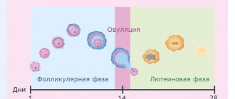
Typically, at least 12 cells mature in two ovaries. Among them stands out a dominant follicle measuring 10-22 mm, from which an egg will be released in the middle of the cycle. If ovulation has already passed, the corpus luteum is visible inside the ovary, secreting hormones necessary for implantation and development of the embryo. If conception does not occur, the corpus luteum dissolves.
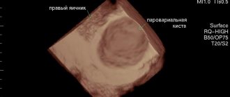
What do the ovaries look like with cysts?
Ultrasound examination allows not only to identify cystic formations, but also to determine their type. Sometimes this requires several ultrasound examinations at different periods of the menstrual cycle:
- A dermoid cyst
has quite dense contents, so it looks lighter than others. Areas of heterogeneous structure are found inside. The walls of the formation are quite thick - 7-12 mm. Since the cyst grows very slowly, repeated ultrasound on any day of the menstrual cycle does not reveal changes in its size and structure. - A paraovarian cyst
is located outside an unchanged ovary. As a rule, it is single-chamber. Its size and appearance do not change depending on the day of the menstrual cycle. - The endometrioid cyst
has a smooth, clear shape. Wall thickness 2-9 mm. Inside there are structures that have a high density and resemble a honeycomb. Therefore, on the screen the cyst looks like a dark formation separated by light areas. The eggs in such an organ do not mature, but the other healthy ovary works normally. Before the critical days, the size of the cyst increases. In parallel, an enlargement of the uterus may be observed. - The follicular ovarian cyst
has thin walls and an even dark color. It is common - these neoplasms account for up to 20% of cases of ovarian tumors. In most cases, it gradually decreases in size, as can be seen on a repeat ultrasound. Sometimes follicular cysts do not resolve, being found even in women with long-extinct menstrual function. - A corpus luteum cyst
is similar in appearance to a follicular cyst. To differentiate them, Doppler ultrasound is performed. On examination, a bright circle is visible around the cystic formation, which doctors call the “ring of fire.” There is no blood flow inside the cystic formation itself.
Possible complications
If left untreated and the ovarian cyst continues to grow, the following complications may develop:
- Hemorrhage into the cyst cavity and subsequent transformation of a functional neoplasm into a hemorrhagic one;
- Torsion of the cyst pedicle . With incomplete torsion (gradual, 90-180 degrees), circulatory disturbances, loss of cyst mobility, and formation of adhesions occur. With complete (acute) torsion (360 degrees), necrosis (tissue death) may occur, leading to the development of peritonitis (inflammation of the peritoneum). This is a dangerous condition for which medical attention must be provided immediately. Complete torsion of the pedicle of the cyst manifests itself with symptoms such as decreased blood pressure and increased body temperature, nausea and vomiting, diarrhea or constipation, sharp paroxysmal pain in the lower abdomen, radiating to the lower back or leg.
- Rupture of the cyst wall (can lead to the development of peritonitis). The patient feels severe unilateral pain in the lower abdomen (depending on the location of the tumor). The abdominal wall is tense. Constipation may occur. Cyst rupture can occur as a result of abdominal trauma, physical activity, sexual intercourse, inflammatory diseases of the pelvic organs, hormonal imbalance, or torsion of the cyst stalk.
- Intra-abdominal bleeding (can be observed when a tumor ruptures).
- Infertility.
What to do if you find a cyst in the ovary
Detection of an ovarian cyst during an ultrasound is not a reason to panic. Small tumors can often be cured by prescribing hormonal drugs and vitamins A, B, and C. Under the influence of medications, the size of the tumor decreases over several menstrual cycles.
It is better not to touch small congenital cysts that are not disturbing or growing. You just need to visit a doctor and do an ultrasound to monitor the tumor. To exclude malignant degeneration, it is necessary to periodically donate blood for tumor markers.
When planning a pregnancy, it is better to get rid of the cyst, because... it may begin to grow or burst during a period when surgery is unsafe. After a properly performed operation, cysts do not recur. But women prone to the appearance of such formations should be observed by a gynecologist.
Expert opinion of a doctor
Butunov Alexey Andreevich
Obstetrician-gynecologist, laparoscopist surgeon, oncologist. Doctor of the highest category.
In advanced cases, with severe complications, oophorectomy is performed according to indications, when the ovary is removed along with the formation, although this is rare in the functional type. Rehabilitation after laparoscopy takes only 2–3 weeks. All this time, the patient is observed by the attending physician, control ultrasound diagnostics, anti-inflammatory and symptomatic therapy are carried out. The patient must follow medical recommendations, avoid physical activity and exercise, do not take a hot bath, refuse other thermal procedures, avoid sex for a while, and adhere to the recommended diet. Control ultrasonography should be performed 2 months after surgical treatment.
Features of treatment of ovarian cysts
The following factors influence which treatment method the doctor chooses: the woman’s age, parameters of the ovarian cyst, the general health of the patient, and concomitant diseases. Treatment of the cyst may initially be hormonal, then laparoscopic.
Functional cysts are treated with hormonal therapy and, if the method does not bring results, the patient is sent for surgery, which can be performed either laparoscopically or laparotomically. If the presence of a cyst is dangerous to life and health, the woman is urgently operated on.
Growing dermoid, paraovarian and endometrioid cysts are removed laparoscopically through small incisions in the anterior abdominal wall. Modern methods make it possible to remove cysts while preserving the ovary, which is important for women planning to have children in the future.
After surgery, the gynecologist prescribes a course of treatment to restore the proper functioning of the woman’s reproductive system. In addition to medications, hirudotherapy (intravaginal) and homeopathic medications are often prescribed. If the patient is overweight, a nutritionist and endocrinologist are involved in the treatment process.
If the patient is planning to have children, surgery to remove the cyst cannot in any way affect the likelihood of conception. In extreme cases, operations are performed even during pregnancy. Sometimes this does not affect the development of the fetus, but, in rare cases, there is a need to terminate the pregnancy, since the threat to the life and health of the mother is extremely high.
Prevention
To reduce the risk of developing ovarian cysts, women are advised to:
- undergo regular preventive examinations with an obstetrician-gynecologist and promptly treat diseases of the genitourinary organs;
- adhere to the principles of rational nutrition;
- use contraceptives to avoid abortions; correct endocrine disorders in a timely manner;
- do not use COCs or take medications without a doctor’s prescription.
If a functional cyst is discovered during the examination, it cannot be ignored and the recommendations of the treating obstetrician-gynecologist must be strictly followed.