Take the first step
make an appointment with a doctor!
For a long time, this disease was not recognized as an independent disease and was considered as a type of endometriosis. There is still debate in the scientific community about the nature, causes and mechanisms of development of uterine adenomyosis. However, it poses a serious threat to women's health. in particular, their ability to conceive and bear a healthy child. In severe cases, it leads to complete and irreversible loss of reproductive function.
Causes of adenomyosis
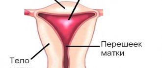
This pathology is an abnormal growth of the endometrium (uterine lining) deep into the muscular layer of the uterus. Previously, adenomyosis was considered an internal type of endometriosis. However, today gynecologists are increasingly inclined to believe that this is a separate, although similar, disease, since the causes and mechanism of its occurrence, as well as the development process and symptoms are significantly different. At the moment, it is not known for certain what factors lead to the appearance of adenomyosis, but there are several factors that are observed in all or most patients suffering from this pathology:
- Genetic predisposition to the formation of benign and malignant tumors of the genital organs;
- Gynecological diseases of an infectious, inflammatory and endocrine nature (genital infections, endometriosis, etc.);
- Excess weight, intestinal pathologies, increased susceptibility to infections, autoimmune diseases;
- Frequent surgical operations on the uterus, including resection, curettage (for example, abortion);
- Ectopic pregnancy, miscarriages and other complications that arise when carrying a child;
- Hormonal disorders caused by dysfunction of the endocrine glands (in particular, the ovaries and thyroid glands), taking hormonal contraceptives or other drugs of the same class;
- Various pathologies of the uterus and adjacent organs - for example, fibroids, erosion of the uterine cervix, etc.
There are also prerequisites that link the risk of developing adenomyosis with worsening environmental conditions or late childbearing. Research shows that this disease most often occurs in women living in developed countries and who prefer to give birth to children closer to 30-35 years of age. However, such a relationship has not been precisely established, because statistics in developing or undeveloped countries are kept much worse, which can distort the results.
The generally accepted view on the mechanism of development of adenomyosis is that there is no single or priority reason for its occurrence. Most likely, the disease appears when exposed to several negative factors at once. Moreover, for each patient the ratio of their roles is distributed individually, which complicates the general systematization of morbidity.
Forecast
Adenomyosis is a chronic disease prone to relapsing. The return of the disease is diagnosed in approximately every fifth fertile patient after conservative therapy or organ-saving surgery. In this regard, gynecologists can prescribe hormonal contraceptives for prevention.
In premenopausal patients, the disease is milder and during menopause it completely disappears.
Book a consultation 24 hours a day
+7+7+78
Types of adenomyosis
Depending on the distribution of pathological structures in the affected tissues, the following forms of adenomyosis are distinguished:
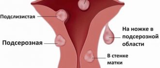
- Diffuse manifests itself as a uniform distribution of small inclusions of the endometrium throughout the muscular layer of the uterus;
- With the nodular type of disease, endometrioid nodes filled with blood are formed in the muscle layer;
- The focal form of adenomyosis is characterized by the formation of small accumulations of endometrial cells in various areas of the uterus.
Take the first step
make an appointment with a doctor!
There is also a cystic form, which is a further development of nodular or focal adenomyosis and is formed when the nodes hemorrhage followed by the formation of cysts. These types of this pathology can form separately or in combination with each other. The most common form is a diffuse nodular form, in which endometrial cells form a node and areas with their uniform inclusion.
There are also several stages of development of this disease, characterized by the degree of damage to the uterine tissue:
- stage 1 - at this stage, endometrial tissue penetrates only the thinnest layer of the myometrium (muscular wall) of the uterus;
- stage 2 - the pathological process penetrates deeper into the muscle wall, consisting of 3 layers, affecting 2 of them;
- stage 3 - the endometrium extends to all 3 layers of the myometrium up to the serous membrane of the uterus, which borders the surface of the bladder;
- stage 4 - endometrial tissue penetrates the uterine myometrium and affects adjacent organs, causing external gynecological endometriosis.
The signs of uterine adenomyosis and its damage to the body depend on the stage of the disease. Determining the stage of development of pathology plays an important role in its diagnosis and treatment. The earlier it is detected, the more effective and gentle the therapy will be.
Endometriosis and infertility
The main complication of this pathology is infertility. There are several mechanisms of reproductive dysfunction:
- Formation of adhesions in the pelvic cavity between the ovaries, fallopian tubes, intestines, abdominal wall and pelvic walls. This causes the fallopian tubes to bend and makes it difficult for the egg to travel to the uterus.
- The formation of endometrioid cysts in the ovaries, which makes ovulation and pregnancy difficult.
- Immune disorders lead to the production of antibodies to all endometrial cells, including normal uterine tissue. As a result, the implantation of the egg to the uterine wall is disrupted, or the pregnancy is terminated in the early stages.
- Hormonal imbalance when estrogen levels exceed progesterone levels, resulting in failure to ovulate.
Infertility treatment tactics for endometriosis most often include the use of assisted reproductive technologies, such as IVF and embryo transfer.
Symptoms of adenomyosis
This disease causes pathological changes in the tissues of the uterus and surrounding organs, which is manifested by the following characteristic symptoms:
- painful sensations appearing in the lower abdomen and, as the disease develops, spreading to the perineum, turning into chronic pain in the entire pelvic region;
- uterine bleeding that occurs before and after menstruation and manifests itself in the form of small brown discharge, the duration of which can reach a week or more;
- disorders of the menstrual cycle - an increase in the duration of menstruation, their pain and intensity, which leads to a decrease in the level of hemoglobin in the blood;
- painful sensations during sexual intercourse or masturbation;
- an increase in the size of the uterus before menstruation due to the growth of endometrial accumulations in its muscular wall under the influence of hormones.
The intensity with which signs of adenomyosis of the uterine body appear depends on the stage of development of the disease. At an early stage, the disease may not manifest itself in any way - the patient does not experience any pain or discomfort, or disturbance of sexual or reproductive function. Mild pain that occurs during the premenstrual period is often attributed by many women to menstruation itself. Only a doctor can identify the initial signs of adenomyosis using diagnostic equipment.
Over time, the intensity of the manifestation of the disease increases - pain in the abdomen and perineum becomes more frequent, the nature of menstrual bleeding changes, discomfort appears during sexual intercourse, etc. But even at this stage, not all patients recognize the disease, attributing these signs to hormonal imbalances and individual characteristics their menstruation, etc. Only when the symptoms of adenomyosis begin to appear outside the menstrual cycle, moving into the chronic stage, do they seek medical help.
Postoperative period
Features of the postoperative period depend on the volume of intervention and the method of its implementation. The easiest way for patients to tolerate laparoscopic or hysteroscopic organ-preserving operations. The pain syndrome in the postoperative period is moderate, activation is possible already on the first day if there are no contraindications from the anesthesiologist.
In the first few weeks, bloody discharge from the genital tract is characteristic, which gradually decreases. During this time, it is recommended to avoid excessive physical activity and sexual activity.
In general, most patients feel satisfactory after surgery and even note an improvement in their quality of life, as pain during menstruation decreases, PMS is easier and the amount of bleeding decreases.
Consequences of adenomyosis
Pathological growth of the endometrium negatively affects the functioning of the woman’s genitourinary system, and in severe stages it also affects the functioning of other abdominal organs - intestines, stomach, liver, etc. Endometrial cells, penetrating into the muscular layers of the uterus, form clusters that, under the influence of hormones, begin to peel off and bleed, causing inflammation and the formation of blood-filled cysts. This leads to the following negative consequences for the body:
- Sexual dysfunction - with the development of the disease, pain begins to appear during sexual intercourse, which makes it difficult not only to obtain satisfaction, but also to carry out the act itself;
- Problems with conception - abnormally overgrown endometrial tissue can affect the ovaries, disrupting the ovulation cycle, and also prevents the penetration of sperm into the genital tract, reducing the likelihood of successful implantation of the embryo;
- Increased risk of oncology - “settled” in foreign tissues, endometrial cells can, in some cases, themselves degenerate into a malignant tumor or provoke hyperplasia of the cervix and other organs;
- Increased risk of infections - blood-filled cavities formed in the tissues of the uterine wall are a favorable environment for the development of pathogenic microorganisms, which can lead to the formation of abscesses.
Even in the absence of such consequences, adenomyosis significantly reduces the patient’s quality of life, causing periodic or constant pain in the lower abdomen, perineum and lower back.
What could this symptom indicate?
For women diagnosed with adenomyosis, it is important to pay attention to the localization and nature of the sensations. This will help the doctor determine the area where the pathological growths are located:
- If the lesions are localized in the corners of the uterus, discomfort will be concentrated in the groin.
- If endometrial cells have affected the muscle tissue of the cervix, adenomyosis causes pain in the intestines and vagina.
If, in addition to pain, a woman is bothered by nausea, vomiting, weakness, or high temperature due to uterine adenomyosis, this indicates severe intoxication and the development of complications. Such symptoms usually require hospitalization and constant medical supervision.
Adenomyosis after menopause
For a long time, it was believed in the medical community that adenomyosis manifests itself mainly in older women before menopause. However, today the statistics of this disease show that it often occurs in girls and even teenagers. After menopause, the likelihood of adenomyosis decreases, since in women during this period the production of estrogens, the sex hormones responsible for the growth of the endometrium, decreases. This mechanism also applies to a method of treating the disease in young patients, such as creating an artificial menopause.
Despite the important role that the menstrual cycle plays in the formation of this pathology, it can also occur at a later age, after menopause. This is most often associated with hormone therapy, which is prescribed to women to compensate for the decline in their own hormones. To completely eliminate the possibility of the occurrence or development of the disease, patients are now often offered a radical option - complete removal of the uterus.
Sources
- Clinical guidelines – Endometriosis – 2021 – Approved by the Ministry of Health of the Russian Federation.
- Unanyan A.L., Sidorova I.S., Kogan E.A. Active and inactive adenomyosis: clinical and morphological development options, differentiated approach to therapy. Scientific article – Journal of Obstetrics, Gynecology and Reproduction, 2012.
- Linde V.A., Reznik M.V., Tarasenkova V.A. Sadikhova E.E. Modern ideas about adenomyosis. Scientific article – Bulletin of the Ivanovo Medical Academy, 2021
Share:
Diagnosis of adenomyosis
Detection of this disease today is carried out using laboratory tests and medical examinations, since external symptoms and a simple gynecological examination are not enough to make an accurate diagnosis. Let's consider what diagnostic procedures are used to determine whether a woman has uterine adenomyosis.
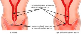
Ultrasound examination (echography). Ultrasound is one of the main ways to detect pathology, as it allows you to study the exact structure of the basal (lower) layer of the endometrium. Sonography is performed transvaginally a few days before and after menstruation. A probe with an emitter and receiver of sound waves is inserted through the vagina into the uterus and illuminates its mucous membrane. Passing through tissues of different densities, sound propagates unevenly, which makes it possible to form a visual image of the structure of the endometrium and the underlying muscle layer based on the received signal. The following echo signs of first degree adenomyosis are distinguished:
- Microchannels extending from the endometrium into the muscular layer of the uterus;
- Defects of the basal layer of the mucosa - unevenness of its thickness, notches, round or oval formations.
Take the first step
make an appointment with a doctor!
These changes in the endometrium persist throughout the development of the disease. Subsequently, they are supplemented by such echo signs of second-degree adenomyosis as thickening of the mucous membrane. Although this symptom may not appear at this stage in all women. In general, ultrasound is a reliable way to diagnose the disease, with an accuracy of 88-96%. With its help, you can establish not only the presence of pathology, but also its form:
- Nodular adenomyosis on ultrasound looks like smooth and round dark formations in the muscular wall of the uterus;
- The focal form of the pathology is characterized by the presence of cavities with uneven edges in the uterine myometrium.
However, despite the high effectiveness of ultrasound, incorrect interpretation of study data is possible. It also provides very limited information if the patient has a diffuse form of adenomyosis, since microscopic inclusions of the endometrium in the muscle layer are poorly visible on echography. Also, ultrasound examination provides unreliable data when endometriosis is combined with myomatosis, in which multiple foci are formed. To make the diagnosis more accurate, ultrasound for uterine adenomyosis is combined with other medical research methods.
CT scan. This method is used mainly as an addition to ultrasound and consists of “transilluminating” tissues with X-ray radiation, followed by computer image processing. To improve the accuracy of the image, contrast agents are injected into the area under study. CT allows you to establish the following signs of uterine adenomyosis:
- The focal form is characterized by the appearance of many small foci of the endometrium in the muscular layer of the uterus, which makes its structure heterogeneous and similar to a honeycomb;
- Signs of diffuse endometriosis of the uterus on CT are unclear contours and increased thickness of the uterine walls;
- the nodular type of pathology is manifested in an increase in the size of the uterus and the presence of foci with clear boundaries in its walls.
Magnetic resonance imaging. Diagnosing adenomyosis using MRI is an alternative to ultrasound and provides the same high diagnostic accuracy (95%). This method is based on measuring the response of hydrogen atomic nuclei or other elements in tissue cells in response to their excitation by electromagnetic waves of a certain frequency.
Magnetic resonance imaging allows you to study in detail the structure of the transition of the endometrium into the myometrium, the number and location of blood vessels, endometrioid foci in the thickness of the muscle wall, etc. Using MRI, you can also track fluctuations in the thickness of the walls of the uterine mucosa, the presence of “tunnels” penetrating the muscles organs when mucosal cells spread into them. This method is especially effective in diagnosing focal and nodular forms of adenomyosis.
The disadvantage of magnetic resonance imaging is its high cost and inaccessibility, which does not allow it to be widely used for diagnosis - only for its clarification, localization of disease foci and formation of an accurate tissue structure, which allows avoiding injury to healthy areas during surgical treatment of pathology.
Hysteroscopy. Another common way to diagnose uterine adenomyosis is to visually examine its cavity using a hysteroscope, which is inserted into the organ through the vagina and cervical canal. This method allows you to study the structure of the surface layer of the endometrium, identify irregularities and other defects in it that indicate a possible pathology.
As a rule, hysteroscopy does not provide comprehensive information, so it is performed in combination with ultrasound, CT or MRI of the pelvic organs. The undoubted advantage of this research method is its simplicity and accessibility, due to which it is often used to make a primary diagnosis.
Laparoscopy. This research method is rarely used to diagnose adenomyosis. It is mainly used in severe stages of the disease, not for the purpose of making a diagnosis, but to clarify the extent of spread of the pathology to other organs. To do this, a small puncture is made in the patient’s abdominal wall, through which a thin flexible device with an optical system or digital mini-camera is inserted, transmitting the image to the monitor.
Laparoscopy allows you to see foci of abnormally enlarged endometrium on other organs and tissues of the abdominal cavity - the peritoneum itself, liver, intestines, bladder, etc. This method can also be used when performing operations to remove endometriotic nodes.
Lab tests. Blood and urine tests are used to indirectly confirm the diagnosis. In particular, adenomyosis may be indicated by an increased level of estrogen in the body, the presence of blood in the urine (if the bladder is damaged) and other signs. As a rule, laboratory tests are a standard procedure when any gynecological disease is suspected.