Tinea versicolor can be transmitted through personal hygiene products, contacts and clothing. At the same time, the most favorable time for the development of lichen will be the summer period, when high temperatures and air humidity are combined. The peculiarity of the course of the disease, which causes the formation of colored spots on the skin, is a wave-like nature, which subsides in the cool season and intensifies at elevated ambient temperatures.
Tinea versicolor most often affects people with reduced immunity, increased sweating, disruption of the physiological processes of the skin and a genetic predisposition to the development of skin diseases. In this case, lichen affects people who have been diagnosed with tuberculosis, diabetes mellitus, metabolic disorders or vegetative neurosis. The development of lichen can be provoked by excessive use of antibacterial hygiene products, excess weight, and long-term use of medications.
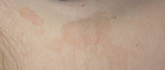
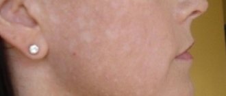
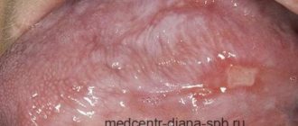
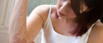
Only a dermatologist can make a correct diagnosis, since the symptoms of pityriasis versicolor are similar to other skin diseases: vitiligo, pityriasis alba and rosea, psoriasis, dermatophytosis, seborrheic dermatitis and others. The main sign of the appearance of tinea versicolor is the presence on the body of colored spots from yellow to brown, which have uneven edges and are not symmetrically located. In this case, colored spots are localized in the back, chest, shoulders, abdomen, and can sometimes be found on the neck, face and scalp.
1.What is pityriasis versicolor?
Pityriasis versicolor
Also called “lichen versicolor” or “sun fungus”. This type of fungal disease is not as common in our climate as in countries with hot, humid climates. However, dermatologists in Russia detect pityriasis versicolor in 5% of the population.
Especially often, the fungus colonizes on the body of people prone to excessive sweating and seborrheic diathesis, which creates a favorable environment for the introduction and development of this skin infection. Pityriasis versicolor lasts for years without treatment, practically without affecting the general condition of a person, but creating psychological discomfort due to cosmetic defects of the skin. Manifestations of the disease are reduced in winter and worsen in the hot season.
A must read! Help with treatment and hospitalization!
Coeval with Hippocrates
The name “lichen” (Lichen) has been known since the time of Hippocrates; it includes many skin diseases, which are characterized by the formation of colored spots and peeling. The name “lichen” itself is very arbitrary, since, for example, herpes is popularly called herpes zoster, psoriasis - scaly, etc., but these skin diseases can only be classified as lichen. Different types of lichen are caused by a wide variety of reasons - fungus, virus, but in many cases, reduced immunity is one of the main provoking factors. In this article we will talk about multicolored or, as it is also called, pityriasis versicolor. The causative agent of pityriasis versicolor was described by G. Robin in 1853, and in 1951 M. Gordon identified round and oval forms of the pathogen both in places of pityriasis versicolor rashes and within healthy skin, classifying it as a yeast-like fungus, and proposed its rounded version call Pityrosporum orbiculare.
2. Causes of the disease
Pityriasis versicolor is caused by the fungi Malassezia furfur and Pityrpsporum orbiculare.
Their habitat is the stratum corneum of the epidermis.
The causative agent of pityriasis versicolor is conditionally pathogenic, that is, it is transmitted from person to person, but does not cause the disease itself in everyone. The likelihood of infection and the onset of the disease increases by a number of factors - external and internal:
- increased sweating and oily skin;
- vegetative-vascular dystonia;
- high blood sugar;
- treatment with corticosteroids;
- decreased immunity;
- hormonal disorders;
- chronic diseases;
- solar radiation;
- hygiene violations;
- stress and metabolic disorders.
It has been noted that the disease affects mainly people of the middle age group. Children under 7 years of age practically do not get pityriasis versicolor.
Visit our Dermatology page
Publications
Versicolor (pityriasis versicolor) (Tinea versicolor) is a mycotic skin infection characterized by widespread distribution and a chronic relapsing course. The disease affects 10% of the population. In hot countries, lichen versicolor is more common.
In mid-latitudes, most cases of the disease occur in the summer. Adults and young people get sick more often; The disease is rare in children and the elderly. Transmission of the pathogen from a patient or carrier: for example, through a shared bed or clothing (underwear), is possible, however, in most cases, the source of infection is endogenous. Therefore, lichen versicolor is not considered a contagious disease. Some authors consider iatrogenic immunodeficiencies, pregnancy, and hormonal contraception to be predisposing conditions. A hereditary predisposition to the disease cannot be ruled out.
In the International Classification of Diseases (ICD-10), lichen versicolor is included in the section “Other superficial mycoses” (B36.0) and has code B36.1. Its causative agents are opportunistic imperfect yeast lipophilic fungi. In recent years, the causative agents of pityriasis versicolor have been assigned to the genus Malassezia [2,4]. Currently, the genus Malassezia has 9 described species of fungi, among which the main causative agents of pityriasis versicolor are M. globosa, M. sympodialis, M. sloolliae |6, 7|. Fungi of the genus Malassezia are imperfect yeast fungi, basidiomycetes. A common characteristic of most Malassezia species is lipophilicity: (requirement of a source of lipids for growth). Malassezia are distinguished by polymorphism, consisting in different cell shapes (round or oval) in representatives of the same genus and even species, and the ability to form mycelial forms.
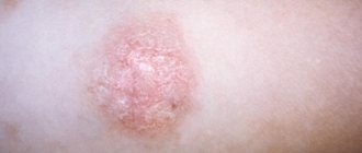
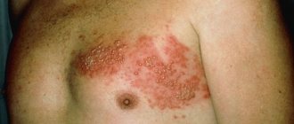
The disease begins with the appearance of small spots with a clear edge, or papules slightly raised above the surface of the skin. Small spots are often located around the hair follicles. The color of the spots ranges from yellowish-pink to brown in different shades. The spots often merge, forming large lesions with scalloped edges. On the surface of the spots you can notice a gentle “pityriasis-like” peeling. Peeling intensifies with scraping (symptom of shavings, or “nail strike” of Beignets). The color of the spots differs from the color of the surrounding skin: in people with fair skin, the lesions look darker, in people with dark or tanned skin - lighter, depigmented (pseudo-leukoderma, pityriasis versicolor alba). The same patient may have both hyperpigmented and hypopigmented macules. The color of the spots is mainly determined by exposure to ultraviolet radiation: unlike the color of the surrounding skin, it does not change after tanning. In rare cases, inflammatory lesions with erythema and slight infiltration occur. Typical localization of lesions in lichen versicolor is areas of the body rich in sebaceous glands: chest, back, neck, shoulders. Less commonly, the axillary, groin, forearms and lower legs are involved. In tropical climates, lesions on the face, abdomen, and scalp are more common. The inverse form (tinea versicolor inversa) is the lesion of large, usually inguinal, folds. Hair and nails are not affected. Changes in the skin, as a rule, are not accompanied by subjective sensations. The disease is characterized by a long-term relapsing course.
The variety of clinical manifestations makes timely diagnosis of mycosis difficult [6]. A long-term recurrent course, significant contamination of the skin and scalp with pathogens, and their penetration into the pilosebaceous follicles cause certain difficulties in the treatment of patients with lichen versicolor [1]. Until recently, keratolytic agents, which have low antifungal activity, are inconvenient to use and do not reduce the number of disease relapses, have been widely used in treatment.
N.V. Kungurov et al. developed an algorithm for the management of patients with lichen versicolor. When detected in patients on the skin in typical localizations (areas rich in sebaceous glands), spots of a round or oval shape, non-inflammatory in nature, not rising above the surface of the skin, with clear boundaries, scalloped edges, of various colors (from yellowish-pink, light brown to brown) and gentle pityriasis-like peeling on the surface for the purpose of diagnosis, a Balser test is performed (when the lesions are smeared with alcohol solutions of iodine or aniline dyes, the affected skin is colored much brighter than healthy), and the symptom of “chips” is revealed - with careful scraping of the spot, a barely noticeable tender pityriasis peeling intensifies, which is due to loosening of the stratum corneum of the epidermis.
The skin of the torso and extremities is examined under a Wood's lamp in order to identify non-visual, atypical forms of the disease. Lesions occupying up to 18% of the area are limited (localized) forms, more than 18% are widespread. The erythematous-squamous form (the most typical) is recorded in 98.5% of cases and is characterized by the clinical manifestations described above. The follicular variant, found in 7% of patients, is characterized by the appearance of perifollicular papules or pustules of a yellowish color, located on an erythematous background, while in the middle of the spots, point-shaped follicular openings are clearly visible. In the inverted variant (3.5%) of the clinical course, the lesions are localized in the inguinal-femoral folds, in the pubic area, on the thighs and legs. Less commonly, rashes of pityriasis versicolor rise above the skin and to the touch create the impression of nodules the size of a lentil or smaller, located in groups (pseudo-papular form). Certain difficulties in making a preliminary clinical diagnosis arise in the presence of hypopigmented lesions on the skin that appear in place of typical spots of pityriasis versicolor, usually after irradiation with ultraviolet rays or insolation. Differential diagnosis is carried out with diseases such as Zhiber's pityriasis rosea, syphilis (primary and secondary), seborrheic dermatitis, chloasma, vitiligo, as well as other skin dyspigmentations.
After making a preliminary clinical diagnosis of lichen versicolor, taking into account the course and extent of the process, it is confirmed in the laboratory. During microscopic examination, scales of the stratum corneum of the epidermis of the skin, obtained by scraping with a scalpel from the lesions, are treated with a 20% solution of potassium hydroxide and, after 30 minutes of exposure, are subjected to microscopic examination. The preparation shows short, slightly curved, septate, thick filaments of pseudomycelium and round spores with a double-circuited shell, located in clusters and singly.
To realize the pathogenic properties of the fungus, its transformation from a non-pathogenic form of blastospore into a pathogenic mycelial, certain conditions are necessary, therefore it is necessary to establish factors contributing to the occurrence and spread of fungal infection, analyzing anamnestic data, prescribing consultations and examinations with specialists. Pathology of the gastrointestinal tract, dysfunction of the endocrine system, diseases and functional disorders of the cardiovascular system, seborrheic processes, foci of acute and chronic infection - these conditions directly or indirectly contribute to hyperhidrosis, which results in changes in the physicochemical properties of sweat and sebum (in side of increasing the alkaline reaction), reducing the rate of physiological peeling of the stratum corneum or its loosening, which contributes to the spread of fungal colonies [3].
The choice of treatment tactics depends on the duration, prevalence and form of the fungal process, and identified concomitant diseases.
Most cases of lichen versicolor can be treated with external remedies. Patients with a limited form of lichen versicolor (the fungal process occupies less than 18% of the body skin area), whose disease duration is no more than 2 years, who have not previously been treated, as well as patients with widespread keratomycosis (affected area more than 18%), who have absolute contraindications for use systemic antimycotics, one of the external antimycotic drugs is recommended. The use of external forms that allow covering a significant surface of the body is of considerable importance in the treatment of lichen versicolor. Even if the lesion occurs in the form of separate small lesions, it is recommended to treat all areas where lichen versicolor usually develops. The shampoo is used once a day for 5 days.
Other imidazole antimycotics in the form of creams ( Fungazol "Farmakar") are used 2 times a day for at least 2 weeks. The use of modern antifungal aerosols is promising.
Lichen versicolor is characterized by relapses after treatment. As a rule, a year after treatment, a relapse is observed in every second patient, and within two years - in almost all. To reduce the likelihood of relapses, longer treatment and treatment of larger surfaces are recommended. For frequent relapses, systemic antimycotics, azole derivatives, are prescribed. Systemic antimycotics are prescribed to the following groups of patients: with the prevalence of the fungal process exceeding 18% of the skin area; with limited forms (less than 18% of the skin area), with a disease duration of more than 2 years, with relapses after treatment; with follicular and pseudopapular clinical variants of the course of keratomycosis, regardless of the area of the lesion; with the lightning-fast form of the fungal process.
Itraconazole ( Mikotrox "Farmakar") is an antimycotic with the widest spectrum of action, also effective in the treatment of lichen versicolor. It is prescribed at 200 mg/day for 1 week. Fluconazole ( Diflox "Farmakar") is prescribed once at a dose of 400 mg/day. These same drugs, with single or short-term use, can be used to prevent relapses.
We observed 24 patients with a common form of lichen versicolor, who were repeatedly treated with external antifungal drugs. To treat these patients, we chose the drug itraconazole ( Mikotrox ), which was prescribed at a dose of 100 mg 2 times a day for 1 week. All patients tolerated the therapy well, there were no side effects or cases of negative attitude of patients towards treatment. The criteria for achieving complete clinical and laboratory recovery are the absence of skin rashes, negative specific clinical tests, and the absence of fungal mycelium on microscopy.
Thus, despite the fact that lichen versicolor is a very common disease, it often causes difficulties in diagnosis and treatment, especially in recurrent forms. The use of the modern systemic antimycotic Mikotrox helps solve this problem and improve the quality of life of patients.
LITERATURE:
- Bragina E. E., Novoselov A. Yu., Stepanova Zh. V. // Esthet. honey. - 2002. - T. I, No. 5. - P. 447-453.
- Dmitriev G. A., Grammatikova N. E., Bibikova M. V. et al. // Vestn. dermatol. - 2002. - No. 2. - P. 7-9.
- Kungurov N.V., Skurikhina M.E., Budumyan T.M. etc. //Russian Journal of Dermatovenerol. - 2004. - No. 4. – P.49-51.
- Novoselov A. Yu., Bragina E. E., Stepanova Zh. V. // Immunopathol., allergol., infectol. - 2000. - No. 4. - P. 95-98.
- Potekaev II. P. // Ross. Journal dermatovenerol. - 2001. - No. 3. - P. 9-10.
- Sergeev L. Yu., Sergeev Yu. V. Fungal infections: A guide for doctors. - M., 2003. - 440 p.
- Crespo E, Ojeda M, Vera. A. et al. // JEADV. — 2000. -Vol. 14. - R. 47.
E.A. Levonchuk
Medical news, 2007 No. 13
3. Signs and diagnosis of the disease
, asymmetrical spots up to 5 cm in size appear
, which merge over time, forming quite large foci. The edges are usually jagged and the color is yellow, pink, brown or white. Sun exposure often changes the color of the spots. Pityriasis-like peeling occurs on the surface, although inflammatory processes are rare. If papules and pustules appear, then most likely this is a consequence of other skin infections superimposing on the affected areas.
Foci of lichen are usually located on the back, neck, abdomen and shoulders, in the groin area.
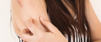
If the head is affected, the hair in these places does not change at all. Sometimes pityriasis versicolor causes itchy skin.
It is used to examine foci of affected skin.
Wood's lamp, microscopic analysis, Balser's iodine test.
About our clinic Chistye Prudy metro station Medintercom page!
How to diagnose lichen
Diagnosis of tinea versicolor consists of conducting a visual examination by a dermatologist and ordering a study of the affected area of the epidermis. When examining the patient, it is necessary to identify: for what reason the lichen occurred, the duration of the process and the degree of damage to the skin by colored spots.
To identify lichen, the following research methods can be used:
- Skin scraping. The study reveals round fungal spores with a double contour.
- Culture method. It is based on placing a sample of cells in a nutrient medium in order to develop colonies of the fungus lichen cauliflower.
- Balser's test (iodine test). The affected area of skin is covered with iodine solution (or 1-2% aniline solution) to change the color of the epithelium. At the same time, the cells that have been affected by tinea versicolor acquire a loose structure and are painted in a rich dark color, while healthy cells are painted in a light shade. After the examination, the skin is cleansed with an alcohol solution.
- Using a Wood's lamp. Examination of skin areas under an ultraviolet lamp. In this case, the areas that are affected by lichen give off a greenish-blue or red-yellow glow.
- Use of mercury-quartz lamps. The study is used to identify areas of skin that are affected by the fungus, but have no visual manifestation. Under the light of a lamp, these areas acquire noticeable color pigmentation.
4. Treatment of pityriasis versicolor
Treatment of pityriasis versicolor should be carried out in a course. Unsystematic measures only reduce the likelihood of a complete cure. Antimycotics, salicylic alcohol, products with selenium sulfide, and imidazole derivatives have an effective effect on the fungus
rumicosis and terbizil
tablets are prescribed The course of continuous treatment must be
at least 14 days.
In addition to external influences on the fungus - ointments, lotions and sprays - the effectiveness of treatment can increase significantly if the patient himself takes a number of measures:
- exposure to ultraviolet light on the skin;
- wet cleaning and disinfection of living space;
- washing clothes at 90-95 degrees;
- ironing towels and bed linen;
- replacing the washcloth with a new one, updating detergents;
- wearing underwear only made from natural fabrics;
- proper nutrition, adequate sleep, immunostimulating procedures.
During the treatment period and for further prevention of relapse of the disease, folk remedies
, aimed at reducing sweating, cleansing the skin of excess fat, antiseptic measures. Skin fungus does not do well in an acidic environment, so lotions with vinegar, lemon juice and sour berries are suitable.
Independence, which can get in the way
As mentioned above, there are many types of lichen, so you should not try to treat strange spots or rashes that suddenly appear on you. Since the symptoms of some types of lichen are similar to the symptoms of other diseases, consult a dermatologist so that in the process of self-medication you do not trigger the true disease and harm yourself even more. Each type of lichen has its own specific treatment. Some can be cured with special antiviral or antifungal ointments, others require a course of immune restoration, and some even go away on their own. Therefore, if you have lichen, follow only the advice of a dermatologist, and do not try to treat yourself.
Using effective strokes we “kill” the fungus
If you choose among the many antifungal drugs widely represented on the modern Russian market, then preference should be given to those that, firstly, are able to accumulate in those layers of the skin where the fungal process develops, and secondly, do not penetrate into those layers where fungal activity is impossible. Usually, treatment of lichen versicolor is carried out with local drugs, and in severe cases with the use of systemic antimycotics, which can significantly reduce the treatment time and prevent the frequency of relapses. But you should always remember that if you find signs of a disease in yourself, under no circumstances try to treat yourself, but immediately go to a doctor who will accurately determine your diagnosis and prescribe the right treatment for you.
What is erythrasma
Erythrasma is a chronic bacterial disease affecting the epidermis layer in the deep folds of the skin. It is characterized by a long course - in some cases, symptoms develop for at least 10 years, without causing significant discomfort to the patient. The clinical picture of erythrasma is similar to a fungal infection of the skin, but modern dermatology classifies it as a group of pseudomycoses.
The following main stages are distinguished in the development of the disease:
- Progression. The first characteristic spots appear on the skin, their size slowly increases, and additional symptoms develop. In some cases, secondary infections occur. The spots gradually merge with each other, forming large areas of damage.
- Stabilization. New spots do not appear, and existing ones stop growing. Peeling of the skin begins. This stage is usually associated with a change in external conditions, for example, cold weather, during which the intensity of sweating decreases and the skin condition stabilizes.
- Exacerbation or relapse. Usually associated with the beginning of the warm season. But in the case of prolonged erythrasma, the disease constantly develops in waves, and after a slight decline its symptoms again actively appear.
- Remission. Occurs with a favorable microclimate, compliance with preventive measures and proper skin care. The color of the affected areas gradually returns to normal, itching and flaking disappear, and the skin is restored.
Without timely, well-chosen treatment, erythrasma can lead to the development of serious complications.
For example, it can provoke eczema and secondary infection in patients with diabetes or obesity. Also, the course of the disease is aggravated by increased humidity and contamination of the affected areas. As a result, its typical symptoms are complicated by burning, itching and pain.
How does infection occur?
The contagiousness of pityriasis versicolor is believed to be very low. But not all dermatologists-mycologists support this opinion. Pityriasis versicolor can be contracted from a loved one through personal contact or through household contacts through objects used by the infected person, for example, bedding, towels, clothing, washcloths. You can also pick up a fungus in the fitting room of a store. The incubation period for pityriasis versicolor ranges from two weeks to several months.
Make an appointment