What does lichen look like?
As the disease progresses, rashes appear - most often they take the form of small red or brown-purple lumps (on the skin) - a characteristic sign is the shine of the affected areas.
- On the scalp, lichen planus may take the form of small itchy bumps.
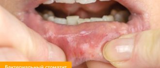
- In the mouth - on the surface of the mucous membrane - it can appear as a reticular change in white color.
- On the nail plate, lichen can form cavities and yellowish grooves, and in extreme cases, lead to atrophy of the nail plate.
Lichen planus is an inflammatory disease. It is worth emphasizing that this is not an infectious disease.
Lichen planus
Lichen planus of the oral mucosa
Lichen planus – medicinal
Lichen planus hypertrophic form
Reasons for appearance
The disease was first identified in 1860, but since then doctors have not been able to determine exactly what the prerequisites for its appearance are.
Today, doctors do not have a common opinion about the origin of lichen. This is partly why there are no therapeutic agents that eliminate the cause of this disease and cure it completely. Scientists can identify only a certain group of factors that contribute to the occurrence of the disease:
- Genetic predisposition (genetically predetermined characteristics of immunity).
- Internal disorders (immune problems, neurological problems).
- External influences (the influence of viruses, allergens, side effects of using certain drugs).
On the surface of the body there are characteristic places that are most susceptible to lichen planus - the joint area of the wrist, forearm, lower leg, sacrum, skin of the penis. Speaking of mucous membranes, plaques appear in the mouth and genitals.
Lichen planus - causes
Although the reasons for the appearance of lichen on the skin or mucous membranes remain a mystery, this disease is often associated with autoimmune diseases. The immunological basis of the disease is indicated by the frequent coexistence of lichens with autoimmune diseases, that is, those in which cells of the immune system attack the body for unknown reasons, destroying “healthy” cells.
Lichen planus may appear in people diagnosed with diabetes, vitiligo, alopecia areata, certain liver diseases (including hepatitis and cirrhosis), and ulcerative enteritis (Crohn's disease). Likewise, lichen planus is more common in people who have had a transplant (especially graft-versus-host disease - GVHD) or are taking certain medications, such as penicillamine, methyldopt.
Lichen planus (LP) is an inflammatory dermatosis with diverse clinical manifestations, involving the skin, its appendages (hair, nails) and mucous membranes. The prevalence of LP is unknown, but according to statistics, patients with LP at an outpatient appointment with a dermatologist in a large clinic account for 1.3-2.4% [1]. The incidence of LP varies depending on the population studied, with a particularly high incidence in India and only 1% in the United States. LLP most often affects middle-aged people, but cases of this disease have also been described in children. Women get sick just as often as men [2]. In more than 50% of cases in patients with the skin form of the disease, resolution of the rash occurs within 6 months; in 85% of cases, regression of efflorescence occurs within 18 months. However, according to an epidemiological study, resolution of the process can last from 1 month to 7 years [2]. Thanks to the use of a number of local and systemic drugs, improvement and regression of the manifestations of LP were observed.
It is currently believed that LP is a specific delayed-type hypersensitivity reaction in response to an unidentified epidermal neoantigen [3].
The initiating moment in the pathogenetic chain of LP is the presentation of an unknown antigen by cytosolic (using the major histocompatibility complex type I - MHC I, located on the surface of keratinocytes of the basal layer) and endosomal (using MHC type II, located on the surface of Langerhans cells of the epidermis) pathways. Both mechanisms are involved in the activation of CD8+ cytotoxic lymphocytes.
In the first case, there is a direct interaction of MHC I with the T-cell receptor of CD8+ lymphocytes, in the second - the interaction of MHC II with the T-cell receptor of CD4+ lymphocytes and their subsequent differentiation into type I T-helpers, which produce a number of cytokines, including IL-2 and interferon-γ. Cytokines interact only with MHC I activated CD8+ lymphocytes, induce their proliferation and are an additional confirmatory factor that triggers cytotoxic mechanisms. The main mechanism of cell death in LP is apoptosis, induced by the interaction of tumor necrosis factor family cytokines produced by CD8+ lymphocytes, such as TNF-alpha and FasL, with the corresponding receptors on the surface of keratinocytes (Fig. 1)
.
Figure 1. Scheme of the pathogenesis of lichen planus [4].
Despite the dominant role of cytotoxic CD8+ lymphocytes in the pathogenesis of LP, CD4+ lymphocytes predominate in the inflammatory infiltrate; CD8+ lymphocytes are located in the epidermis and along the dermis-epidermal junction.
Morphological changes in LP are a consequence of a combination of processes of damage and regeneration of keratinocytes (Fig. 2)
.
Figure 2. Vacuolization of the cytoplasm of cells of the basal layer;
apoptosis of keratinocytes, melanophages in the dermis. Hematoxylin and eosin staining. Uv. 400 (provided by M.A. Bobrov). A fully formed element of a skin rash is characterized by: - in the epidermis
— orthohyperkeratosis, wedge-shaped hypergranulosis in the zone of intraepidermal components of sweat glands and hair follicles (clinically represented by Wickham’s grid), acanthosis in the form of “saw teeth”
(Fig. 3)
, vacuolization of the cytoplasm and apoptosis of cells of the basal layer;
Figure 3. Epidermis with orthohyperkeratosis, hypergranulosis, acanthosis in the form of “saw teeth”.
In the upper parts of the dermis there is a band-like lymphohistiocytic infiltrate. Hematoxylin and eosin staining. Uv. 100 (provided by M.A. Bobrov). - in the upper dermis
- strip-like lymphohistiocytic infiltrate, melanophages, colloidal bodies
(see Fig. 2)
.
Characteristic signs of LLP are polygonal, flat-topped, purple papules, prone to crowded arrangement, grouping with the formation of rings, garlands, lines ( lichen ruber anularis, marginatus, striatus
and etc.).
lichen ruber corymbiformis
appear , which is explained by the “push-like” nature of the appearance of rashes in this dermatosis
(Fig. 4)
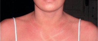
.
Figure 4. Typical shape and location of lichen planus. The elements are located symmetrically on the skin of the flexor surfaces of the wrist joints and forearms, the anterior surface of the legs, torso, genitals, and quite often on the mucous membrane of the oral cavity. Rarely the palms, soles and face are affected. Unilateral skin lesions (zosteriform LP) may also be observed (Fig. 5)
. Figure 5. Zosteriform localization of lichen planus (photo courtesy of M.A. Kochetkov, Ph.D.). The number of rashes varies: from single ones (in the mouth, on the genitals) to multiple ones, covering large areas of the body so that the impression of a complete skin lesion can be created, sometimes like erythroderma. Subjectively, patients are bothered by itching [5].
On the surface of the formed relatively large (0.5 mm in diameter) nodules, one can find the Wickham grid, pathognomonic for the disease, characterized by opalescent white or grayish dots and stripes. Its formation is explained by uneven granulosis. The mesh becomes more visible if the surface of the papules is moistened with water or oil during dermatoscopy [6].
In the progressive stage of the disease, a positive Koebner phenomenon is observed (the appearance of rashes in the area of even minor skin trauma). Patients are bothered by intense itching (Fig. 6)
[7].
Figure 6. Koebner phenomenon in lichen planus.
When the process regresses, secondary hyperpigmentation usually remains in place of the papule. Changes in the nails are often observed, especially when multiple rashes occur. Changes in the nails may precede skin rashes and be the only manifestation of dermatosis. The rash usually lasts a long time, the process can worsen, but over time it regresses, usually without a trace. With a long course, especially with irrational treatment, spinalioma can develop in the foci (mainly in erosive-ulcerative and verrucous forms).
In addition to the described typical form of LP, there are a number of varieties.
Clinical variants of lichen planus
The classification of clinical variants of LP is based on the characteristics of morphology and localization of rashes. Some systems take into account the etiological factor [5].
Anular (ring-shaped) lichen planus (lichen ruber anularis)
(Fig. 7)
is a common variant of damage not only to the skin, but also to the mucous membranes [8].
Figure 7. Anular form of lichen planus (photo courtesy of M.A. Kochetkov, Ph.D.).
The most typical localization of rashes is the skin of the trunk, limbs, genitals (scrotum) and mammary glands [9]. The color of the plaques varies from pink to purple. When their central part is reabsorbed or due to the peripheral growth of individual nodules, elements in the form of rings are formed. After regression of the elements, an area of atrophy is noted in the center [10]. Even after regression of the rash, brownish pigmentation remains, which is also found in subtropical LP, found in the Middle East. Hypertrophic lichen planus (lichen ruber hypertrophicus, s. verrucosus)
(Fig.
also known as verrucous or hyperkeratotic form.
Figure 8. Hypertrophic form of lichen planus.
Mostly, the rashes are localized on the skin of the anterior surface of the legs, rarely on the skin of the upper extremities and torso [11]. Rashes in the form of red, gray-yellow, dark red infiltrated hyperkeratotic papules or plaques are formed as a result of the fusion of individual small plaques and nodules [12]. The rash is hard on palpation. Most rashes are intensely itchy. Individual isolated hyperkeratotic lesions resemble senile keratosis or basal cell carcinoma. It is possible to develop squamous cell carcinoma at the site of long-existing rashes [13, 14]. Atrophic lichen planus (lichen planus atrophicus)
(Fig. 9)
is a rare form localized on the skin of the lower extremities [11].
Figure 9. Atrophic form of lichen planus (photo courtesy of M.A. Kochetkov, Ph.D.).
Clinically, the rashes are presented as round or oval, with a depression in the center, brown or purple papules and plaques [9, 15]. In place of the regressing nodular elements, atrophy develops. Ulcerative lichen planus (lichen ruber ulcerosus)
(syn.: erosive) is most often located on the feet, namely in the interdigital spaces
(Fig. 10)
[16-18].
Figure 10. Ulcerative form of lichen planus.
Only a few cases of localization of this form on the trunk and in the perianal region have been described [9, 19]. Single or multiple rashes are sharply demarcated, with a bizarre configuration, forming small ulcers and erosions with a raised edge or mesh areas of leukoplakia. As a rule, there is a pain syndrome that seriously impairs walking to the point of impossibility. They are often preceded by bullous rashes. Dystrophic changes in the nails are expressed, up to their rejection; paronychia, which, like bullous rashes, may be the initial manifestation of the disease. Bullous lichen planus (lichen ruber planus bullosus)
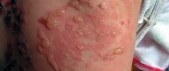
(Fig. 11)
is characterized by typical blisters.
Figure 11. Bullous form of lichen planus.
The skin of the lower extremities is affected in the form of numerous small, sometimes large grouped blisters with a tense tire; some blisters are cellular and contain serous or serous-yellow fluid [20-22]. In lichen planus pemphigoides
(Fig. 12),
the clinical manifestations, histopathology and immunofluorescence are similar to those of bullous pemphigoid [23].
Figure 12. Pemphigoid form of lichen planus.
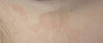
Characteristic of this form is the development of vesicles and blisters not only on existing rashes, but also on unaltered skin [24]. In the latter case, the possibility of developing bullous pemphigoid cannot be excluded. Along the periphery of the rash there are tense flat multi-chamber blisters. The rash predominates on the extremities, most rarely on the torso; located in groups or disseminated [25]. This form of LP has not yet been classified pathophysiologically. When the elements are localized on the scalp, cicatricial alopecia may develop. In some cases, this form is a manifestation of toxicoderma or paraneoplasia. Atrophic changes such as anetoderma and hyperpigmentation may remain on the skin. Pigmented spotted or papular elements are characteristic of pigmented lichen planus (lichen planus pigmentosus)
(Fig. 13, 14)
.
Figure 13. Pigmented form of lichen planus (photo courtesy of M.A. Kochetkov, Ph.D.).
Figure 14. Pigmented form of lichen planus.
In addition to the widespread perifolicular linear or encircling nature of the location, rashes can be localized along Blaschko's lines, along the vessels of the lower extremities and on the skin of the extensor surfaces of the extremities. Intertriginous areas are also most characteristic [26]. Rashes have also been repeatedly described in the lumbar region. Pigmentation ranges from grey-brown, violet to dark brown. Patients with fair skin are most susceptible to this form of LP [26]. Pigmentation may also appear during the period of regression of typical nodular elements, and sometimes changes similar to poikilodermic ones develop. It is possible that one of the variants of pigmented LP is erythema dyschromicum perstans
. It is also assumed that the formation of foci of pigmentation may precede the appearance of a common papular rash; in some cases, pigmentation on the skin may be combined with typical LP rashes in the mouth.
Erythrodermic variant (erythrodermic lichen planus)
is the rarest [11]. Morphologically, red or purple papules and extensive infiltrated erythematous plaques with or without a tendency to grow are detected. In addition to erythroderma, typical elements of LP, blisters or erosions may be detected. The rash is accompanied by intense itching, which reduces overall well-being.
Inverse lichen planus (lichen planus inversus)
(Fig. 15)
typically affects the skin of intertriginous areas - the armpits, inguinal folds, flexor surfaces of the extremities and the submammary region.
Figure 15. Inverse form of lichen planus.
Clinically, the rashes are localized, erythematous, with blurred boundaries and areas of lichenification. In addition, keratotic (keratinized) papules and erosions with bizarre outlines can be detected. Pigmentation is also typical for this form [27]. Linear form (lichen ruber planus linearis)
most common in children and adolescents. Its clinical manifestations are stripes that can be limited to several centimeters or segmentally, have a girdling character, or follow Blaschko's lines. Individual rashes may look like typical flat papules, but they are itchy; rashes can also be cellular, hyperkeratotic or ring-shaped [28].
Follicular or genital lichen planus (lichen planopilaris)
(Fig. 16)
is localized on the skin of the scalp, armpits, groin, scrotum and flexor surfaces of the extremities.
Figure 16. Follicular form of lichen planus (Little-Lassueur syndrome). Clinically, they are grouped or disseminated, follicular raised or hemispherical erythematous papules with a horny plug [15]. On the surface of individual nodules there are horny spines ( lichen ruber spinulosis
).
If they are localized on the scalp, the process often ends with atrophy and baldness ( lichen ruber follicularis decalvans
), which in combination with alopecia in the armpits and pubic area, spiny horny papules on the trunk and limbs are designated as
Little-Lassuer
. The onset of the disease may be onychodystrophy.
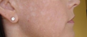
Actinic or subtropical lichen planus (lichen planus subtropicus)
(Fig. 17)
typically appears in autumn or summer on open areas of the skin exposed to sunlight (face, arms, shoulders).
Figure 17. Actinic form of lichen planus.
Children and adolescents with dark skin living in the tropics and subtropics are most susceptible to the disease. Clinical variants of the course are possible. Lichenoid or annular hyperpigmented papules and erythematous infiltrated plaques with varying boundaries and distribution are noted [20]. Cases of a combination of signs of LP and discoid lupus have been described. In this case, the lesions are located on the distal parts of the extremities, have a bluish-red or purple color, with hypo- and hyperpigmentation, telangiectasia, atrophy, and sometimes with warty, bullous-ulcerative changes. In other areas, including on the oral mucosa, typical LP rashes or atrophic lilac-reddish plaques may be detected. Pronounced dystrophic changes in the nails up to anonychia are also observed; Some patients develop cicatricial alopecia. Histological and immunofluorescence (direct reaction) studies also reveal signs of both diseases. One of the reasons for this combination is the commonality of pathogenetic mechanisms. Apparently, for accurate diagnosis, in addition to a complex of histological, serological, clinical, immunofluorescence parameters, the indirect immunofluorescence reaction proposed by R. Olsen et al. may be useful. (1984) to identify the specific antigen of LLP. This antigen was found in 80% of patients with LP and in no patients with lupus erythematosus.
Differential diagnosis.
LLP should be distinguished from papular syphilide, toxicoderma, amyloid and myxedematous lichen, lichenoid and warty tuberculosis of the skin, small nodular form of sarcoid, nodular prurigo, chromomycosis, diffuse and limited neurodermatitis, pityriasis rubra pilaris, callus, granuloma annulare, bowenoid nogo papulosis, sclerotic white lichen , pemphigus, toxic melasma, Darier's disease, psoriasis, lichenoid parapsoriasis, flat juvenile warts, Kaposi's sarcoma, lichen planus, Fox-Fordyce disease, Kirle disease, papular acrodermatitis, malignant atrophying papulosis, vulvar kraurosis, pretibial myxedema.
Differential diagnosis of common forms of LP is usually not difficult.
Lichen planus of mucous membranes
The most common localization of LP is the oral mucosa (Fig. 18)
.
Figure 18. Damage to the oral mucosa in LLP (photo courtesy of A.V. Tereshchenko, Ph.D.). Almost half of the patients have single lesions; multiple lesions are very rare. The topography of the rashes is very diverse, and the morphology of the rashes is even more diverse. There are six morphological types of lesions of the oral mucosa: reticular, plaque-like, atrophic, papular, erosive and bullous (Fig. 19)
.
Figure 19. Damage to the oral mucosa in LLP. Erosive-ulcerative form (photo courtesy of A.V. Tereshchenko, Ph.D.).
Reticular and erosive forms are the most common [15, 20]. The combination of individual morphological forms with a specific localization on the oral mucosa has not been described, with the exception of the plaque form with damage to the apex and lateral part of the tongue.
The erosive or atrophic forms are often associated with burning pain, which can be triggered by eating sour, spicy or hot foods.
In other forms, these symptoms do not appear or are less pronounced. Damage to the oral mucosa is an optional precancerous lesion (according to various sources, 0.4-5.3% of cases). A recent review describes a case of esophageal involvement. Moreover, damage to the esophagus was described without characteristic lesions of the oral mucosa.
The most common location for lesions of the genital organs is the glans penis in the form of ring-shaped rashes (Fig. 20)
.
Figure 20. Lesions of the skin of the glans penis in LP. In women, the vulva is most often affected compared to the vagina (Fig. 21)
.
Figure 21. LLP of the vulva (photo courtesy of M.A. Kochetkov, Ph.D.). The risk of developing squamous cell carcinoma cannot be reliably estimated. Estimates range from "low" with a frequency of up to 2.4%. Penile malignancy is very rare [29].
In 80% of women, a syndrome occurs that combines damage to the mucous membranes of the oral cavity, vulva and vagina, and HLA DQB1*0201
a gene indicating a predisposition to this syndrome. There is a risk of developing scars and strictures. Damage to the mucous membrane of the urethra, nasopharynx and esophagus is also possible [30].
Lichen planus of the nail plates
LLP affects the nail matrix, which can lead to a variety of morphological changes in the nail plate that are not pathognomonic (Table 1)
.
The first sign of isolated nail damage is thinning of the nail plate and longitudinal striations [31].
Characteristic features of LP are dorsal pterygium and trachonychia. Pterygium develops due to the adhesion of the eponychium and matrix, which leads to cracking of the nails and, sometimes, complete loss of the nail plate. Trachonychia is characterized by roughness of the nail plate, loss of transparency and gray color.
Characteristic changes usually develop simultaneously on several nails.
It is not uncommon for all 20 nails to be affected. This may be preceded by rashes on the skin or mucous membranes [31]. With isolated nail lesions, the diagnosis must be confirmed by pathological examination.
Treatment
Phototherapy.
Disseminated rashes with an acute onset and severe itching require effective and timely treatment
(Table 2)
.
Phototherapy can be considered as the therapy of choice.
Thus, UVB (narrowband 311 nm and broadband 280-320 nm) and PUVA therapy (psoralen + UVA 320-400 nm) have proven to be highly effective in the treatment of common forms of LP. The duration of PUVA therapy is on average 10-12 weeks, for narrowband phototherapy - 8-9 weeks. However, LP may recur in approximately half of patients treated with phototherapy. The long-term results of PUVA therapy are comparable to the results of narrow-band phototherapy. In most cases, a combination of phototherapy with topical corticosteroids (prednicarbate or mometasone furoate) for 4 weeks is recommended. It is the treatment of choice for the treatment of common forms of LP.
First-line systemic therapy.
Systemic therapy is considered if the above phototherapy methods have failed. There are a number of drugs with a high evidence base for the treatment of patients with LP, which have their own advantages and disadvantages. Therefore, an individual and personalized approach to the choice of therapy is important.
Systemic retinoids
— Acitretin is prescribed at a dose of 0.5-0.7 mg/kg per day until remission is achieved, then at 0.3-0.5 mg/kg per day as monotherapy or in combination with topical or systemic corticosteroids. This method is most indicated in cases of acute onset of the disease with severe inflammation, intense itching or bullous/erosive-ulcerative rashes.
— Isotretinoin at a dose of 0.3-0.5 mg/kg per day for up to 2 months. Effective in the treatment of LP of the oral mucosa and cutaneous forms, it causes fewer side effects compared to acitretin.
The combination of PUVA therapy with retinoids (re-PUVA) improves efficacy and reduces side effects in severe cases by reducing the cumulative radiation dose. However, there is a high risk of hyperpigmentation, especially in patients with dark skin. The combination of narrowband phototherapy with retinoids may be an alternative to re-PUVA therapy with comparable efficacy and a reduced risk of side effects.
— Methotrexate is prescribed at a dose of 15-20 mg once a week for 10 weeks, while most patients achieve complete remission within a month after starting treatment, and only 5% of patients experience a relapse during the observation period (6 months). Low doses of methotrexate (10 mg subcutaneously once a week) are most effective and safe compared to high doses.
Systemic corticosteroids.
Although systemic corticosteroids have not been thoroughly tested in clinical studies in traditional LP therapy, their undisputed effectiveness has been described in long-term follow-up. Durant corticosteroid injections given at 2-week intervals resulted in complete remission in 61 (83.6%) of 73 patients with the generalized form. An oral corticosteroid (prednisolone) at a dose of 0.5 mg/kg per day was as effective as its injectable form. The course is usually 2-4 weeks.
Hydroxychloroquine (Plaquenil)
prescribed 200 mg or 6.5 mg/kg per day in rounds: 5 days with a break of 2-5 days; up to 5 rounds per course. It is possible to prescribe hydroxychlorine daily at a dose of 200 mg/day for up to 2 months. This is a safe and effective method in the treatment of the generalized form as monotherapy or in combination with other drugs.
Dapsone
Prescribe 50-150 mg per day for 1-2 months. It should be noted that this drug was one of the first systemic agents in the treatment of torpid forms of LP. Many studies indicate the high effectiveness of dapsone, especially in severe cases in children and in the treatment of bullous form of LP. However, side effects such as dose-dependent hemolysis and methemoglobinemia limit the use of this drug in elderly patients (over 65 years of age) and in patients with anemia or heart disease.
Second-line systemic drugs
used for torpid course of the disease. Quite high effectiveness in the treatment of severe generalized forms of LP was recorded with the use of cyclosporine (2.5-5 mg/kg body weight per day for up to 2 months). Their combination with phototherapy is not recommended, as there is a high risk of skin cancer.
Alternative methods used in treatment include:
— azathioprine
(up to 2.5 mg/kg per day) in combination with topical corticosteroids can be used for a long time (up to 6 months);
— mycophenolate mofetil
use 2 g per day, or in small dosages (500 mg/day), combination with plaquenil is possible. Usually the course is 1-2 months;
— pulse therapy with dexamethasone
at a dose of 100 mg 3 days every 4 weeks. There are 4-6 rounds per course.
Other drugs:
- griseofulvin 7.5-10 mg/kg for 3-6 months;
— itraconazole 200 mg/day for 1 week of each month; course - 3 months;
— metronidazole 500 mg/day for 20-60 days, both as monotherapy and in combination with PUVA therapy;
— thalidomide 150 mg/day for 4 months. Due to possible side effects, including irreversible neuropathy, thalidomide should be used with caution.
Lichen planus - symptoms
Symptoms of the disease are well expressed:
- red or purple papules;
- skin lesions are usually shiny;
- formations that tend to stick together and appear in groups;
- whitish lines on the surface of the skin (the so-called Wickham grid);
- itchy skin.
Itchy skin
Symptoms of lichen planus usually appear suddenly - skin lesions during lichen planus are usually limited, the rash does not increase after the first symptoms appear.
Folk remedies
Many examples are given that promise an unprecedented result and tell how to quickly treat lichen ruber in humans. Unfortunately, none of these methods are scientifically proven and often people can only observe a cure as a placebo effect.
“Dummy” and the power of human persuasion sometimes really work psychosomatically and the mechanism of healing by persuasion has not yet been studied. Nevertheless, in medical practice, even if only a little, similar examples still occur.
Try to avoid aggressive treatments such as applying hot objects or products containing acids and other similar substances. This will only make the situation worse, causing injury to the affected areas of the skin.
Popular folk remedies include:
- sea buckthorn oil – has a wound-healing effect;
- birch tar is a remedy actively used in the treatment of psoriasis and other dermatitis;
- laundry soap - its natural composition does not damage the skin.
When using this or that technique, you need to focus on your own feelings. Allergic reactions and individual intolerance can appear at any time.
Types of lichen planus
There are many varieties of lichen planus, including: lichen planus, pigmented, nodular, pemphigoid, linear, follicular, erosive.
The type of lichen can also be determined by the place of its appearance: lichen planus in the mouth, lichen on the nails, lichen of the perianal area, lichen on the genitals: in men, lichen planus is found on the foreskin and on the head, in women, lichen can be found on the genitals lips - and resemble leukoplakia.
Complications and consequences
In most cases, the disease does not pose a life-threatening danger. The patient may experience discomfort from severe itching, which will increase without medical attention. This can significantly affect the human psyche, and also cause some problems in communicating with society.
In addition, recent studies suggest that it is extremely rare (in 1 case out of 100) that seemingly harmless rashes of pityriasis rubra may degenerate into malignant neoplasms. This once again confirms the need to see doctors to receive quality medical care.
Treatment of lichen planus
In case of lesions characteristic of lichen planus, you should consult a dermatologist. Differentiation of lichen planus is based on the exclusion of diseases with a similar course, for example, psoriasis, scabies. In some cases, histopathological examination is required to make a more accurate diagnosis.
Treatment of lichen planus
Lichen planus is a condition that usually goes away on its own – it is estimated that in 90% of people the lichen planus will disappear within 2 years. The main treatment for this disease is to relieve its symptoms.
The dermatologist prescribes medications:
- glucocorticoid ointments - the purpose of using this type of ointment for lichens is to reduce exanthema and itching;
- glucocorticosteroids, corticosteroids, cytostatics - used in the general treatment of lichens;
- cyclosporine, a powerful immunosuppressant used in treatment-resistant lichens;
- retinoids - derivatives of vitamin A, including tretinoin, isotretinoin, are used mainly in the case of lichen, which affects the mucous membranes.
Pharmacological treatment of lichens can be supplemented with photochemotherapy, which involves taking drugs that increase the sensitivity of the skin to radiation, and then irradiating the skin formations with long-term irradiation.
ONLINE REGISTRATION at the DIANA clinic
You can sign up by calling the toll-free phone number 8-800-707-15-60 or filling out the contact form. In this case, we will contact you ourselves.
If you find an error, please select a piece of text and press Ctrl+Enter
Diagnostics
Diagnostic procedures to determine the disease are carried out by dermatologists. To do this, they evaluate the patient's symptoms and compare them with those of lichen. If it is difficult to make a specific diagnosis, a histological examination is prescribed. In addition, symptoms of similar dermatoses, in particular psoriasis, syphilis, and other pathologies, are excluded.
When the skin is covered with a characteristic rash, this symptom is sufficient to make a diagnosis. If the patient has some kind of atypical or rare form of the disease, it will be much more difficult to differentiate this type of lichen from others. The problem is also that conventional laboratory tests do not simplify the task, since with this lichen there are practically no deviations in blood counts. And only occasionally eosinophilia, leukocytosis, and a slightly increased ESR are observed.
In some cases, a biopsy is performed to determine the disease. The method involves examining skin tissue for hyperkeratosis, the presence of inflammation, hypergranulosis, hydropic degeneration of the basal part of the epidermis, stripe-like infiltration of the upper part of the dermis, and the presence of colloidal Sevatt bodies between the epidermis and dermis.