General information
Due to the high pace of life, working with equipment, driving vehicles, constant migration and active travel of people, the risk of injuries, including severe ones, increases.
In case of severe injuries and polytrauma, victims develop a traumatic disease - this is a set of changes and reactions that occur in the body from the moment of injury until its outcome. Traumatic illness occurs in several periods. The first of these is traumatic shock (ICD-10 code T79.4). This is a severe, critical condition, characterized by severe disturbances in vital functions, especially blood circulation - a critical decrease in blood flow in the tissues develops. Pulmonary and tissue gas exchange and metabolism in tissues are also disrupted, and endotoxicosis (poisoning of the body with tissue decay products) increases.
Hemodynamic disturbances arise due to hypovolemia (decreased circulating blood volume) that develops against the background of blood loss. In patients with severe shock, the volume of the liquid part of the blood also decreases due to its transfer into tissues (interstitial edema). As a result, the blood thickens sharply, the pressure decreases, and under these conditions the function of the heart as a pump decreases almost twice (small output syndrome). Thus, in shock, hypocirculation syndrome with impaired tissue perfusion in response to mechanical damage comes first. A type of traumatic shock is burn shock . They have much in common in pathogenesis and basic approaches to treatment.
The period of acute shock is followed by a period of multiple organ failure lasting 3-7 days. This is followed by a period of infectious complications (up to 1 month) and a period of delayed convalescence. Thus, disturbances in important body functions caused by injury are long-lasting. If in case of shock all measures are aimed at saving life (eliminating the causes of shock, correcting blood circulation and breathing), then in the future the traumatic disease requires long-term treatment until the outcome.
The processes occurring in later periods are associated with the initial hours of the disease. The relevance of the problem is that the state of shock not only poses a threat to life if assistance is provided incorrectly and untimely, but also has adverse consequences regarding the physical and mental health of a person.
Types of shock
| Hypovolemic | Cardiogenic | Redistributive |
1. Blood loss:
2. Plasma loss:
3. Fluid loss:
| 1. Acute myocardial infarction (true cardiogenic). 2. Heart rhythm disturbances. 3. Pulmonary embolism (obstructive) 4. Cardiac tamponade | 1. Anaphylactic shock. 2. Septic shock (infectious-toxic). 3. Spinal shock (neurogenic) |
Pathogenesis
The pathogenesis of traumatic shock can be represented as follows: a deficiency of circulating blood and plasma due to blood loss and the release of the liquid part of the blood into the tissue, which leads to a decrease in venous return and a decrease in the stroke volume of the heart with the development of changes in vital organs and disruption of their function.
The trigger for any shock is the discrepancy between the volume of circulating blood and the volume of the vascular bed. As a result, the pressure decreases, to which the baroreceptors of the vascular wall react. Impulses from them enter the brain and the sympathoadrenal system is activated: adrenaline and norepinephrine and accumulate in them. Catecholamines , affecting the heart and blood vessels, cause increased frequency and intensification of heart contractions, vascular spasm (skin, kidneys, gastrointestinal tract). Centralization of blood circulation occurs. Catecholamines do not affect the blood vessels of the brain. This mechanism maintains pressure at a sufficient level until the supply of catecholamines is exhausted. These phenomena correspond to the erectile phase of shock.
When catecholamine reserves are depleted, the second phase of shock (torpid) develops. All spasmodic vessels lose their tone (dilate) and the cardiac output decreases. By this time, non-oxidized products have accumulated in the damaged tissues, which enter the bloodstream, which worsens the situation. , multiple organ failure quickly develops .
Doctors' actions
The arriving team of doctors begins to immediately provide medical assistance to the victim. If necessary, resuscitation (cardiac or respiratory) is performed, as well as blood loss replacement using saline and colloid solutions. If required, additional anesthesia and antibacterial treatment of wounds are performed.
Then the victim is carefully transferred to the car and transported to a specialized medical facility. While moving, blood loss replacement and resuscitation efforts continue.
Classification
According to the etiology (reason), traumatic shock occurs:
- As a result of mechanical injuries (fractures, severe injuries, tissue compression).
- As a result of burns.
- From exposure to low temperatures.
- As a result of exposure to electricity.
By time of occurrence:
- Primary - develops immediately after injury (1-2 hours) and is the result of injury.
- Secondary - appears after 5-24 hours as a result of additional trauma.
If we consider the clinical stages of traumatic shock, then two phases are distinguished:
- Erectile phase. In case of injury, impulses entering the central nervous system cause short-term excitation.
- Torpid phase. Short-term excitation is replaced by inhibition and true shock develops with inhibition of all functions. Hemodynamic disturbances prevail - a drop in arterial and venous pressure, a decrease in cardiac output, impaired gas exchange in tissues and metabolic disorders. As a result of circulatory and respiratory hypoxia , all organs suffer.
True shock is classified according to the severity of its manifestations. The following degrees of traumatic shock are distinguished:
- First degree (this is compensated shock). The victim is a little lethargic, the skin is pale, the extremities may be cold, breathing and cardiac activity are increased. Tachycardia up to 100 beats. Systolic pressure 100-90 mmHg. Art.
- Second degree (or subcompensated shock). The victim is slow and does not move. There is pallor and coldness of the skin, as well as a marbled pattern of the skin. Heart rate increases to 110-120, and pressure decreases to 80-75 mm Hg. Art. There is also a decrease in diuresis. The first two degrees have a favorable outcome, since protective-adaptive reactions and assistance prevent the development of hypoxia and deepening of shock.
- Third degree (decompensation). As a result of prolonged spasm of small vessels, hypoxia and cell damage occurs when assistance is delayed. The patient is lethargic, does not respond to external stimuli, and has an earthy-colored skin. Heart rate increases even more (130-140 beats), pressure progressively decreases (60 mm Hg and below), and diastolic pressure is often not determined. The patient develops anuria . This condition is typical for prolonged (hours) traumatic shock. However, properly performed resuscitation measures are often effective and the patient is brought out of shock. However, after removing patients from this state, 70% develop severe complications, the treatment of which is more complex than recovery from shock.
- Fourth degree (terminal, irreversible). Changes in the body reach the point where all measures taken are unsuccessful. Destructive processes in the body continue, deep decompensation of homeostasis and the victim dies.
Burn shock is one of the types of traumatic shock that develops with deep burns occupying more than 15% of the body. It is the first stage of burn disease. Its severity is influenced by the total area of the burn and its depth. The pathogenesis of burn shock is associated with pain from the burn area, which causes overexcitation of the central nervous system, and then hemodynamic and microcirculatory disorders occur. Generalized spasm of the arteries in the periphery in the next 1-2 hours maintains the pressure and functions of the life support organs, but after this the volume of circulating blood decreases due to loss of plasma, which increases over the course of two days. Victims experience increased blood coagulation and thrombosis . Oliguria appears associated with arterial spasm.
Large loss of plasma, blood thickening and the formation of toxic substances in the necrosis zone after a burn leads to disturbances in the acid-base state and electrolyte balance. A peculiar clinical picture is noted: at first the patient is agitated, talkative, fidgety, and inadequately assesses the condition. After this, excitement gives way to inhibition, as the patient develops hypovolemia - its degree depends on the severity of the burn. There are also 4 degrees of severity, which depend on the area of the burns.
- A mild degree develops with superficial or deep burns, the area of which is up to 10% of the surface. Consciousness is preserved, pale skin, muscle tremors, nausea, and sometimes vomiting. Moderate tachycardia , blood pressure within normal limits. The victims can be brought out of shock by the end of the day.
- Moderate severity of shock occurs with superficial burns, occupying 20-40% of the surface. This stage is characterized by excitement turning into inhibition. The patient's consciousness remains intact, the skin is pale and cold. The victim is worried about thirst and nausea, breathing is rapid, blood pressure is reduced, oliguria and kidney function is impaired (residual nitrogen increases, blood and protein appear in the urine). The victims can be brought out of this state within two days.
- Severe degree accompanies superficial burns of 40-60% of the surface. The patient's condition is extremely serious, confusion and lethargy are noted. Patients are pale gray in color and have severe thirst, vomiting, seizures , tachycardia and shortness of breath . Oliguria is noted , and in older patients anuria . The level of residual nitrogen increases to 51-56 mmol/l. Treatment is not always effective.
- An extremely severe degree is observed with burns affecting 60% of the surface. The condition is very serious, there is no consciousness. The victim's temperature is reduced, there is severe shortness of breath, and the pulse is thready. The vomiting of “coffee grounds” is disturbing, intestinal paresis progresses, and metabolic acidosis intensifies. Patients have anuria, blood and protein are detected in the urine, residual blood nitrogen is above 60 mmol/l. Victims in this condition die within the first or second day.
In clinical practice, we encounter not only post-traumatic shock associated with various injuries, but also shock that develops against the background of various diseases, for example, the cardiovascular system. Thus, arrhythmic shock (another name is “cardiogenic”) occurs in 4-5% of patients with large-focal transmural myocardial infarction . Its development is associated with a decrease in cardiac output due to disturbances in heart rate. It occurs in the form of tachysystole (rapid contractions of the heart) or bradysystole (excessive slowdown of contractions).
Tachysystolic shock develops in patients with atrial fibrillation. In this condition, diastole is shortened, therefore the filling of the heart decreases and its minute volume decreases. However, the leading role in the development of shock is given to ventricular tachysystole, in which it develops in the first hours of the disease. The condition of the patients is serious, since the pressure decreases significantly and diuresis decreases. Patients are given adequate pain relief (narcotic analgesics) and antiarrhythmic drugs are administered.
With intractable tachysystole, pulmonary congestion and right ventricular failure develop. The prognosis for tachysystolic shock is unfavorable. Mortality is 40% and its main cause is progressive heart failure. Bradysystolic shock develops with atrioventricular block , junctional rhythm and Frederick's syndrome . The course is severe, and the mortality rate reaches 60%.
Among the complications of acute pancreatitis is pancreatogenic shock , which occurs in 20% of patients. It most often develops with necrotizing pancreatitis and is characterized by unstable hemodynamics. The cause of the development of this type of shock is endotoxemia due to necrotic damage to large volumes of pancreatic tissue. The volume of pancreatic necrosis determines the likelihood of endotoxic shock. The timing of its appearance is different, and therefore there are early (appears in the first week of the disease) and late (develops in the third week) shock.
The main cause of shock of this nature is a decrease in the volume of circulating blood. This can occur due to swelling of the gland tissue, saturation of the retroperitoneal space with fluid, accumulation of hemorrhagic fluid in the abdominal cavity and intestinal loops, as well as due to stagnation of blood in the portal system of the liver. This loss of extracellular fluid leads to hypovolemia and shock.
In early shock, the patient experiences tachycardia (or bradycardia), signs of peritonitis , decreased blood pressure, cold extremities and facial cyanosis . Late shock occurs against the background of generalized sepsis (fever, leukocytosis ), which is accompanied by hemodynamic instability, which, in fact, is shock.
Providing first aid for traumatic shock
In medicine, there is a concept of the “golden hour”, during which it is necessary to provide assistance to the victim. Its timely provision is the key to preserving human life. Therefore, before the ambulance team arrives, it is necessary to take measures to eliminate the causes of traumatic shock.
Algorithm of actions
1. Elimination of blood loss is the first step in providing assistance. Depending on the complexity of the case and the type of bleeding, tamponing, applying a pressure bandage or tourniquet is used.
2. After this, the victim must be helped to get rid of pain by using any painkillers from the analgesic group
- ibuprofen,
- analgin,
- ketorol, etc.
3. Ensuring free breathing. To do this, the wounded person is laid on a flat surface in a comfortable position and the airways are cleared of foreign bodies. If clothing restricts breathing, it should be unbuttoned. If there is no breathing, artificial ventilation is performed.
4. In case of fractures of the limbs, it is necessary to perform primary immobilization (ensuring the immobility of the injured limbs) using available means.
In the absence of these, the arms are wound to the body, and the leg to the leg.
Important! If the spinal column is fractured, it is not recommended to move the victim.
5. It is necessary to calm the injured person and cover him with some warm things to prevent hypothermia.
6. In the absence of abdominal injuries, it is necessary to provide the victim with plenty of fluids (warm tea).
Important! Under no circumstances should you adjust injured limbs yourself unless absolutely necessary to move the wounded person. Without eliminating the bleeding, you cannot apply a splint or remove traumatic objects from the wounds, as this can lead to death.
Symptoms
Symptoms of pain shock differ in the erectile and torpid phases. The first is very short, it occurs immediately after the injury, does not happen to all victims and is characterized by excitation of the sympathoadrenal system . The patient retains consciousness, there is arousal (motor and speech), rapid breathing, satisfactory pulse filling, not accelerated, blood pressure is normal or increased. The victim's behavior resembles a state of alcohol intoxication.
In the torpid phase, signs of traumatic shock include lethargy, mental depression, an indifferent attitude towards everything, and lack of response to pain come to the fore. The temperature is low, the skin is cold, clammy sweat, breathing is rapid, pulse is increased, blood pressure is reduced, urination is impaired, the severity of which depends on the stages of shock.
- In the first degree (considered as a mild degree), the general condition is satisfactory, the patient is conscious, there may be slight lethargy, pulse up to 100 beats, pressure 95-100 mm Hg. Art. The prognosis is favorable. If help is not provided or additional trauma occurs, shock of this degree may progress to the second degree.
- In the second degree, severe lethargy is noted, pressure (90-75 mm Hg) and temperature decrease, pulse 110-120 beats, breathing becomes rapid. Pale skin with a bluish tint. The prognosis for this degree is serious. Recovery from shock is possible, but requires long-term (possibly several days) anti-shock therapy.
- Stage 3 pain shock is severe. In the general serious condition of the victim, a sharp lethargy appears, a decrease in pressure of 70 mm Hg. Art. (below a critical level), increased heart rate, which has very weak filling and tension. Decreased diuresis and anuria. If the causes are not eliminated in a timely manner and assistance is not provided, it enters a terminal state.
- Shock of the 4th degree is a preagonal state. The patient's pressure and pulse are not determined in the radial arteries, but a weak pulsation is still detected in the large arteries, heart sounds are barely audible, and rare shallow breathing is noted. Reflexes are not evoked, anuria. In the agonal state, respiratory disturbances ( Chine-Stokes breathing ) are more pronounced. Clinical death is considered from the moment of cardiac arrest and the last breath. The functions of the central nervous system are completely absent, but metabolic processes in the brain continue for another 5-6 minutes.
Trauma of any location is accompanied by blood loss. There is a relationship between it and the severity of the shock. In the first degree, blood loss is 500 ml, in the second 1,000 ml, and in the third - 1,500 ml or more.
Patients who have suffered severe trauma and stress develop post-traumatic syndrome, which is characterized by changes in the psycho-emotional sphere. The patient develops unmotivated vigilance, he monitors what is happening around him and does not leave him with a feeling of threatening danger. Patients have an explosive reaction when they throw themselves to the ground at unexpected loud noises. It becomes difficult for such patients to establish contacts with others and they experience dullness of emotions. From time to time, aggressiveness and the desire to solve all problems with the help of force arise, getting one’s way.
The most important symptom is uninvited memories. They are the ones who give the right to say that the patient has post-traumatic syndrome . Scenes associated with a trauma, disaster, fire or other event emerge in his memory. Memories arise during sleep, and when awake they appear in cases where the situation is somewhat reminiscent of what happened (this could be a smell, sound or sight). The patient is worried about anxiety, constant worry, a feeling of fear, fear of persecution, constant preoccupation. An important factor in this condition is depression , when a person feels that everything is useless, there is a negative attitude towards life and apathy. It is also possible to develop a tendency to abuse alcohol and drugs. In some patients, post-traumatic syndrome is very pronounced and requires psychiatric correction.
Tests and diagnostics
In the diagnosis of shock, examination, physical examination and instrumental examination methods are of primary importance. The patient is continuously monitored for vital signs to monitor the severity of shock and the effectiveness of treatment.
- Physical examination gives a general idea of the severity of the condition.
- The general condition ranges from moderate to very severe. Impaired consciousness from confusion to coma.
- Characteristic appearance: pale face, cold sticky sweat, cyanosis of the extremities, decreased temperature.
- Frequent weak pulse, low blood pressure, rapid and shallow breathing.
- Damage to internal organs.
- Fractures, crushed tissue.
At the hospital level, examinations, depending on conditions and indications, include:
- General radiography (extremities, skull, pelvis, chest, abdominal cavity).
- Ultrasound of the abdominal and pleural cavity.
- Carrying out laparoscopy .
- Measurement of central venous pressure.
- Conducting thoracoscopy and bronchoscopy .
- Computed tomography.
- MRI.
Causes
Traumatic shock is a consequence of fractures of the skull, chest, pelvic bones or limbs. And also as a result of injuries to the abdominal cavity, which led to large blood losses and severe pain. The appearance of traumatic shock does not depend on the mechanism of injury and can be caused by:
- accidents on railway or road transport;
- violations of safety regulations at work;
- natural or man-made disasters;
- falls from height;
- knife or gunshot wounds;
- thermal and chemical burns;
- frostbite.
Who is at risk?
Most often, those who work in hazardous industries, have problems with the cardiovascular and nervous systems, as well as children and the elderly can suffer traumatic shock.
Diet
The patient’s nutrition depends on his condition and the presence or absence of injuries to the abdominal organs for which surgery was performed. If the victims are in serious condition after burns or in a comatose state due to polytrauma, they are provided with parenteral nutrition, which is included in the complex of treatment measures. Before it is carried out, the victim’s condition must be stabilized and hypoxia eliminated. Therefore, in terminal conditions and shock, only glucose solutions with insulin . After the glucose infusion, an amino acid preparation or protein hydrolysate . Amino acid mixtures: Polyamine , Panamin , Levamin-80 , Levamin-normo , Alvezin , Aminoplasmal , Aminofusin , Moriamin , Trofisan , Okhamin , Friamin . Protein hydrolysates: Casein hydrolysate , Aminofusin , Aminonorm , Aminoplasmal , Aminomel , Aminovenoz , Aminon , Amigen , Izovac .
Then amino acid mixtures or protein hydrolyzate are administered with vitamins, electrolytes and glucose. Fat emulsions are poured together with amino acid mixtures and hydrolysates. They should not be administered with electrolytes, since electrolytes enlarge fat particles, which significantly increases the risk of fat embolism. Fat emulsions include Lipofundin 10% , Intralipid , Lipomul 15% , Lipofundin 15% , Lipifizan 15% . Preparations based on soybean oil are also produced: Lipofundin-S 20% , Emulsan , Venolipid , Infusolipol . The main components of parenteral nutrition are distributed as follows: proteins make up 10-15% of the total daily caloric intake, carbohydrates - 50%, and fats - 35-40%. Vitamins are also an integral component of parenteral nutrition, they are administered separately, and there are also multivitamin complexes for parenteral administration - these are Vitafuzin , Protavit , Soluvit , which contain the daily requirement of essential vitamins.
As the patient's condition improves, the patient is transferred to enteral nutrition with formulas. This is especially indicated if there were abdominal injuries with damage to the gastrointestinal tract or if operations were performed on these organs. Semi-element mixtures are fully balanced with nutrients; proteins are represented by peptides and protein hydrolysates. The most commonly used are Peptamen and Nutrien Elemental - these are specialized medical nutrition products that can be used as the only source of nutrition. Available in powder form, which is diluted with warm boiled water and taken orally.
When switching to a normal diet, the patient must gradually expand the food load, starting with Dietary Table No. 1 with the transition to the standard Diet No. 15 with the physiological content of proteins, fats and carbohydrates, vitamins and minerals.
Particular attention is paid to nutrition in case of burn disease, in which there is an increased breakdown of muscle and visceral proteins. For such patients, the following scheme is used: parenteral nutrition - transition to enteral nutrition - siping (consumption of specialized mixtures in small sips) - transition to specialized diets with a high protein content.
Prevention
The following preventive measures can prevent the development of shock or reduce its severity:
- Timely stopping of bleeding.
- Reliable immobilization for fractures.
- Carrying out analgesia (pain relief).
- Knowledge of the rules for providing self- and mutual assistance and the ability to apply them in the right environment.
- Because serious injuries often occur in the workplace, increased worker vigilance and safety monitoring by management can significantly reduce injuries and related complications.
Consequences and complications
Complications of shock include:
- Increasing respiratory failure ( respiratory distress syndrome ).
- Coagulopathic disorders with possible transition to disseminated intravascular coagulation.
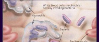
- Traumatic endotoxicosis . Endogenous intoxication develops due to the accumulation of tissue destruction products and is caused by bacterial toxins and vasoactive substances. The effect of these factors manifests itself 48-72 hours after injury.
- Development of fat embolism with bone injuries and extensive destruction of soft tissues.
- Hepatic-renal failure (development of “shock kidney” and “shock liver”).
- Heart failure and central hemodynamic disorders.
- Infectious and septic complications.
- Delay of reparative processes at the site of damage.
The consequence of trauma and shock is post-traumatic syndrome. It develops in people who have experienced life-threatening events (transport accidents, plane crashes, natural disasters) and received injuries as a result. These events deeply affect a person’s psyche, and he loses a sense of security, anxiety, fear appear, sleep disturbances occur, suicidal tendencies and alcohol or drug abuse occur. If the trauma and stress suffered were severe, the painful mental reaction can persist for many years.
Forecast
The prognosis depends on the degree of shock: in the first degree it is favorable, in the second it is doubtful, in the rest it is not favorable. Despite the improvement of anti-shock therapy methods, mortality in severe injuries has decreased slightly. As stated above, shock is a life-threatening condition, but death does not occur from painful shock. The main cause of death from injuries is blood loss in the absence of medical attention. Pain aggravates shock and is not its main cause, much less the cause of death.
Death can also occur after recovery from shock due to multiple organ failure. The immediate cause of death is heart, kidney and liver failure , increasing respiratory failure and coagulopathic disorders.
If death is prevented during the first week, then the threat of generalization of wound infection ( wound sepsis , pneumonia ) is of decisive importance in the patient’s condition. It is infectious complications that occur at a later date that affect the survival of patients. This threat persists for several weeks, as victims of the trauma experience severe metabolic disorders and impaired immunity .
Treatment
Providing first aid for painful shock can save a person’s life. If an accident occurs before your eyes, a person is injured and shows all the signs of painful shock, you must urgently call an ambulance and begin providing first aid to the victim. First of all, it is necessary to warm him up using warm clothes, blankets, heating pads; if there is no vomiting, you can give water and hot tea. However, it is forbidden to give liquid if you are wounded in the stomach. Cold is applied to the damaged area. You cannot remove foreign objects from the victim’s body - such an operation can only be performed by a qualified doctor. The patient can be given analgesics and sedatives to relieve stress.
It should also be taken into account that some time after the pain shock has been eliminated, neuritis may occur. If the victim does not receive first aid, his condition may worsen and even lead to death.
List of sources
- Bagnenko S. F., Lapshin V. N., Shah B. N. Hemodynamic depression in victims with combined trauma in the acute period of traumatic illness is the basis for subsequent hypoxic changes and reperfusion injuries // Efferent Therapy, 2004. Vol. 1, No. 5 . pp. 23–34.
- Shestopalov A. E., Pasko V. G. Volume replacement therapy for acute blood loss in victims with severe combined trauma // Difficult Patient. 2005. No. 4. pp. 17–23.
- Gural K.A., Klyuchevsky V.V., Dambaev G.Ts. Human traumatic shock // International Journal of Experimental Education. – 2011. – No. 12. – P. 15-16.
- Singaevsky, A.B. Current problems of modern severe trauma // Abstracts of the All-Russian Scientific Conference. - St. Petersburg, 2001. - pp. 106-107.
- Sheiko V.D. Surgery of injuries during polytrauma in peacetime and wartime / Textbook. — Poltava ASMI LLC. — 2015. 558 p.