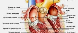
Structure, localization, anatomy of the tricuspid heart valve
The tricuspid heart valve is located in the area of the atrium and ventricle on the right side.
Its structure is represented by the following elements:
| Name | Description |
| Fibrous ring | Consists of a large number of elastic fibers. Attached to the interatrial septum. A large number of cardiac conduction channels pass through the annulus fibrosus. Some part of this element is represented by muscle fibers and has a loose structure. As the annulus fibrosus moves away from the interatrial septum, its thickness gradually becomes smaller. |
| Doors | In most cases, the normal development of the heart valve involves 3 main leaflets (anterior, posterior and septal). Sometimes their number increases, according to experts, due to the bifurcation of the rear valve. They all vary in size. The largest door is the main one. The structure of these elements provides for the presence of a base, a body and a closure section. |
| Chordae tendons | There are 3 main elements, each of which performs its own specific functions. |
| Papillary musculature | The muscle is located on top of the interatrial septum. The chordae tendineae emerge from it. |
Tricuspid and other heart valves
The condition and functioning of the tricuspid valve is important for normal blood circulation. It prevents the backflow of blood into the right atrium.
Causes of acquired heart defects
In most cases, the defects are caused by rheumatic diseases, in particular rheumatic endocarditis (about 75% of cases). The cause may also be the development of atherosclerosis, systemic connective tissue diseases, trauma, sepsis, infections, overload, and autoimmune reactions. These pathological conditions cause disturbances in the structure of the heart valves.
Classification of acquired heart defects
The human heart has four chambers: the left and right atria and ventricles, between which are the heart valves. The aorta emerges from the left ventricle, and the pulmonary artery emerges from the right ventricle.
Between the left heart chambers is a bicuspid valve, the mitral valve. And between the right sections there is a tricuspid valve, another name is tricuspid. In front of the aorta there is the aortic valve, in front of the pulmonary artery there is another valve, the pulmonary valve.
The performance of the heart muscle depends on the functioning of the valves, which, when the heart muscle contracts, allow blood to flow into the next section without obstruction, and when the heart muscle relaxes, do not allow blood to flow back. If the function of the valves is impaired, the function of the heart is also impaired.
Based on the reasons for their formation, defects are classified as follows:
- degenerative, or atherosclerotic, they occur in 5.7% of cases; More often, these processes develop after forty to fifty years, calcium is deposited on the leaflets of the damaged valves, which leads to the progression of the defect;
- rheumatic, formed against the background of rheumatic diseases (in 80% of cases);
- defects that arise as a result of inflammation of the inner lining of the heart (endocarditis);
- syphilitic (in 5% of cases).
Based on the type of functional pathology, defects are divided into the following types:
- simple – valve insufficiency or stenosis;
- combined – insufficiency or narrowing of two or more valves;
- combined - both pathologies on one valve (stenosis and insufficiency).
Depending on the location, the following pathologies are distinguished:
- mitral valve disease;
- tricuspid defect;
- aortic defect.
Hemodynamics can be impaired to varying degrees:
- insignificant;
- moderately;
- expressed.
The mitral valve is affected more often than the aortic valve. Pathologies of the tricuspid valve and pulmonary valve are less common.
Work of the organ
The function of the tricuspid heart valve is to maintain normal blood circulation throughout the human body. Passing through all cells and internal organs, the blood gives off oxygen and receives carbon dioxide back. It is also saturated with decay products, due to which it acquires a dark color.
The work of the tricuspid valve is necessary for the separation of jointly functioning elements (left and right ventricles, atria). Venous blood returns through these sections to the pulmonary circulation. Saturated with oxygen through the left atrium, the ventricle flows into the systemic circulation.
Tricuspid valve insufficiency
Hemodynamics
During right ventricular systole, part of the blood returns to the right atrium, causing its hypertrophy and dilatation. Constant volume overload leads to eccentric hypertrophy, then dilatation of the right ventricle. Congestion develops in the systemic circulation (enlarged liver, ascites, swelling in the legs).
Diagnostics
There are no complaints characteristic of this defect. They are usually caused by the presence of concomitant pathology of the mitral or aortic valve. Patients note weakness, heaviness in the right hypochondrium, increased abdominal volume (in the presence of ascites).
On examination, swelling of the neck veins and their systolic pulsation are noted. Upon palpation, pulsation of the entire region of the heart and the epigastric region is determined. In the area of the right hypochondrium, systolic pulsation of the liver and its increase are detected. Sometimes there is swelling in the legs and ascites.
During auscultation, a systolic murmur is found at the xiphoid process of the sternum, which intensifies with inspiration (Rivero-Carvallo symptom), which is explained by an increase in the volume of regurgitation. The first tone is usually weakened. The magnitude of the second tone on the pulmonary artery usually decreases, which is associated with a decrease in stagnation in the pulmonary circulation.
X-ray examination reveals a significant increase in the right atrium and right ventricle, expansion of the shadow of the superior vena cava. The ECG shows signs of hypertrophy of the right atrium and right ventricle. Incomplete blockade of the right bundle branch may also indicate damage to the tricuspid valve. The presence of atrial fibrillation is characteristic.
Echocardiography
In case of organic damage to the tricuspid valve, sealing of the leaflets is found. With relative insufficiency, the valves are not changed, regurgitation of blood into the right atrium is determined, and an increase in the size of the right ventricle and atrium is noted.
Cardiac probing demonstrates increased pressure in the right side of the heart. The pressure waveform in the right atrium has a characteristic V-wave curve. Pulmonary artery pressure is usually elevated (> 30 mmHg) due to concomitant mitral valve disease.
Indications for surgery
For a long time, surgical correction of tricuspid valve defects was not given serious importance. This was due to the fact that in the vast majority of cases, changes in the tricuspid valve were secondary. With more experience, the situation changed, since the unresolved tricuspid defect did not allow patients to achieve an optimal quality of life after correction of the pathology of the left heart. In case of tricuspid valve stenosis, the effective orifice area is < 1.5 cm2, and in case of insufficiency, regurgitation of blood into the right atrium 2-4 cm above the valve (II-III degree) are an indication for correction. In case of grade I regurgitation, the tricuspid valve need not be corrected.
Operation technique
Correction of the tricuspid valve is carried out after surgery on the mitral and aortic valves. Artificial blood circulation is carried out in a standard mode. The AIK is connected according to the scheme: vena cava – ascending aorta. The vena cava on the cannulas is clamped with tourniquets. Given the need for mitral valve repair or a labyrinth procedure, the left atrium and mitral valve may be accessed through the right atrium and interatrial septum.
Access to the tricuspid and mitral valve
Valve functions
The tricuspid heart valve is located on the right side and is an important element in the functioning of the pulmonary circulation.
It performs the following functions:
- During heart contractions, it prevents blood flow from the lower right ventricle to the atrium.
- Takes direct part in the blood circulation process. The tricuspid valve is responsible for the delivery of venous blood to the vessels located in the bronchi.
- Supports gas exchange in the alveoli of lung tissue and heat transfer.
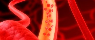
Continuous blood circulation in the human body throughout his life is responsible for the rapid and timely delivery of oxygenated blood to all internal organs and tissues. Through venous outflow, carbon dioxide and decay products leave the body. In medicine, this mechanism is called the cardiovascular system.
The systemic circulation begins from the left ventricle and ends in the right atrium. The pulmonary circulation is directed from the right ventricle to the left atrium.
Tricuspid valve stenosis
Tricuspid valve stenosis is caused by a narrowing of the atrioventricular opening, which impedes the flow of blood from the right atrium to the right ventricle. Overload of the right atrium leads to hypertrophy and stretching of the walls of the right half of the heart and insufficient filling of the right ventricle. In most cases, this heart defect is benign and does not require special therapy, but when it is combined with other cardiac anomalies, which is observed in most cases, or in the presence of a pronounced clinical picture, surgical treatment may be required.
Kinds
Tricuspid stenosis can be:
- congenital: provoked by hereditary diseases;
- acquired: provoked by various pathologies that developed at birth.
Causes
The most common cause of tricuspid valve stenosis is the consequences of rheumatic fever. Much less often it can be provoked by:
- congenital pathologies;
- myxoma of the right atrium;
- systemic lupus erythematosus;
- metastatic tumor;
- infectious pericarditis;
- carcinoid syndrome.
Symptoms
Patients with tricuspid stenosis may feel heaviness and pulsation in the right hypochondrium.
With severe tricuspid valve stenosis, patients experience the following symptoms:
- pronounced pulsation and discomfort in the neck and jugular vein;
- dark complexion of facial skin;
- swelling of the veins of the head;
- peripheral edema;
- fatigue;
- exhaustion;
- skin that is cold to the touch;
- feeling of discomfort and heaviness in the liver area;
- sensation of pulsation in the liver area;
- liver enlargement;
- ascites.
When listening to heart sounds, a soft tone of valve opening is determined. In some cases, a click is heard during diastole. A characteristic sign of tricuspid stenosis is an increasing-decreasing grinding presystolic murmur, which can be heard in the IV-V right intercostal space in the epigastric region or to the right of the sternum with the patient sitting, leaning forward and lying on the right side. When listening to the noise in a standing position, it becomes softer, and after attempting physical exercise, inhalation or Müller's test, it lengthens and becomes more intense. When tapping (percussion) of the heart, a shift of its borders to the right is noted due to an increase in the size of the right sections.
In many patients, symptoms of tricuspid valve stenosis are combined with signs of mitral stenosis (mitral-tricuspid stenosis).
Normal valve performance
The tricuspid heart valve is located between the right ventricle and the atrium. When the heart muscle relaxes, it opens. When the myocardium contracts, the valve closes. The valves close tightly due to the presence of chordae and muscles. During normal operation of the tricuspid valve, venous blood passes from the right side of the heart to the left. It is then sent to the lungs for gas exchange.
The backflow of blood into the heart cavity from large vessels is not observed due to the correct functioning of this valve. With the development of pathological processes, the entire mechanism is disrupted, as evidenced by the emerging clinical symptoms.
Tricuspid valve insufficiency
Tricuspid valve insufficiency (or tricuspid regurgitation) is a heart defect that develops due to the backflow of blood from the right ventricle into the atrium during ventricular systole (contraction) and insufficient closure or perforation of the valve leaflets. Due to constant regurgitation of blood, diastolic volume and pressure in the right atrium increase, which leads to hypertrophy and dilatation of its walls. When compensatory mechanisms are exhausted, the patient develops signs of blood stagnation in the systemic circulation, which manifest themselves as symptoms of heart failure.
Kinds
Tricuspid regurgitation can be:
- congenital: the defect is formed in the prenatal period as a result of hereditary diseases;
- acquired: the defect is formed after birth due to various pathologies;
- primary: provoked by heart pathology and does not cause pulmonary hypertension;
- secondary: provoked by pulmonary hypertension.
Causes
The reasons for the development of primary tricuspid insufficiency can be:
- rheumatic fever;
- infective endocarditis;
- tricuspid valve prolapse;
- Marfan syndrome;
- Ebstein's anomalies;
- right ventricular infarction;
- myxomatous degeneration;
- heart injuries;
- carcinoid syndrome;
- taking certain medications (Ergotamine, Rigetamine, Minifage, Fenfluramine).
Secondary tricuspid insufficiency can be caused by the following pathologies and diseases:
- right ventricular dysfunction;
- pulmonary hypertension;
- mitral stenosis;
- cardiopathy;
- anomalies of the interatrial septum;
- decompensated left ventricular failure.
Symptoms
When examining a patient with tricuspid insufficiency, the doctor identifies pathological noise and arrhythmia during auscultation.
The severity of symptoms depends on the degree of damage to the valve leaflets. During the course of heart disease, the following stages are distinguished:
- I – the reverse flow of blood from the right ventricle into the atrium is minimal;
- II – the reverse jet reaches 20 mm from the valve flaps;
- III – the reverse flow reaches 2 cm from the valve flaps;
- IV – the reverse jet is thrown more than 2 cm from the valve flaps.
At stage I of tricuspid regurgitation, the heart defect does not manifest itself. Sometimes, with significant physical exertion, the patient’s neck veins may pulsate. As the disease progresses, the following symptoms appear:
- pulsation to the left of the sternum, increasing with inspiration;
- pulsation in the right hypochondrium;
- heartbeat;
- dyspnea;
- chilliness of the limbs;
- decreased tolerance to physical activity;
- an increase in the size of the right chambers of the heart;
- swelling in the legs;
- discomfort and pain in the right hypochondrium;
- expansion of the boundaries of the liver;
- yellowness of the skin;
- frequent urge to urinate;
- ascites;
- signs of pulmonary hypertension;
- pulsation in the right atrium (rare).
When listening to heart sounds, the following are determined:
- pansystolic murmur;
- atrial fibrillation;
- increased pulsation of the pulmonary artery during inspiration to the left of the sternum;
- splitting tones;
- protodiastolic or mesodiastolic murmur (in severe forms of the defect).
With the rheumatic nature of tricuspid regurgitation, the patient almost always has symptoms of aortic or mitral heart disease.
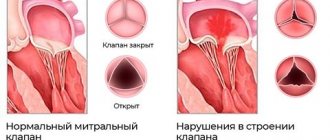
Pathologies of the 3-leaf heart valve, causes and symptoms
In most cases, pathological changes in the functioning of the tricuspid valve provoke heart failure or the development of stenosis. Degenerative processes impair blood circulation, resulting in characteristic clinical signs.
| Name | Description |
| Failure | A disease characterized by insufficient closure of the tricuspid valve. In this situation, the blood returns back to the atrium. |
| Stenosis | The pathological condition is characterized by narrowing of the holes located in the tricuspid heart valve. In most cases, problems arise against the background of rheumatism, congenital defects, or after prolonged mechanical impact on the chest. |
| Congenital defects | Pathological changes occur during the period of intrauterine development of the human body, often against the background of hereditary diseases in the mother’s body. |
Pathological processes in the area of the tricuspid heart valve develop as a result of the following provoking reasons:
- complication of rheumatism;
- severe thyrotoxicosis;
- development of myocarditis;
- infective endocarditis;
- cardiomyopathy;
- carcinoid syndrome;
- mechanical trauma to the chest, resulting in damage to the heart;
- surgical intervention in the area of the mitral valve, due to which its leaflets enlarged;
- myocardial infarction;
- increase in diameter of the fibrous ring.
Congenital defects in most cases develop together with other hereditary abnormalities (patent foramen ovale, atrial septal defect).
Tricuspid valve dysfunction is also provoked by tumor growths that have appeared on various internal organs. In this situation, disruption of the functioning of the cardiovascular system occurs as a result of the toxic effects of malignant processes.
Developing pathological changes, taking into account the main reason for their appearance, provoke the appearance of certain clinical signs. Symptoms will help the cardiologist determine the degree of damage to the cardiovascular system and assess its ability to function.
Tricuspid valve dysfunction is accompanied by the following symptoms:
- the shade of the skin on the face changes;
- shortness of breath appears;
- breathing is impaired;
- the face swells;
- general weakness occurs in the body;
- the patient is bothered by vomiting; the discharge contains blood impurities;
- metabolism in the intestinal area is disrupted;
- a person gets tired quickly;
- Painful sensations appear in the area on the right side under the ribs.
The liver also enlarges, not only the face, but also the lower and upper limbs swell.
Excess fluid accumulates in the lungs. The intensity of the clinical picture and the presence of certain signs depends on the degree of development of pathological processes. It is important, if any symptom appears, to go to the hospital and undergo a full examination to establish an accurate diagnosis. Properly selected therapy will prevent serious consequences.
Symptoms of acquired heart defects
Mitral stenosis
It manifests itself as compaction or fusion of the leaflets, a decrease in the area of the mitral valve opening. As a result, the flow of blood from the left atrium to the left ventricle is obstructed, and the left atrium begins to work with increased load. This leads to enlargement of the left atrium. The left ventricle receives less blood.
Due to the decrease in the area of the mitral orifice, the pressure in the left atrium increases, and then in the pulmonary veins, through which oxygenated blood flows from the lungs to the heart. Typically, the pressure in the pulmonary arteries begins to increase when the diameter of the opening becomes less than 1 cm, compared to the normal 4-6 cm, and a spasm occurs in the arterioles of the lungs, which aggravates the process. Thus, so-called pulmonary hypertension is formed, the long-term existence of which leads to sclerosis of arterioles with their obliteration, which cannot be eliminated even after eliminating the stenosis.
With this defect, first of all, the left atrium hypertrophies and expands, and then the right parts of the heart.
At the beginning of the formation of this defect, the symptoms are little noticeable. Subsequently, shortness of breath, cough during physical activity, and then at rest come first. Hemoptysis, persistent pain in the heart, and rhythm disturbances (tachycardia, atrial fibrillation) may occur. If the process goes far, then pulmonary edema may develop during physical activity.
There are physical signs of mitral stenosis: diastolic murmur in the heart, a trembling of the chest corresponding to this noise is felt (“cat purring”), and the boundaries of the heart change. An experienced specialist can often make a diagnosis after a careful examination of the patient.
Mitral regurgitation
Valve insufficiency is expressed in the ability of blood to return back into the atrium during contraction of the left ventricle, since a message remains between the left atrium and the ventricle, which is not closed by the valve leaflets at the time of contraction. Such insufficiency is caused either by deformation of the valve as a result of a tissue-changing process, or by its sagging (prolapse), due to stretching of the chambers of the heart when they are overloaded.
Compensated mitral regurgitation usually lasts for several years; in the affected heart, the work of the left atrium and left ventricle increases, first hypertrophy of the muscles of these parts develops, and then the cavities begin to expand (dilatation). Then, due to a decrease in stroke volume, the minute ejection of blood from the heart begins to decrease, and the amount of blood returning (regurgitation) to the left atrium increases. Blood stagnation begins in the pulmonary circulation (pulmonary), the pressure in it increases, the load on the right ventricle increases, it hypertrophies and expands. This leads to rapid decompensation of cardiac activity and the development of right ventricular failure.
If compensatory mechanisms do not have time to develop in acute mitral valve insufficiency, the disease can begin with pulmonary edema and lead to death.
Clinical manifestations of mitral regurgitation in the compensated stage are minimal and may not be noticed by the patient. Beginning decompensation is characterized by shortness of breath, poor tolerance to physical activity, then, when stagnation in the pulmonary circulation increases, attacks of cardiac asthma appear. In addition, pain in the heart area, palpitations, and interruptions in heart function may bother you.
Right ventricular heart failure leads to stagnation of blood in the systemic circulation. The liver becomes enlarged, cyanosis of the lips and limbs appears, swelling in the legs, fluid in the abdomen, heart rhythm disturbances (50% of patients have atrial fibrillation).
It is not difficult to make a diagnosis of mitral regurgitation at present with the available instrumental research methods: ECG, ECHO-CG, radiation diagnostic methods, ventriculography and others. However, an examination by an attentive cardiologist based on anamnesis, auscultation, percussion, and palpation will allow you to draw up the correct examination algorithm and timely take measures to prevent further development of the process of formation of the defect.
Aortic stenosis
This defect among PPS is detected quite often, in 80-85% of cases it is formed as a result of rheumatism, in 10-15% of cases it is acquired against the background of an atherosclerotic process, with subsequent deposition of calcium in atherosclerotic plaques (calcinosis). There is a narrowing of the aortic orifice at the site of the semilunar valve of the aorta. For many years, the left ventricle works with increasing tension, however, when the reserves are depleted, the left atrium, the pulmonary circle, and then the right parts of the heart begin to suffer. The pressure gradient between the left ventricle and the aorta increases, which is directly related to the degree of narrowing of the opening. The ejection of blood from the left ventricle becomes less, the blood supply to the heart deteriorates, which is manifested by angina pectoris, low blood pressure and weak pulse, cerebrovascular insufficiency with neurological symptoms, including dizziness, headache, loss of consciousness.
The appearance of complaints in patients begins when the area of the aortic mouth decreases by more than half. When complaints appear, this indicates an advanced process, a high degree of stenosis and a high pressure gradient between the left ventricle and the aorta. In this case, treatment should be discussed taking into account surgical correction of the defect.
Aortic insufficiency
- this is a valve pathology in which the outlet from the aorta is not completely blocked; blood has the ability to flow back into the left ventricle during its relaxation phase. The walls of the ventricle thicken (hypertrophy) as more blood has to be pumped. With ventricular hypertrophy, insufficiency of its nutrition gradually manifests itself. More muscle mass requires more blood flow and oxygen supply. At the same time, due to the fact that part of the blood in diastole returns to the left ventricle, the aortic-left ventricular gradient decreases (it is what determines the coronary blood flow) and, as a result, less blood enters the arteries of the heart. Angina occurs. There are sensations of pulsation in the head and neck. Neurological manifestations are characteristic, such as: lightheadedness, dizziness, sudden fainting, especially during physical exertion, when changing body position. The hemodynamics of the systemic circulation with this defect are characterized by: high systolic pressure, low diastolic pressure, compensatory tachycardia, increased pulsation of the croup
ny arteries, including the aorta. During the decompensation stage, dilatation (expansion) of the left ventricle develops, the efficiency of systole decreases, the pressure in it increases, then in the left atrium and pulmonary circulation. Clinical signs of stagnation in the pulmonary circulation appear: shortness of breath, cardiac asthma.
A thorough examination by a cardiologist may allow the doctor to suspect or even diagnose aortic insufficiency. Well-known symptoms such as “carotid dance” - increased pulsation of the carotid arteries, “capillary pulse”, which is detected by pressing on the nail phalanx, de Musset’s symptom - when the patient’s head sways in time with the phases of the cardiac cycle, pulsation of the pupils and others are detected already at the stage a far advanced process. But palpation, percussion, auscultation and careful history taking will help identify the disease at earlier stages and prevent the progression of the disease.
Tricuspid stenosis
This defect rarely occurs as an isolated pathology. It is expressed in the narrowing of the existing opening between the right ventricle and the right atrium, which are separated by the tricuspid valve. Most often it occurs with rheumatism, infective endocarditis and other systemic connective tissue diseases; sometimes there is a narrowing of the opening as a result of the formation of a myxoma-tumor formed in the right atrium, less often there are other reasons. Gamodynamics are disrupted as a result of the fact that not all the blood from the atrium can enter the right ventricle, which normally should occur after atrial systole. The atrium becomes overloaded, stretched, blood stagnates in the systemic circulation, the liver enlarges, swelling of the lower extremities and fluid appear in the abdominal cavity. Less blood flows from the right ventricle to the lungs, which causes shortness of breath. In the initial stage, there may be no symptoms; these hemodynamic disorders arise later - heart failure, atrial fibrillation, thrombosis, cyanosis of the nails, lips, yellowness of the skin.
Tricuspid insufficiency. This pathology most often accompanies other defects and manifests itself in the form of tricuspid valve insufficiency. Due to venous stagnation, ascites gradually develops, the liver and spleen increase in size, high venous pressure is noted, liver fibrosis develops and a decrease in its function.
Stages of dysfunction
The tricuspid heart valve is located on the right side of the organ. Pathological changes develop gradually. In medicine, disorders of the functioning of the tricuspid valve are distinguished, taking into account the complexity of pathological changes and the possibility of their reversal.
There are the following stages of development of organ damage:
| Name | Description |
| Stage I | At this stage of pathological processes, minor disturbances occur that affect the movement of the pulmonary and systemic circulation. |
| Stage II | Pathological processes progress, but if treatment is started in a timely manner, degenerative changes can be reversed. |
| Stage III | Significant problems in the functioning of the tricuspid valve provoke the appearance of specific symptoms, for example, increased blood pressure. |
| IV stage | At the last stage of the disease, the valve structure is affected. The person’s condition is deteriorating, progressive symptoms require urgent hospitalization and strict supervision by the attending physician. |
At the last stage of valve damage, pathological processes are irreversible. Properly selected therapy will improve the patient’s quality of life and prevent complications.
Tricuspid valve repair
The main method for correcting relative tricuspid valve insufficiency is annuloplasty. Methods for reducing the diameter of the tricuspid valve ring include plication of the posterior leaflet (bicuspidization), purse-string plasty (DeVega technique) and the use of rigid or flexible corrective rings. The degree of pulmonary hypertension, right ventricular dilatation and systolic function, together with the size of the right atrium, should be taken into account when deciding on the type of annuloplasty. Minimal enlargement of the right atrium and regurgitation of I or I+ degrees usually do not require correction, since after correction of the mitral valve, pulmonary hypertension decreases, which also reduces regurgitation at the tricuspid valve. In all other cases, tricuspid valve repair is indicated.
Our experience has shown that the determining factor in choosing the type of plastic surgery is the degree of pulmonary hypertension. Pulmonary artery pressure > 45 mm Hg. before surgery does not decrease in the postoperative period after correction of mitral valve disease, since it is largely due to sclerosis of the pulmonary vessels. When choosing a method for correcting a tricuspid defect, one should also take into account the presence of other predictors of residual pulmonary hypertension in the patient: RV wall thickness >7 mm, LA diameter >55 mm, RV EF <30%. For patients with mitral regurgitation, an additional independent prognostic factor is LVEF <40%. Taking into account the data of correlation and variance analysis, when predicting the dynamics of pulmonary hypertension, one should also take into account the patient’s age (>50 years) and the duration of onset of symptoms of circulatory disorders in a large circle (>24 months)
If the level of residual pulmonary hypertension is ≤45 mmHg. good long-term results were obtained using both ring and suture annuloplasty. At higher pressure in the pulmonary circulation system (>45 mm Hg) in the long-term period, more stable results were obtained after the use of ring techniques for eliminating tricuspid insufficiency.
Diagnostic methods
A comprehensive examination will help the doctor establish the correct diagnosis and select the most effective treatment regimen.
Patients with damage to the tricuspid heart valve are prescribed the following diagnostic methods:
| Name | Description |
| Electrocardiogram (ECG) | An examination method that will assess the functioning of the heart and identify existing pathological abnormalities. Also determine the volume of the atria. |
| Phonocardiogram | During the examination, the specialist identifies systolic murmurs. |
| X-ray examination | Using the images, the doctor will determine the dilatation of the affected ventricle or atrium, the size of the organ itself, and identify congestion. |
| Echocardiography | The cardiac septa and their movements are observed. Deformation of the valves and the appearance of new formations are also revealed. |
| Spiral computed tomography (CT) | The specialist evaluates the functioning of the main muscle in the human body. |
| Catheterization | The diagnostic method is carried out to determine the pressure in the right side of the heart. |
An important determining point in the diagnosis is the systolic murmur during exhalation. Its presence indicates the development of tricuspid valve insufficiency. Additionally, or before surgery, patients are prescribed coronocardiography. A diagnostic method that will allow you to evaluate blood circulation and detect pathological changes.
Signs of tricuspid valve insufficiency
Prevalence
Tricuspid valve insufficiency in most cases is a complication of other acquired and congenital heart defects. Registered in 33% of patients with rheumatic mitral heart defects.
Causes of tricuspid valve insufficiency
According to Doppler echocardiography, the tricuspid valve normally does not close completely during systole. Regurgitation from the right ventricle into the right atrium is found, according to various sources, in 24-96% of healthy people, but is not detected by auscultation, since the volume of regurgitation is insignificant. In heart disease, pathological regurgitation occurs. The cause of pathological tricuspid valve insufficiency can be diseases leading to dilatation of the right ventricle, or primary lesions of the tricuspid valve apparatus. More often, relative tricuspid valve insufficiency is observed as a result of right ventricular failure with severe pulmonary hypertension against the background of mitral disease.
Diseases leading to dilatation of the right ventricle and annulus fibrosus include all heart lesions with pulmonary hypertension (mitral and aortic defects, left ventricular failure, VSD, ASD, patent ductus arteriosus), primary pulmonary hypertension, PE.
Primary diseases of the tricuspid valve: rheumatism, rheumatoid arthritis, trauma, myxoma, infective endocarditis, eosinophilic myocarditis, right ventricular myocardial infarction, tricuspid valve prolapse.
Most often, tricuspid valve insufficiency develops with mitral orifice stenosis as a result of pulmonary hypertension.
Hemodynamics of tricuspid valve insufficiency
Pathological reflux of blood from the right ventricle into the right atrium leads to an increase in pressure in the latter. An increase in pressure in the right atrium causes a retrograde increase in pressure in the vena cava and stagnation of blood in the systemic circulation. During diastole, excess blood from the right atrium enters the right ventricle, causing it to dilate.
Signs of tricuspid valve insufficiency
With mitral orifice stenosis, the addition of tricuspid valve insufficiency leads to some improvement in the condition of patients. This occurs due to a decrease in the severity of pulmonary hypertension, since part of the blood begins to be deposited in the right side of the heart and in the systemic circulation. At the same time, swelling of the legs and pain in the right hypochondrium appear due to an enlarged liver. When right ventricular failure occurs, the central venous pressure increases, which leads to swelling of the veins of the neck and their pulsation.
Methods of treating diseases
The tricuspid heart valve (located between the right atrium and the ventricle) is treated depending on the degree and stage of its damage, how extensively the pathological processes develop, and the area of their localization. During the period of therapy, it is important to strictly follow all the doctor’s recommendations, without exception, since the disease is life-threatening for the patient.
Drug therapy for damage to the tricuspid heart valve involves the use of the following drugs:
| Group of drugs | Name | Application |
| Diuretics | Britomar, Hydrochlorothiazide | Medicines reduce congestion in the patient’s body and remove excess fluid. The tablets are taken orally, regardless of food, and washed down with plenty of water. Adult patients are prescribed 10-20 mg once a day. If necessary, the dosage is increased to 20-40 mg. |
| Potassium preparations | Panangin, Asparkam | The drugs prevent the accumulation of excess fluid in the patient’s body. The medicine is taken orally 1-2 tablets 3 times a day after meals. The minimum course of therapy lasts 2-3 weeks. In severe situations, the medicine is administered intravenously through a drip. |
| Venous dilators | Corvaton, Nitrosorbide | The drugs reduce the load on the heart and help it accumulate a certain amount of blood. It is recommended to take the tablets at regular intervals and wash down 0.5 tbsp. water. Adult patients are prescribed 1 tablet 1-2 times a day. |
| Anticoagulants | Warfarex, Warfarin | The standard dosage of the drug is 5 mg 1-2 times a day. It is preferable to take the medicine before or after a meal. The course of therapy lasts 6-12 months. |
| Cardiac glycosides | Digoxin, Corglicon | The drugs help eliminate arrhythmia. The medicine should be taken before or after meals. Swallow the tablets whole and take them with plenty of liquid. Take the first dose in a volume of 0.5-1 mg, then every 6 hours at 0.25-0.75 mg for 2-3 days. The maintenance dosage is 0.125-0.5 mg 1-2 times a day. |
| Beta blockers | Diltiazem, Carvedilol | Medicines reduce the contraction of the left ventricle. Adult patients are prescribed 1 tablet 1-2 times a day. The drug is swallowed whole and washed down with a small amount of water. |
During specially selected therapy, patients should also follow the strict recommendations of the attending physician:
- Completely give up bad habits (alcohol, cigarettes).
- Always dress appropriately for the weather to prevent hypothermia or overheating.
- Avoid emotional situations, nervous tension, and severe stress.
- Follow a specially selected diet to reduce the load on a diseased heart.
- Reduce the intensity of physical activity.
Compliance with these recommendations will help not only achieve a positive result in treatment, but also prevent the occurrence of serious complications.
Tricuspid valve stenosis
Hemodynamics
With tricuspid valve stenosis, the pressure in the right atrium increases, which leads to its hypertrophy and dilatation with the development of congestion in the systemic circulation (the liver enlarges, ascites and edema occur).
Diagnostics
Patients complain of shortness of breath, heaviness and pain in the right hypochondrium. There may be dyspeptic disorders - heaviness in the epigastric region, belching. On examination, swelling and pulsation of the neck veins are noted. Their pulsation is synchronous with the contraction of the atria. Liver pulsation is also often observed. In the presence of ascites, the abdomen is enlarged in volume, and in sloping areas there is a dullness of percussion sound (ascites). There is swelling in the lower extremities.
On auscultation, a diastolic murmur is heard at the base of the xiphoid process, which intensifies at the height of inspiration. Much less often, the click of the opening of the tricuspid valve is heard here. The second tone on the pulmonary artery is usually weakened.
X-ray shows a significant increase in the right atrium and the shadow of the superior vena cava.
The ECG reveals signs of right atrial hypertrophy and prolongation of the P-Q interval, as well as mild right ventricular hypertrophy. Various complex forms of rhythm disturbance are characteristic.
Echocardiography reveals thickening of the tricuspid valve leaflets and a decrease in the area of the atrioventricular orifice. The size of the right atrium is sharply increased. Pressure gradient between the right atrium and ventricle > 5 mm Hg.
Indications and types of operations
The indication for surgical intervention is the second degree of pathological changes in the area of the tricuspid heart valve. The same applies to the lack of positive dynamics after drug therapy.
The tricuspid heart valve, depending on the location of the pathological foci, is operated on using the following methods:
| Name | Description |
| Annuloplasty | During medical procedures, the fibrous ring of the tricuspid valve is mobilized. U-shaped sutures are placed on the expanded commissures. |
| Semicircular annuloplasty | The operation is performed in case of severe expansion of the fibrous ring of the tricuspid valve. The surgeon uses a purse string suture. |
| Bicuspidization | Suture annuloplasty of the tricuspid valve, which involves placing U-shaped sutures on the fibrous ring in the area of the posterior leaflet. Teflon gaskets are used during this operation. |
| Prosthetics | A method of surgical treatment that is most often used for stenosis or insufficiency of the tricuspid valve. During surgery, the surgeon excises all the main valves and installs artificial prostheses in their place. |
Surgical treatment is carried out using an artificial device that maintains blood circulation in the patient’s body. The surgical method is selected taking into account the anatomical characteristics of the patient’s body and the provoking factors that caused the development of the pathological mechanism.
Etiology of tricuspid valve defects
Dysfunction of the tricuspid valve is usually secondary to increased pressure in the pulmonary circulation. Congenital anomaly (Ebstein's disease) is a fairly rare pathology. Cases of isolated tricuspid valve pathology are associated with systemic diseases (lupus erytematosus, scleroderma), cor pulmonale, and inferior myocardial infarction are rarely encountered in surgical practice.
The most common cause of tricuspid valve insufficiency is severe pathology of the mitral valve, leading to pulmonary hypertension, failure and dilatation of the right ventricle. The circumference of the fibrous ring increases mainly in the area of the anterior and posterior leaflets. Since the base of the septal valve is fixed between the fibrous triangles, expansion of the ring in this part does not occur. Dilatation of the right ventricle additionally leads to translocation of the papillary muscles and tension of the leaflets. This combination prevents reliable co-optation of the tricuspid valve leaflets, resulting in its incompetence. Direction of tricuspid valve annulus dilatation
The same effect underlies tricuspid valve insufficiency in Eisenmenger syndrome and primary pulmonary hypertension. Myocardial infarction leads to destruction of the papillary muscle or akinesia of the wall of the right ventricle, preventing normal closure of the valves. Marfan syndrome and myxomatous degeneration cause elongation of the chordae and prolapse of the valves. Infective endocarditis or blunt or penetrating chest trauma can destroy the structural components of the tricuspid valve. Dilated cardiomyopathy in the last stage also leads to tricuspid valve insufficiency. Rheumatic lesions of the tricuspid valve cause both stenosis and organic insufficiency of the tricuspid valve.
Prognosis for pathologies
In most cases, progressive pathological processes with damage to the tricuspid valve lead to serious complications within 5-10 years.
Among the dangerous consequences it is necessary to highlight:
- pulmonary embolism;
- fatal atrial fibrillation;
- congestive heart failure;
- the occurrence of ascites;
- damage to the liver or blood vessels of internal organs;
- development of severe pneumonia;
- the occurrence of gastrointestinal bleeding due to portal hypertension.
As a result of secondary infectious damage to the human body against the background of defects, the risk of complete cardiac arrest increases. Life expectancy for patients with tricuspid heart valve disease is 20-30 years. It is important to constantly be under the supervision of a doctor.
Timely diagnosis of the location of the pathological focus and treatment of the tricuspid heart valve is the main prevention against complications and consequences.
At an early stage of dysfunction, drug therapy will help to cope with disorders. Late stages of disease require surgical intervention. Modern medicine offers enough methods to avoid death and prolong a person’s life.
conclusions
The basis of prevention for tricuspid valve pathology is timely examination and treatment. Drug therapy can be effective only in the early stages. If valvular dysfunction is detected late, surgical intervention is indicated. As a rule, the life expectancy of a patient with tricuspid stenosis is no more than 20 years, with insufficiency - 25-30. Currently, successful implantation, plastic surgery, correction or prosthetic valve replacement is carried out surgically, which makes it possible to prevent dangerous complications, death and prolong the patient’s life.