Significant enlargement (hypertrophy) of the nasopharyngeal tonsil, making nasal breathing difficult, is called adenoids. This is a childhood disease, as it most often affects children 3-10 years old. In adolescence and adults, the disease is rare, due to the fact that the nasopharyngeal tonsil is very developed in children, and in adults it significantly decreases in size or atrophies.
Hypertrophy of the palatine tonsils can cause hearing loss, the development of inflammatory processes in the ear and throat, speech disorders, in especially severe cases, delayed speech and mental development, the formation of malocclusion, and pathological changes in the voice.
The nasopharyngeal tonsil is part of the lymphatic and hematopoietic system, and also belongs to the peripheral organs of the immune system. Its main function is to prevent pathogenic microorganisms (bacteria, viruses) from entering the body; it delays the entry of microbes into the body. In a healthy state, the nasopharyngeal tonsil is small in size.
Penetration of infection causes increased production of leukocytes and, as a result, an increase in the size of the tonsils with the development of adenoids. At the same time, prolonged inflammation leads to the fact that the adenoids themselves become a source of chronic infection in the body and lead to the development of various complications. The pathology is characterized by several stages of development:
- Grade 1 is characterized by minor breathing problems, mainly at night; the child breathes normally during the day.
- Stage 2 is characterized by breathing problems, both at night and during the day. The child breathes mainly through his mouth.
- 3rd degree. There is practically no breathing through the nose; the child breathes only through the mouth.
Symptoms of adenoid development
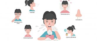
The development of the disease is accompanied by a constant complete or partial absence of nasal breathing, a constant runny nose with copious serous discharge. The child becomes easily excitable, restless, normal sleep is disturbed, he often has nightmares, and he snores in his sleep. Voice changes occur, the baby speaks through his nose. Due to enlarged tonsils, hearing is impaired. A chronic inflammatory process in the adenoids leads to the development of permanent inflammation of the nose, rhinitis or adenoids (adenoiditis), accompanied by fever and general weakness.
Diagnosis of adenoids
Diagnosis of adenoid hypertrophy and chronic adenoiditis includes a thorough collection of anamnesis of the disease and examination of the child by a pediatric ENT doctor using video endoscopic technology. If necessary, in addition to endoscopy of the nasal cavity and nasopharynx, tympanometry is performed to exclude pathology from the middle ear, which very often complicates the course of chronic adenoiditis. Laboratory diagnostics also play a very important role in determining the type of microbial pathogen or identifying the allergic nature of the disease. As a rule, a nasal culture is taken for microflora and sensitivity, and a smear of nasal mucus is taken for cytology to identify allergies. In addition, a clinical blood test is required to assess the general condition of the body and a detailed analysis of stool for scatology, dysbacteriosis, and worm eggs.
Complications of the disease
First of all, adenoids negatively affect the breathing mechanism. It is common for a person to breathe through the nose, this is how complete and high-quality air exchange occurs in the body. Breathing through the mouth creates, albeit a small, chronic lack of oxygen in the body, and also leads to a number of other health problems. Children with adenoids are more likely to suffer from viral and respiratory diseases, inflammation of the nose, ear and throat. The child is characterized by increased fatigue, appetite disturbances and frequent headaches.
Constant mouth breathing leads to many disorders in the body:
- Brain hypoxia caused by oxygen deficiency. Because of this, mental and physical development delays and chest deformation may occur.
- Changes in the shape of the skull and face. A constantly open mouth leads to deformation of the facial skull, lower jaw, and hard palate. The face acquires features characteristic of adenoids.
Clinical picture
Patients with hypertrophy complain of:
- changes in diction and pain that increases with swallowing;
- difficulty in respiratory function (when, along with the palatine tonsils, the nasopharyngeal tonsil enlarges).
As a result of breathing problems, sleep problems may occur in the form of frequent awakenings and snoring. The patient also suffers from coughing at night. With the development of hypoxia, neuropsychic disorders are observed.
Diagnosis of the disease
If symptoms of persistent nasal breathing disturbance appear, consultation with an otolaryngologist is necessary. Diseases can be identified and the degree of its development can be determined through a physical examination and instrumental studies.
- Palpation. The doctor feels the nasal passages and determines the severity of the pathology.
- Pharyngoscopy – examination of the organs of the nasopharynx using special ENT equipment, a spatula, a frontal reflector, a laryngeal and nasopharyngeal mirror, etc.
- Anterior and posterior rhinoscopy - examination of the nasal passages visually and using mirrors.
- Endoscopy is the examination of the nasal passages using an endoscope.
- Radiography is an effective way to determine the degree of development of pathology and the size of the adenoids.
- Audiometry – examination of a child’s hearing, etc.
Treatment
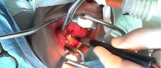
Treatment of enlarged tonsils, leading to sleep and respiratory problems, is only surgical. The operation is called tonsillotomy and involves their partial removal. Under local anesthesia (application anesthesia with lidocaine solution), the doctor cuts off those parts of the tissue that protrude beyond the arches. During the intervention, the patient is in a sitting position.
Classic tonsillotomy requires the use of a Mathieu tonsillotome, which has a special fork to hold the removed tissue. An alternative technique is performed using the Bahon loop. In this case, the separated part of the lymphadenoid tissue is fixed with a Kocher clamp. Our clinic also uses laser techniques for this purpose.
Treatment of the disease
The method of treating the disease is selected based on several criteria: depending on the severity of the pathology, the duration of the disease, the age and general health of the child, existing complications, etc. To a greater extent, treatment will depend not on the size of the adenoids, but on the severity of complications of the disease.
With the initial stage of the disease and minor breathing problems, in the absence of other pathological symptoms, conservative therapy can be used. It includes anti-inflammatory treatment, local use of drops and ointments, physiotherapy, breathing exercises, nasal rinsing, taking antihistamines, immunomodulating drugs, etc.
Adenoids of 2-3 degrees, which have led to significant breathing problems, and also with the progression of the pathology and the ineffectiveness of conservative therapy, the adenoids are removed - adenotomy. It is performed under general anesthesia in young children and under local anesthesia in older children. The operation can be performed classically or endoscopically, as well as using a laser or radiofrequency method.
More than 5% of children under 10 years of age have adenoids and suffer from complications of the disease. If you have symptoms of the disease, contact multidisciplinary clinics that provide a wide range of treatment and diagnostic services in the field of otorhinolaryngology. If a child’s mouth is constantly open, or he breathes mainly through his mouth, or has a chronic runny nose, then be sure to consult an otolaryngologist.
With timely treatment, complications of the disease can be avoided and conservative treatment will be sufficient. In medical diagnostics and treatment, only highly qualified specialists with extensive experience are able to make the correct diagnosis and prescribe effective treatment for adenoids for each case individually. You can make an appointment by calling the center or leaving a request on the website.
Adenoid hypertrophy
Adenoid hypertrophy (adenoid vegetations/growths, hypertrophy of the nasopharyngeal/pharyngeal tonsil) is a persistent enlargement of the nasopharyngeal tonsil. It can occur either alone or in combination with hypertrophy of the palatine tonsils.
The nasopharyngeal tonsil (adenoids) is a collection of lymphoepithelial tissue in the upper part of the nasopharynx. The result of its increase is called adenoid hypertrophy, and with inflammation the process is called adenoiditis. In combination with the palatine, tubal and lingual tonsils, they form the Pirogov-Waldeyer ring, located at the entrance to the upper respiratory tract and digestive tract and acting as a local immune barrier.
Adenoid hypertrophy is most often observed between the ages of 2 and 6 years, but can also occur at a later age.
There are several classifications of adenoids according to the degree of enlargement. The most popular division is based on the doctor’s subjective assessment of the size of the tonsil during examination:
- I degree – the lumen of the choanae is blocked by 1/3
- II degree – the lumen of the choanae is blocked by 1/3–2/3
- III degree – the choanae are completely closed by adenoids.
However, just assessing the size of the tonsil is not enough; a careful history taking is an important criterion.
Causes
Hypertrophy of grade I-II adenoids at the age of 2 to 6 years is often a physiological process that is associated with the fact that the child begins to actively contact the outside world and the amygdala is subjected to constant antigenic stimulation.
Severe (or pathological) hypertrophy, which leads to the symptoms described below, can occur due to an infectious or non-infectious cause. The infectious diseases include the following:
- viral (adenovirus, Coxsackie virus, cytomegalovirus, Epstein-Barr virus, herpes simplex virus, parainfluenza virus, rhinovirus, etc.)
- bacterial (Streptococcus, Haemophilus influenzae, Moraxella catarrhalis, Staphylococcus aureus, Neisseria, Mycoplasma pneumoniae, Fusobacterium, Peptostreptococcus, Prevotella, etc.)
Non-infectious causes include gastroesophageal reflux, allergies, and prolonged exposure to cigarette smoke.
Symptoms
Symptoms of hypertrophy occur due to an increase in the size of the tonsil and/or the presence of chronic inflammatory changes in it (chronic adenoiditis). Although these processes are often related (prolonged inflammation leads to hypertrophy), it must be borne in mind that this does not always happen. For example, with adenoid vegetations of the 1st degree, there may be pronounced signs of chronic inflammation, and with vegetations of the 3rd degree there may be no clinical manifestations at all. With physiological hypertrophy, the process occurs without any symptoms or with minor manifestations.
With severe hypertrophy, the child has difficulty breathing through his nose, and therefore he breathes through his mouth, snores during sleep, sometimes with pauses in breathing (obstructive sleep apnea syndrome). Impaired ventilation of the nasal cavity leads to mucous discharge and frequent rhinosinusitis. Characterized by sneezing and bad breath. Large adenoids put pressure on the soft palate, phonation and articulation are impaired in children, children have a nasal sound. If the adenoids block the auditory tubes, this can manifest as recurrent acute otitis media, hearing loss associated with the accumulation of fluid in the middle ear (exudative otitis media). Over time, due to oxygen starvation, the child becomes lethargic, apathetic, and irritable. Memory deterioration, decreased attention, and learning difficulties occur. Sleeping with your mouth open for a long time leads to disruption of the development of the facial skeleton. The hard palate becomes high and narrow, the lower jaw becomes narrow and elongated, the bite is disturbed, which may require orthodontic treatment in the future. An experienced otolaryngologist will immediately recognize the typical “adenoid” appearance of a child.
There are acute and chronic inflammation of the mucous membrane of the nasopharyngeal tonsil:
- Acute adenoiditis is an acute inflammation caused by a viral or bacterial infection. It manifests itself as a cold: fever, difficulty in nasal breathing, serous or purulent discharge from the nose. The duration of the disease varies and does not exceed one month. In the presence of adenoid hypertrophy, phenomena characteristic of hypertrophy are added to the listed manifestations.
- Chronic adenoiditis is a chronic polyetiological disease, which is based on the disruption of local immune processes and the formation of stable microbial biofilms. It manifests itself as difficulty in nasal breathing, serous or purulent discharge from the nose, mucus running down the throat and coughing. The causes of the disease are the same as those of hypertrophy. There is no consensus on the duration of adenoiditis at which it can be considered chronic. According to various sources, at least 12 weeks must pass from the onset of the disease. This condition can occur independently and over time lead to adenoid hypertrophy.
Diagnostics
The gold standard for diagnosis in global practice and the standard of our hospital is an endoscopic examination of the nasal cavity. We carry out this study with a thin flexible fiberscope under local anesthesia.
Lateral x-rays are a common way to assess hypertrophy but can be misleading. For example, an X-ray may show a picture of grade III adenoids due to enlargement of the tubal tonsils (they are located on the sides of the nasopharyngeal tonsil), due to swelling of the mucous membrane or accumulation of thick mucus. This method is used only if endoscopy of the nasopharynx is impossible.
It is important to carry out differential diagnosis with diseases that may have similar clinical manifestations (juvenile angiofibroma, choanal atresia, polyposis, enlargement of the posterior ends of the inferior turbinates, hypertrophy of the tubal tonsils).
Treatment
Physiological hypertrophy without any manifestations or with minimal symptoms does not require treatment, but endoscopic examination is necessary for differential diagnosis.
Treatment may be conservative. It includes eliminating factors that can cause hypertrophy: allergies, gastroesophageal reflux, passive smoking. Accordingly, in some cases a multidisciplinary approach of an otolaryngologist, allergist, gastroenterologist and pediatrician is necessary. A positive effect can be had by changing the climate to a more favorable one, for example, sea climate. Topical corticosteroids have been used as an adjunctive treatment in children over the age of three, with some studies showing some benefit, but overall evidence on the effectiveness of these drugs is mixed. In acute and exacerbation of chronic adenoiditis in the case of a bacterial infection, treatment is carried out with antibiotics. Irrigation of the nasopharynx with saline solution is also used in treatment. The use of various oils, mucolytics, local antibacterial drugs, silver solutions, herbal remedies, and physiotherapy currently do not have a high-quality evidence base.
In some cases, surgical treatment cannot be avoided. Not every child needs surgery and it requires specific indications. When determining them, doctors at Ilyinskaya Hospital follow the principles of evidence-based medicine, relying on global experience. Indications include failure of conservative treatment, recurrent infections, acute otitis media and sinusitis, and an “adenoid” face. Parents should understand that long-term refusal to undergo surgery if indicated, or an attempt to “wait out” or “outgrow” adenoid hypertrophy can lead to complications, the treatment of which in the future will require a lot of time and can negatively affect the development of the child. According to the recommendations of the American Academy of Otolaryngology and Head and Neck Surgery, adenotomy should be performed in the following cases:
- In children under 4 years of age with symptoms of difficulty in nasal breathing, signs of chronic adenoiditis, as well as exudative otitis media
- In children 4 years of age and older with exudative otitis media
- If you have confirmed obstructive sleep apnea (periodic sudden decrease or cessation of breathing during sleep)
In the presence of pathology of the palatine tonsils, surgery for their resection/removal is often performed simultaneously (adenotonsillotomy/adenotonsillectomy), and in case of exudative otitis media, shunting is performed at the same time.
Adenotomy has undergone significant changes over time. Previously, this operation was performed under local anesthesia using a special knife (adenotome) under the control of a small mirror, and sometimes blindly. During the operation, the child experienced stress, and the surgeon may not have fully performed the operation. Nowadays, this approach is practically not used, but it is preserved in some clinics.
Adenoid treatment programs at the ENT Clinic in Chertanovo
“ENT Clinic in Chertanovo” offers its patients comprehensive programs for the treatment of chronic adenoiditis and the prevention of exacerbations, including examinations by a doctor using endoscopic equipment, tympanometry, sanitation of the nasopharynx using the Cavitar device, therapeutic manipulations for complications of the ears, therapeutic laser and a visit to the salt cave for conducting halotherapy sessions. The cost of the programs, depending on the procedures included, is 30,000 - 35,000 rubles. When treated as part of a comprehensive program, you receive significant savings (up to 30%) from the cost of services according to the clinic’s regular price list.
In severe, long-term chronic adenoiditis, against the background of severe concomitant pathology, when courses of conservative treatment do not have a positive effect, the question of surgical treatment and removal of the adenoids is raised. In the postoperative period, to prevent relapses of the disease, regular monitoring by an ENT doctor and courses of preventive treatment are necessary.
Remember that the health of your baby is in your hands! And with regular preventative care, your baby will develop and grow healthy and happy!
Modern diagnostic methods (endoscopic examination of the ear, nose and throat by an ENT doctor)
The gold standard for diagnosis (and the only correct one) is an endoscopic examination of the nasopharynx by an ENT doctor with good skills and experience working with children from birth. An endoscopic examination is an examination using a video camera, with additional powerful lighting and magnification and display on a television screen, on which both the doctor and the child’s parents will clearly see not only all the structures of the nose, but also the nasopharynx with the mouths of the auditory tubes and adenoid vegetations. In this case, the doctor will evaluate the degree of their increase, inflammation of the mucous membrane, the nature of the discharge, the presence and size of the respiratory lumen. The examination is carried out with a special children's nozzle - a tube less than 3 millimeters in diameter, the examination is non-contact, absolutely painless, does not require special preparation (the ENT doctor only drips vasoconstrictor drops in an age-appropriate dosage before the examination) and has no contraindications and is used in children from birth.
Of course, with such a diagnosis, digital examination is not required.
It is very important that during an endoscopic examination of the ears, such a significant complication as exudative otitis (accumulation of fluid behind a healthy, non-inflamed eardrum) will not be missed. Thanks to good lighting and magnification, the eardrum is transparent during endoscopic examination, so the liquid and air bubbles behind it are clearly visible. Whereas when examined in the usual way, the eardrum, if it is not inflamed, reflects the light of a flashlight and looks gray and opaque, the liquid behind it cannot be seen.
Comparative characteristics of traditional examination of ENT organs and endoscopic examination of ENT organs
| Criteria | Traditional ENT examination | Endoscopic examination of ENT organs |
| Price | Low inspection costs due to the use of tools and a reflector | High cost due to the use of expensive equipment |
| Painless | The contact method can be unpleasant due to the instrumental expansion of the entrance to the nose for examination; the use of an ear mirror is also contact. Digital examination is traumatic and can be painful | The method is non-contact, painless, as nozzles of less than 3 mm are used. in diameter |
| Diagnostic reliability | The examination is superficial, the structures of the nose are examined only in the anterior sections, the nasopharynx is inaccessible for inspection, the adenoids and the mouths of the auditory tubes are not visible, it is impossible to assess the condition of the middle ear behind a healthy eardrum | The reliability of diagnosis of adenoiditis, adenoid hypertrophy, and complications in ENT organs is 100%. All structures of the nose, nasopharynx, ear and throat are examined on a screen with additional magnification and lighting. A diagnostic error has been ruled out. |
| Accuracy of the diagnosis | The diagnosis is presumptive | Reliable diagnosis with assessment of possible risks and complications |
| Purpose of treatment | Overdiagnosis, high probability of prescribing antibacterial therapy “just in case” | Prescribing adequate therapy |
| Assessment of dynamics during treatment | Assessment of only external signs and symptoms (presence of runny nose, cough, etc.) | Assessment of both the dynamics of symptoms and mucus and size of the adenoids, development or relief of complications. |
| Recommendations for adenoid removal | It is possible to prescribe removal of adenoids if they can be preserved and monitored. | Appointment of adenoid removal only if there are indications for removal and an objective assessment of the lack of effect of treatment. |
| Who can carry out diagnostics | Pediatrician, ENT doctor with any experience and qualifications | ENT doctor with practical skills and experience in endoscopic examination of ENT organs in children from birth. |
Laboratory diagnostic methods (the goal is to clarify the cause of chronic adenoiditis and/or adenoid hypertrophy)
To establish the cause of enlarged adenoids, as a rule, a detailed blood test is prescribed, a nasal smear for cytology (cellular composition of nasal discharge), blood for total immunoglobulin E (to exclude an allergic nature or allergic background), feces for worm eggs and scraping for enterobiasis ( adenoids often enlarge due to helminthic lesions), a PCR test for the Epstein bar virus is possible (if mononucleosis is suspected)
Traditional diagnostic methods (examination by an ENT doctor or pediatrician)
In a regular clinic, to diagnose chronic adenoiditis and adenoid hypertrophy, an examination of the nose, throat and ears is used using metal instruments for examining ENT organs: nasal and ear mirrors and a spatula illuminated with reflected light, using a conventional head reflector (head mirror) or a head lamp in the form flashlight. In this case, the ENT doctor (or pediatrician) only sees the area of the nasal vestibule and the surrounding parts of the nose. The nasopharynx, the anatomical location of the adenoids, is not visible. The examination is contact (when examining the nose, the wings of the nose in the vestibule area are expanded with a nasal speculum; when examining the ear, an ear speculum is inserted into the ear canal)
In addition, it will be possible to perform a digital examination of the nasopharynx for a subjective assessment of the size of the adenoids (the doctor feels the nasopharynx and the surface of the adenoids through the mouth, trying to determine their size). This method is not reliable, is unpleasant for a small patient and is traumatic for adenoid vegetations.
Causes of enlarged tonsils in children
The increase is caused by the following infectious agents: pneumococci, staphylococci, herpes, streptococci, chlamydia, Haemophilus influenzae, adenovirus, influenza virus. They can be located in the lacunae of the tonsils without manifesting themselves in any way (for example, remain after treatment for a disease), but at a favorable moment they begin to actively multiply and cause an enlargement of the organ. Such triggers can be:
- decreased immunity;
- allergies;
- bad ecology;
- hormonal disorders;
- severe hypothermia;
- lack of vitamins;
- infections.
Causes and course of the disease
Hypertrophic changes in the tonsils are considered as an immunoreactive condition that occurs when the body adapts to changing conditions and mobilizes compensation that has arisen in the lymphoid pharyngeal ring.
This is facilitated by breathing through the mouth, which is caused by hypertrophy of the adenoids, especially in the cold season. If adenoiditis provokes the formation of mucus, which forms in the nasal cavity, infects the palatine tonsils. Hyperplasia (increased cell proliferation) is promoted by infectious diseases and repeated inflammation in the oropharynx and nasal cavity.
Also, this process is influenced by poor living conditions, insufficient nutrition, as well as other factors that can reduce immunity. The size of the tonsils is affected by various disorders in the endocrine system, especially hypofunction (weakening of activity) of the adrenal cortex, exposure to small doses of radiation over a long period of time, hypovitaminosis (lack of vitamins).
The basis for too large an increase in the lymphoid tissue of the tonsils is the active proliferation (proliferation of tissue by cell multiplication) of immature T-lymphocytes and T-helper deficiency, which does not allow the production of full-fledged antibodies. The immunopathological predisposition of the child’s body to lymphatic diathesis, which is caused by hereditary deficiency of the lymphoid system, is influenced. Allergic reactions also influence the formation of hypertrophy of the palatine tonsils.
However, do not forget that hyperplasia is a reversible process. During adolescence, involution (reverse development) of lymphoid tissue occurs.
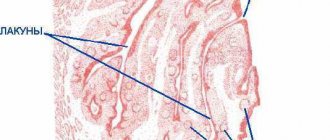
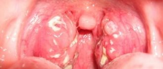
Hypertrophy of the palatine tonsils is often combined with a pathological enlargement of the entire pharyngeal lymphadenoid ring, especially often with hypertrophy of the pharyngeal tonsil (adenoids). Enlarged palatine tonsils generally have a dense and fairly elastic consistency, but in some cases they are spread out and soft on palpation. The palatine tonsils have no signs of inflammation and are not fused to the palatine arches. They have a developed lower pole and a triangular fold below, and the lacunae have a normal structure.
From a histological point of view, there is a predominance of hyperplasia of lymphoid tissue, in which there is an increase in the area of the follicles and the number of mitoses (indirect division), but plasma cells and macrophages are absent.
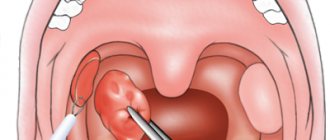
Hypertrophy of the palatine tonsils has 3 degrees:
- 1st degree - the palatine tonsils occupy 1/3 of the distance from the arch to the center of the pharynx (if measured along the midline of the pharynx);
- 2nd degree – already 2/3 of the distance mentioned above;
- 3rd degree – the tonsils are almost touching.
A microscopic examination reveals a large number of follicles with very frequent areas of mitosis, which indicates a very high activity of lymphoid tissue.
Friends! Timely and correct treatment will ensure you a speedy recovery!
The degree of enlargement of the tonsils in a child
There are 4 stages of hypertrophy of the glands:
- At the first stage, the inflamed tissue of the tonsils covers no more than 30% of the lumen between the pharynx and the sky. In this state, it causes almost no discomfort, so it is difficult to detect at this stage.
- At the second stage, hypertrophied tissue covers half of the lumen. The child begins to have difficulty swallowing and breathing freely, and a sensation of a foreign body in the throat.
- At the third stage, there is severe difficulty in breathing and swallowing.
- On the fourth, the pharynx is almost completely blocked by inflamed tonsil tissue. It is difficult to bring the condition to this stage, since already at the third stage the child’s well-being requires immediate medical attention.
Introduction
How often, having a preschooler child, we get tired of the seemingly endless runny nose, daily noticing small changes in the amount, thickness and color of “snot”: sometimes liquid and colorless, sometimes thick, green and plentiful. It is not for nothing that popular rumor, with its inherent irony and worldly wisdom, assigned its caustic stamp to preschool age, long before the emergence of the institution of preschool education: the capacious and simple-minded “brat.” And it becomes completely alarming when, despite careful treatment, an obsessive dry cough at night, snoring during sleep, and significant difficulty in nasal breathing do not go away. Rare and such short “bright” intervals of health, and again a runny nose, cough, and sometimes congestion and pain in the ears. And the natural question arises: “Who is to blame? So what should I do? And of course, a trip to the doctor, and maybe more than one, to make a diagnosis: chronic adenoiditis, adenoid hypertrophy . And again persistent treatment and the thought: “Can I remove them already and forget about the problem?” What choice to make: Delete or save? So is it worth fighting and how to do it in modern conditions of medical development? And what is the effectiveness of this fight? We will talk about all this in this article. But let's start with the very basics:
Chronic adenoiditis is a chronic inflammation of adenoid vegetations (accumulations of lymphoid tissue) located in the nasopharynx, accompanied by their mucus (which is manifested by a runny nose, cough, mucus flowing down the back wall of the pharynx), swelling and/or hypertrophy (enlargement) (which is manifested by difficulty in nasal breathing up to to complete absence of breathing through the nose, sometimes snoring during sleep). All or some of these manifestations can occur periodically up to 4-6 times a year or more, or be present constantly for several months.
Treatment of adenoids
Treatment of adenoid hypertrophy and chronic adenoiditis must begin as early as possible, when the first signs of the disease appear, avoiding complications from the middle ear and other organs and systems of the child’s body.
First of all, it is necessary to restore nasal breathing, which is achieved by thorough toileting of the nasal cavity, rinsing the nose using an electric suction or hardware rinsing with drugs using ultrasound on the Cavitar apparatus. Nasal corticosteroids and oral antihistamines are prescribed to reduce swelling and inflammation of the nasal mucosa. Vitamins must be prescribed according to the child’s age, as indicated by drops in the ears (if there are complications from the ears). If there are signs of intoxication of the body, the use of antibacterial drugs locally in the form of drops in the nose and orally for a general effect is indicated.
To sanitize and improve the lymphadenoid tissue of the nasopharyngeal tonsil, the use of homeopathic remedies is justified.
It must be remembered that the treatment of chronic adenoiditis must be comprehensive. A very important role in the conservative treatment of chronic adenoiditis, along with generally accepted drug treatment, is given to physiotherapeutic procedures, which significantly speed up the healing process and play a major role in the prevention of exacerbations in the future. Only such an integrated approach to the treatment of chronic adenoiditis is justified and gives good results.
Degrees of hypertrophy (enlargement) of adenoids
I degree - when adenoid vegetations during endoscopic examination cover less than 1/3 of the lumen of the choana (the outlet from each half of the nose into the nasopharynx)
II degree - when adenoid vegetations during endoscopic examination cover 1/2 of the lumen of the choana (the outlet from each half of the nose into the nasopharynx)
III degree - when adenoid vegetations during endoscopic examination cover the entire lumen of the choana (the outlet from each half of the nose into the nasopharynx)
Often, ENT doctors assign grade II-III - in the case when more than ½ of the lumen of the choanae is closed, but the lumen for breathing still remains, and is not completely closed.