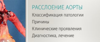
A peritonsillar abscess is a purulent cavity located in the tissues at the level of the soft palate above the palatine tonsil. Most often, such purulent inflammation occurs against the background of a previous sore throat or exacerbation of chronic tonsillitis. A purulent lesion can be on the right or left in the pharynx, or it can be bilateral. Treatment of peritonsillar abscess is usually surgical. Conservative therapy is carried out at the initial stage of inflammation. A peritonsillar abscess often results in removal of the tonsils. The causes, symptoms and methods of treating abscess are the topic of our new article.
A peritonsillar abscess is a festering cavity. It is localized in the tissues, at the level of the soft palate above the palatine tonsil. These tissues are called paratonsillar tissue. The concept of “peritonsillar abscess” is already a purulent process. It is preceded by diagnoses such as paratonsillitis and phlegmonous tonsillitis. All three of these diagnoses describe the progression of one condition into another, more severe one.
The disease is the highest degree of inflammation of the tonsils and one of the most severe forms of purulent lesions of the pharynx. One third of cases of paratonsillitis development occur as complications of the disease “chronic tonsillitis”. Also, phlegmon can form in the mediastinum - the diagnosis sounds like mediastinitis. This can happen if treatment for the disease is ignored.
Most often, this diagnosis is made at a young age - up to thirty - thirty-five years.
Inflammation can occur in several forms depending on its location. It can also be unilateral (with a lesion on the left we are talking about a left-sided paratonsillar abscess, with a lesion on the right - about a right-sided paratonsillar abscess), less often - bilateral.
Treatment of a left-sided throat abscess, as well as treatment of a right-sided abscess, should be carried out only under the supervision of an otolaryngologist. Attempts to treat the disease at home are not only ineffective, but also dangerous, as they can provoke serious complications of the disease.
The causes, signs, symptoms and treatment of peritonsillar abscess are the topics of our new article.
Causes of the disease
Peritonsillitis occurs when pathogens enter the tissues around the tonsils.
The causative agents of the disease are streptococci, staphylococci, and fungi.
How do pathogenic microorganisms get into the paramygdaloid tissue? There may be several reasons for the start of the inflammatory process:
- untreated tonsillitis, when bacteria from the tonsils penetrate into nearby tissues;
- exacerbation of chronic tonsillitis, when pathogenic microflora also penetrates from the tonsils;
- dental diseases - for example, caries (infection comes from the oral cavity), although it is not necessary that the tonsils themselves become inflamed;
- injury to the tissue around the tonsils, during which infection occurs;
- infection through the bloodstream;
- chronic accumulations of infection in the body (for example, chronic sinusitis);
- diabetes;
- reduced immunity;
- smoking.
Typically, purulent inflammation acts as a complication of chronic or acute tonsillitis (tonsillitis) - this is the most common cause of the disease.
Why is it necessary to open and drain a peritonsillar abscess?
Peritonsillar abscess is an urgent condition in otolaryngology. The proximity of vital structures (windpipe, large vessels of the head and neck) requires immediate diagnosis and treatment. In the initial stages, when an abscess has not yet formed, the pathology responds well to drug therapy. A course of antibiotic therapy, anti-inflammatory and painkillers is prescribed. If conservative treatment is ineffective, the abscess is opened surgically.
Peritonsillar abscess is dangerous due to its complications, fraught with the development of phlegmon, mediastinitis and even sepsis. The Otolaryngology Department at the GMS Clinic has everything you need to accurately diagnose and treat peritonsillar abscesses.
The opening of the abscess is carried out using modern surgical equipment. During the operation, the potential of laser and radio wave surgery is actively used, which allows the procedure to be performed in the most gentle manner, eliminating the development of surgical complications. Opening abscesses in children is performed under anesthesia, which significantly increases the comfort of the intervention.
Types of disease
The inflammatory process can occur on the left or right in the pharynx. Such inflammation is called one-sided - respectively, a left-sided or right-sided abscess. If inflammation occurs on both the left and right at the same time, this is a bilateral type of inflammation.
If we take the location of the purulent “pocket” as the basis for the classification, the following types of disease are distinguished:
- anterior - the most common type of diagnosis; the tissues above the tonsils become inflamed;
- posterior, when purulent inflammation occurs between the posterior palatine arch and the tonsil; such inflammation can spread to the larynx;
- lower, when inflammation is located at the lower pole of the tonsil, while the anterior palatine arch moves forward and downward;
- external (lateral) - a less common, but most severe type, in which a suppurating cavity appears between the edge of the tonsil and the wall of the pharynx. With this form of the disease, pus can break into healthy tissue in the neck.
Causes and course of the disease
The development of a paratonsillar abscess occurs in several stages.
First, paratonsillitis is formed, then a phlegmonous (purulent diffuse focus) process and abscess formation follows, a process of spontaneous emptying of pus into the space near the tonsils or into the pharyngeal cavity occurs. The cause of the development of this disease may be suppuration of the tonsil during tonsillitis or exacerbation of chronic tonsillitis. In addition, a peritonsillar abscess can occur due to the spread of a purulent process that has developed in the gums due to dental caries, as well as when it is difficult for the eighth tooth to erupt in the lower jaw. Depending on the location of the lesion, by analogy with paratonsillitis, the abscess in location to the palatine tonsil can be anterior, posterior, inferior and external.
Symptoms of paratonsillar throat abscess
The main symptom of the disease is acute, unbearable pain in the throat: signs of an abscess on the right are pain sensations concentrated in the right side of the pharynx, on the left - in its left side, respectively. Only in ten percent of cases does the pain occur on both sides of the throat. At first the pain is felt during swallowing, then it develops into a constant pain. It can “give” to the ear or teeth.
Then other symptoms of the disease appear: an increase in body temperature to high levels (40℃), body aches, headaches.
In some cases, specific symptoms of the disease may be observed:
- spasms of the masticatory muscles, due to which the patient has difficulty opening his mouth;
- sensations of the presence of a foreign object or coma in the throat;
- enlarged lymph nodes;
- the appearance of nasality in the voice;
- foul odor from the mouth;
- pain when turning the head;
- tilting the head towards the affected area or forward;
- difficulty breathing, shortness of breath.
Sometimes spontaneous opening of the purulent cavity occurs, which leads to the subsidence of the signs of the disease and improvement of the patient’s condition.
Clinical picture
There is pain in the throat, which increases with swallowing, turning the head and coughing.
The patient experiences difficulty opening his mouth, which is accompanied by severe pain. Trismus (inability to fully open the mouth) of the masticatory muscles is noted. The patient is forced to refuse to eat and drink, the pain interferes with his normal sleep. Body temperature is elevated, patients complain of weakness and malaise. When performing pharyngoscopy, hyperemia (pronounced redness) of the mucous membrane and infiltration of one half of the soft palate and palatine arches on the affected side are determined.
The palatine tonsil is slightly shifted to the healthy side and is tense. The lymph nodes located under the jaw and on the neck become somewhat swollen. Due to the fact that the soft palate is almost motionless, the voice becomes nasal. The most common are peri-almond abscesses with anterior and upper anterior localization.
Diagnostics
Timely and, most importantly, correct diagnosis is an important step on the path to recovery. The diagnosis, as well as the treatment of the throat with an abscess, is carried out by an otorhinolaryngologist. The clinical picture of the disease, as a rule, is so indicative that making a correct diagnosis is not particularly difficult.
At the initial consultation, the ENT doctor collects the patient’s medical history and complaints: the presence of recent sore throat or exacerbation of chronic tonsillitis, dental problems or mechanical damage to the walls of the pharynx is clarified. The ENT doctor finds out how the inflammation of the tonsils was treated (in the case of tonsillitis), and when the patient’s well-being worsened.
Then a direct examination of the patient is performed: in some patients, an involuntary tilt of the head towards the inflamed area of the pharynx is immediately noticeable, an unpleasant odor is felt from the oral cavity, and enlarged lymph nodes are clearly palpable.
The most obvious method of research for this condition is pharyngoscopy, which allows you to determine the area of localization of the abscess.
If we are dealing with a lateral form of phlegmon location or if complications are suspected, the patient is prescribed an ultrasound and CT scan of the neck.
If the problem is not diagnosed in time and the throat is not treated, dangerous complications may develop:
- phlegmon of the neck (purulent inflammation of the tissues of the neck);
- mediastinitis - inflammation of the mediastinum;
- blood poisoning;
- laryngeal stenosis, which can lead to suffocation.
Such complications of a left-sided abscess, as well as a right-sided one, can be fatal.
Treatment
For peritonsillar abscess, surgical intervention is indicated.
The opening of the abscess is performed through the anterior palatine arch, moving 1-1.5 cm outward from the free edge in the place where the greatest softening and protrusion is observed. If it is very difficult to determine the location of the softening, it is recommended to make an incision in the middle of the line that connects the last molar and the base of the tongue. The mucous membrane is treated with an anesthetic and an incision is made with a scalpel at the site of the greatest bulging of the infiltrate, no more than 1 cm long. Using a forceps, the soft tissues are moved apart to a depth of 1 to 2 cm. After the abscess is opened, treatment is prescribed, the same as for follicular and lacunar angina. If a person has suffered a peritonsillar abscess, then this is an indication for surgery to completely remove the tonsils (bilateral tonsillectomy).
Make an appointment right now!
Call us by phone or use the feedback form
Sign up
Surgical treatment of this disease is complemented by drug therapy, which includes all the components used in the treatment of paratonsillitis.
If paratonsillar abscesses often recur or paratonsilitis does not resolve for a long time (does not go away), then abscessonsillectomy is performed (complete removal of the tonsils).
Indications for opening an abscess
The possibility of developing this kind of complications can be assumed if the following symptoms are present:
- tonsillitis, the duration of which exceeds 5-6 days: during this time an abscess may form;
- severe, severe pain in the throat that is felt when swallowing or moving the head;
- temperature increase over 39 degrees;
- significant enlargement of one or two tonsils;
- signs of intoxication and fever (weakness, malaise, muscle pain, apathy and drowsiness);
- enlarged lymph nodes;
- increased heart rate and breathing.
The development of an abscess, which is accompanied by such symptoms, is a direct indication for opening the abscess.
Usually the operation is performed 4-5 days from the onset of the inflammatory process - by this time the abscess is already fully formed. To check the readiness of the cavity for removal, the doctor may prescribe a diagnostic puncture - puncturing the cavity with a thick needle in the place that protrudes the most above the tonsil. The progress of the procedure is most often monitored by endoscopic or ultrasound monitoring. If pus is found in the cavity of the syringe used for puncture, it means that an abscess has formed and is ready for removal.
The autopsy is performed on an outpatient basis, that is, to perform it, the patient does not need to go to the ENT department of a medical institution.
results
A method has been developed to improve the technique of percutaneous drainage with a stylet catheter under ultrasound guidance by infiltrating the interloop tissue (intestinal mesentery, omentum) with an anesthetic solution. Injecting fluid into the adjacent tissue allows you to increase the space for acoustic access, displacing the intestinal wall, and outline the drainage path, bypassing large vessels and the intestinal wall and thereby avoiding their damage.
Drainage was performed in 103 patients. The duration of the manipulation was 20±8.2 minutes. In 97 of 103 patients, one intervention was required to adequately drain the cavity. In 6 cases, additional drainage was performed on days 3–11 due to the insufficient effectiveness of primary drainage. In 3 patients, repeated drainage was performed on the 60th day after surgery due to failure of the intestinal anastomosis and failure to close the internal fistula. Cure occurred in 101 (98%) patients within 10–73 days. Mortality rate was 1.9% ( n
=2). A 53-year-old patient died from pulmonary embolism 2 months after undergoing surgery (laparoscopic gastrectomy for stomach cancer, gastric bleeding). The second patient, 74 years old, died due to adhesive intestinal obstruction, which required surgical intervention - laparotomy, adhesiolysis. The course of the postoperative period was complicated by multiple intestinal fistulas, abdominal abscesses, and suppuration of the postoperative wound. In none of the cases was repeated open drainage of the abscess required. In 2 cases of the formation of external intestinal fistulas in the postoperative period, obstructive intestinal resection was required.
The results of dynamic ultrasound and fistulography were analyzed. It was revealed that in 67% the data coincided, which makes it possible to abandon fistulography at the final stage of treatment if the clinical course is positive. Therefore, only ultrasound is sufficient to assess the residual cavity. In addition, ultrasound-guided drainage is an effective independent method of treating abscesses associated with hollow organs without additional interventions to close the fistula.
Material and methods
We analyzed 103 clinical observations from 2012 to 2017 that required percutaneous drainage of intra-abdominal abscesses under ultrasound guidance. Patients who underwent drainage of intraorgan and retroperitoneal abscesses due to pancreatic necrosis were excluded from the study. The average age of the patients was 54±16 years (minimum age - 21 years, maximum - 85 years), there were 59 (53.7%) men, 48 (44.3%) women.
The distribution of abscesses by etiology is presented in Table. 1.
Table 1. Distribution of abdominal abscesses depending on the disease. In 75 (73.5%) patients this is a complication of surgical diseases of the abdominal organs.
Postoperative abscesses were noted in 28 (26.5%) patients (Table 2).
Table 2. Distribution of postoperative abscesses by type of operation. Distribution by location is presented in Table. 3.
Table 3. Distribution of abscesses by location On average, the volume of the abscess was 35±7 ml (minimum volume 5 ml, maximum 300 ml). In 3 (3%) patients, multiple abscesses were identified, localized in more than one anatomical area (subhepatic, subphrenic, interloop), which required simultaneous separate installation of several drains.
Abdominal ultrasound was performed in all patients. A Siemens Acuson Antares ultrasonic device with a 3.5 MHz convex probe and a 5-7 MHz linear probe was used. In acute surgical diseases of the abdominal organs, sonography was performed upon patient admission to the hospital. Indications for ultrasound in the complicated course of acute surgical diseases of the abdominal organs were late admission to the hospital (3-7 days from the onset of the disease or complication), the presence of clinical and laboratory signs of an inflammatory reaction - low-grade or hectic temperature, local pain syndrome, palpable infiltrate abdominal cavity. In the postoperative period, dynamic ultrasound control was carried out from the 2-3rd day after surgery. In patients who have undergone surgery on the abdominal organs, signs of inflammatory complications include slow progress in recovery, temperature reaction, non-decreasing leukocytosis and a shift in the leukocyte count to the left.
Appendiceal abscess in our study is the most common complication of acute surgical disease of the abdominal cavity - 45 (59.85%) cases. Sonographic signs of an appendiceal abscess appeared on days 3–20 of the disease (on average on days 5–9) in the form of the presence of a fragment of a destructive vermiform appendix in the cecum with a diameter of more than 0.7 cm with a thickened (>0.3 cm) wall , in some places its wall was not differentiated, and during compression the process was rigid. In 7 cases the process was not visualized. When an abscess formed at the stage of phlegmon, hypo- or anechoic accumulations of irregular shape, without a clear capsule with heterogeneous contents, gas inclusions, and suspension, were located in the destructively altered appendix. The transformation of phlegmon into a mature abscess in the form of a hypoechoic heterogeneous accumulation with a pyogenic capsule with a volume of 5 to 90 ml was observed in 12 patients over 3-6 days. The wall of the cecum in all observations was involved in the inflammatory infiltrate, thickened (>0.4-0.5 cm), and the layers of the wall were differentiated. The abscess was identified in 39 patients in the right iliac region, in 1 - in the subhepatic space, in 3 - in the pelvic cavity, in 1 - in the retroperitoneal space. The surrounding tissue, depending on the location of the appendix (mesentery of the appendix, cecum, small intestine, paracolytic, perirenal fat, greater omentum) was usually dilated, increased echogenicity, and had a heterogeneous structure.
Paravesical abscess as a complication of acute destructive cholecystitis was observed in 2 (2.66%) patients on days 6–14 of the disease in the form of a hypoechoic liquid heterogeneous accumulation in the tissue measuring 3–6 ml, connecting to the lumen of the gallbladder through a wall defect. The tissue of the bladder bed and hepatoduodenal ligament was found to be thickened, with increased echogenicity (infiltrate).
Paracolytic abscess as a complication of diverticular disease of the colon due to perforation of diverticula was observed in 4 (6.1%) patients on days 6–14 of the disease. Its ultrasound signs: visualization in most cases in the left iliac region, in the hypogastrium of a fragment of the sigmoid colon with an uneven outer contour due to diverticula, the wall is thickened from 0.5 to 1 cm, the differentiation of layers is preserved, pronounced swelling of the submucosal, muscular layer, adjacent paracolytic tissue is thickened and has increased echogenicity. When a diverticulum is perforated, small hyperechoic inclusions with a reverberation effect (gas bubbles) are located in the tissue; hyperechoic inclusions with an acoustic shadow (coprolites) are often found; when an abscess is formed, hypoechoic, heterogeneous round-shaped accumulations with a pyogenic capsule with a volume of 5-100 ml are found (in most cases located in the tissue along the mesenteric edge).
An abscess due to perforation of a colon tumor was observed in 7 (9.33%) patients on days 2–7 of the disease. A characteristic feature of the echographic picture in this case is a change in the intestinal wall over 3-10 cm in the form of significant (0.9-2.5 cm), uneven thickening, unclear external contours, lack of differentiation of the layers of the intestinal wall, strengthening and deformation of the vascular pattern in color Doppler mapping mode, narrowing of the intestinal lumen. In addition, visualization of enlarged (>1.5 cm) regional lymph nodes, decreased echogenicity, round shape with impaired corticomedullary differentiation, as well as distant metastases helps to establish a tumor lesion of the intestine.
A feature of the abscess during perforation of the colon against the background of a tumor is its large (200-300 ml) volume, which indicated the connection of the cavity with the intestinal lumen, i.e., the presence of an internal intestinal fistula. Differential diagnosis of a complicated form of diverticular disease is helped by the absence of obvious positive dynamics on the part of the intestinal wall within a week after drainage of the abscess against the background of antibiotic therapy, which is typical for diverticular disease.
Sonographic signs of an abscess during perforation of a gastric ulcer ( n
=3) or duodenum (
n
=1) were detected in 4 (6.1%) patients on the 8-21st day from the onset of the disease. Upon admission to the hospital, they had a clinical and instrumental picture of a covered perforation of the ulcer - a pronounced local pain syndrome. But upon palpation, the abdomen remained soft; an X-ray examination of the abdominal organs revealed free gas. Dynamic ultrasound revealed the absence of an increase in free fluid in the abdominal cavity and detected an abscess with a poorly defined capsule, surrounded by an infiltrate with gas and suspension in the center, volume 10-150 ml, localized in the subhepatic space, omental bursa, subphrenic space involving the walls of the duodenum or stomach. The fragment of the intestinal (stomach) wall adjacent to the abscess is thickened to 0.5-0.7 cm, reduced echogenicity, the layers of the wall are differentiated, the fiber along the vessels, the tissue of the lesser and greater omentum, paravesical tissue (depending on the location of the perforation) are expanded, increased echogenicity. The infiltrate is limited to the omentum, gall bladder, and pancreas. Patients with a clinical and instrumental picture of a covered perforation did not undergo surgical intervention; they received a nasogastric tube with constant aspiration of the contents.
An abscess as a result of perforation of the small intestine by a foreign body (according to the patient, animal bone) was observed in 1 (1.5%) patient on the 6th day of the disease. At the same time, nonspecific echographic signs of inflammatory changes in the walls of a fragment of the intestine were noted; an abscess in the form of a heterogeneous accumulation with gas, a suspension of 15 ml without a clear capsule was located in the infiltrated mesentery of the small intestine, the foreign body was not clearly visualized.
The development of clinical manifestations of abscess formation as complications of surgical intervention in the abdominal cavity often occurred slowly against the background of antibacterial, analgesic, and anti-inflammatory therapy. Abdominal abscesses were diagnosed on average 5.3±2.2 days (minimum on the 3rd, maximum on the 10th day) after surgery. Abscesses after operations on the stomach were detected in 8 (28.6%) patients on days 7-20 in the area of anastomosis or at the site of suturing the wall of a hollow organ; they were located in the subdiaphragmatic space, on the left above the spleen or in the bed of the removed spleen, along the anterior slope of the diaphragm, in the omental bursa, in the subhepatic space in the form of a limited liquid accumulation of irregular shape with suspension and gas with a volume of 10-150 ml.
After videolaparoscopic cholecystectomy, the abscess, diagnosed on days 4–6 after surgery, was drained in 4 (14.2%) patients. In the bed of the removed gallbladder, ultrasound revealed tissues of increased echogenicity with an irregularly shaped fluid accumulation and a suspension with a volume of 6-75 ml.
The period of formation of abscesses (5–30 ml) after videolaparoscopic appendectomy, according to our data, is 5–20 days after surgery; they were drained in 5 (17.85%) patients. Typically, such abscesses were located in the area of the stump of the process interloop in the small pelvis.
After right-sided hemicolectomy, resection of the colon with anastomosis, end colostomy, an abscess (15-50 ml) was formed in all cases in the area of the anastomosis or at the site of suturing of the intestine in the adjacent tissue on days 8-30. In 3 patients, re-formation of an abscess was noted on the 60th day after surgery during re-hospitalization due to incomplete closure of the internal intestinal fistula due to failure of the anastomotic sutures.
In 2 cases after surgery, on the 5th and 10th day due to adhesive disease, interloop abscesses with a volume of 15-70 ml were drained. In all patients, fistulography revealed a connection with a hollow organ; however, the internal intestinal fistula closed on its own after drainage of the abscess.
The totality of the clinical and instrumental picture of the intra-abdominal abscess was an indication for drainage of the fluid collection (Fig. 1, 2).
Rice. 1. Sonogram of the pelvic cavity (an abscess is visible after perforation of the sigmoid colon diverticulum).
Rice. 2. Sonogram of a pelvic cavity abscess after drainage under ultrasound guidance. 1 - abscess cavity, 2 - drainage in the abscess cavity, 3 - tumor of the sigmoid colon. The absence of a safe acoustic window was a contraindication.
Minimally invasive puncture-drainage treatment was performed in the operating room. A Siemens Acuson Antares ultrasonic device with a 3.5 MHz convex probe and a 5-7 MHz linear probe was used. The manipulation was carried out using a sensor with a puncture nozzle and the free-hand method. The interventions were performed under local infiltration anesthesia with a 0.5% solution of novocaine or lidocaine in a volume of 5-40 ml with preliminary premedication with a narcotic analgesic.
Approaches through the abdominal wall and transpleural were used. In the latter case, no purulent complications from the pleural cavity were observed. When the abscess was located interloop, the trajectory for puncture or drainage passed through hollow organs, large vessels of the mesentery and anterior abdominal wall; for this purpose, color Doppler mapping technology was used. To increase acoustic access in the interloop space, the adjacent tissue was infiltrated with an anesthetic solution (hydraulic preparation) in 17 (16.5%) patients. When the abscess is located in the subdiaphragmatic space, the passage of a puncture needle or drainage through the pleural cavity, hollow and parenchymal organs, and large vessels was avoided. When draining paravesical abscesses, the puncture channel passed through the liver tissue (about 2-3 cm from the edge), bypassing large vessels and ducts of the liver, simultaneously draining the abscess cavity and the gallbladder cavity.
For puncture, needles of 16-18 G were used, for long-term drainage, Pig Tail type drainages of 6-16 F were used. In all cases, the stylet-catheter technique and drainages were used. The method of simultaneous drainage with a stylet-catheter, in contrast to the Seldinger method, allows for manipulation 3-4 times faster and does not require mandatory x-ray control of the position of the conductor and an additional set of instruments (bougies, strings). The drainage was fixed with a U-shaped suture to the skin and several knots on the drainage itself using a special clamp from the drainage kit.
Treatment was carried out using the puncture method when the size of the purulent cavity was minimal, with homogeneous contents, an unformed capsule and a formed perifocal infiltrate. In case of large sizes of liquid accumulations, their irregular shape, with spurs, with heterogeneous contents (sequestra, clots, thick pus), drainage measures were carried out using several drainages, followed by aspiration and lavage method of treatment according to N.N. Kanshin. The largest volume of abscesses (200-300 ml) was detected in 7 patients with malignant tumors of the colon, which indicated a connection between the abscess cavity and the intestinal lumen, i.e., the presence of an internal intestinal fistula. Using sonography, the internal intestinal fistula was located in 6 patients (3 cases with perforation of diverticula of the sigmoid colon, 2 cases after left-sided hemicolonectomy, 1 case after appendectomy). With our intervention, we transformed the internal intestinal fistula into an external tubular one, which closed independently in 91 (97.8%) patients.
A number of patients with paracolytic abscesses diagnosed by ultrasound, such as a complication of diverticular disease of the colon, did not undergo surgical treatment. Ultrasound in this group of patients identified the collapsed abscess cavity, which suggested spontaneous emptying of the abscess contents into the intestine.
After installing the drainage/puncture, the cavities were repeatedly washed fractionally in small portions with an aqueous solution of chlorhexidine until clean rinsing water was obtained. Subsequently, patients were prescribed antibacterial therapy (cefazolin 1 ml 2 times intramuscularly), which, if necessary, was adjusted taking into account the data of bacteriological examination of the obtained contents. Anti-inflammatory drugs (diclofenac, ketonal) were used as concomitant therapy. The abscess cavity was washed 2-4 times a day with antiseptic solutions. The drainage was removed, being sure to gradually unscrew the tip of the drainage so as not to damage the wall of adjacent organs or vessels.
To assess the adequacy of the drainage performed, the dynamics of the cavity size, as well as to exclude communication with the abdominal cavity and hollow organs, dynamic ultrasound was performed immediately after the intervention (to exclude complications) and the next day, then dynamic monitoring was carried out every 2-3 days.
Fistulography of the abscess cavity (Fig. 3)
Rice. 3. Fistulogram of the residual abscess cavity. 1 — remains of the contrast agent in the cavity. after drainage, it was performed in 71 (76.3%) patients, including in the first 3 days in 30 (42%) people, in subsequent days in the period 4-36 days in the rest, which is associated with the inappropriateness of contrasting large cavities immediately after drainage . In 44 (61.9%) patients, no connection was found between the abscess cavity and the hollow organs and free abdominal cavity; connection with the cecum was established in 14 (19.7%), with the appendix - in 6 (8.4%) patients. Moreover, the appendage and the cecum were simultaneously contrasted in 2 cases; a connection with the ileum was detected in 8 (11.4%) patients, including 2 after adhesolysis. In addition, a connection between the abscess cavity and the duodenum was found in 2 (2.8%) patients, with the gallbladder cavity in 1 (1.4%), with the colon in 3 (4.2%), and with the rectum lumen. intestines - in 2 (2.8%).
When assessing the dynamics of the condition, we used the cure criteria proposed by A.V. Gavrilin in 1999 and D. Nakamoto in 2004, namely:
— ultrasound and X-ray fistulography data;
— almost complete disappearance of the residual cavity according to ultrasound data;
— almost complete disappearance of the residual cavity, filling defects and leaks of contrast material according to X-ray contrast fistulography and CT fistulography;
— normalization of body temperature without the use of antibiotics;
- a persistent tendency towards normalization of laboratory parameters and especially leukocytosis;
— absence of purulent discharge when washing the drains;
— the amount of serous discharge through the drainage is less than 20 ml;
- disappearance of associated effusion in the abdominal cavity.
Opening a hematoma (abscess) of the nasal septum
Traumatic injuries to the nose are often accompanied by external bleeding of varying degrees of intensity, which stops on its own or with the help of nasal tamponade. With normal blood clotting and the absence of severe concomitant pathology, bleeding is not dangerous and the body very quickly compensates for this blood loss.
It is very important to monitor a patient with a nasal injury in the first days after the injury, since complications in the form of a hematoma of the nasal septum and its subsequent abscessation are possible. According to statistics, in 1 out of 100 cases of nasal injuries we encounter such complications. Considering the abundant blood network in the nasal cavity and the direct connection with the vessels of the brain, the high importance and timeliness of the primary diagnosis of these nasal diseases for the prevention of serious intracranial complications becomes obvious.
Causes of hematoma of the nasal septum
The immediate cause of a hematoma of the nasal septum is a trauma to the nose, which results in damage to the vessels of the perichondrium in the cartilaginous part of the nasal septum and hemorrhage into the cavity between the mucous membrane of the nasal cavity and the cartilage. Predisposing factors are disorders of the blood coagulation system, acute respiratory diseases, in which a hematoma of the nasal septum can appear even with the slightest trauma to the nose.
Symptoms of nasal septum hematoma
The main and first symptom of the development of a hematoma of the nasal septum is difficulty in nasal breathing, which develops several days after the injury to the nose, on one or both sides, increasing in intensity every day. The addition of a headache, increased body temperature, and malaise indicate the addition of pathogenic microflora and the formation of an abscess of the nasal septum.
Diagnosis of nasal septum hematoma
Due to the high risk of developing intracranial complications, diagnosis of nasal septum hematoma and its possible abscess formation should be timely and based on characteristic complaints, medical history and rhinoscopy data.
Treatment of nasal septum hematoma
Treatment of hematoma of the nasal septum consists of emptying the hematoma cavity, leaving drainage and tamponade of the nasal cavity on both sides for 1-2 days. General treatment consists of prescribing antibiotics and hemostatic agents for changes in hemostasis. In case of an abscess of the nasal septum, the abscess is opened and emptied, drainage is introduced into the abscess cavity and systemic antibiotic therapy is performed.
As a rule, with timely diagnosis and adequate treatment, recovery occurs within 5-7 days.
The ENT Clinic in Chertanovo provides comprehensive care to patients after nasal injuries complicated by the development of a hematoma of the nasal septum and its abscess formation.
Treatment after the procedure
After the operation, the doctor prescribes a set of medications to completely destroy bacteria and maintain the body. Discharge home occurs within a couple of hours or the next day (for tonsillectomy). Some patients are recommended to remain under medical supervision for 2-3 days.
Treatment after opening the abscess includes:
- intramuscular or oral antibiotics for 5-10 days;
- taking painkillers and antiallergic drugs, multivitamin complexes;
- gargling with antiseptic compounds.
After a few days, you need a follow-up visit to an ENT doctor for examination. If necessary, the doctor will suggest removing the tonsils.