- Home /
- Branches /
- Otorhinolaryngology (ENT) /
- Peritonsillar abscess
02.11.2021 The article was checked by an otolaryngologist, Ph.D. Dubtsova E.A. , is for general informational purposes only and does not replace specialist advice. For recommendations on diagnosis and treatment, consultation with a doctor is necessary.
Diagnosis/exclusion of peritonsillar abscess (PA) at the Yauza Clinical Hospital is carried out using traditional and the most modern high-precision research methods.
- The high qualifications and extensive experience of our otolaryngologists in most cases allow us to make an accurate diagnosis already at the initial consultation based on the results of a patient interview and visual examination.
- A conversation with the patient and medical history is complemented by laboratory tests, pharyngoscopy and laryngoscopy. In some cases, the patient may be referred for an ultrasound and computed tomography scan of the neck.
- Our hospital has all the necessary diagnostic equipment, which allows us to carry out examinations in the shortest possible time and, without wasting time, proceed to treatment (conservative, surgical, combined).
- In 17% of adult patients with paratonsillitis (inflammation that precedes an abscess), the disease becomes chronic. Every tenth patient hospitalized with ENT pathology is diagnosed with peritonsillar abscess.
- With late diagnosis or lack of adequate treatment, the disease can lead to sepsis, infectious-toxic shock, purulent mediastinitis, bleeding from the vessels of the neck and other life-threatening complications
- Prevention of the development of PA, first of all, is the timely treatment of sore throat and chronic tonsillitis
Sign up for a consultation
About the disease
A peritonsillar abscess is a purulent process in the peritonsillar tissue, in which inflammation first occurs (peritonsillitis), and then a cavity filled with pus is formed. In most cases, it develops as a complication of tonsillitis and chronic tonsillitis and is often localized on one side. This is one of the most common infectious and inflammatory diseases of the pharynx. Most often found in people with chronic tonsillitis and those with weakened immune systems.
With late diagnosis or lack of adequate treatment, it can lead to sepsis, infectious-toxic shock, purulent mediastinitis, bleeding from the vessels of the neck and other life-threatening complications.
Depending on the location of the process, it is customary to distinguish the following types of peritonsillar abscess: anterior, posterior, inferior, external - in relation to the palatine tonsil. In 90% of cases, anterior-superior location of the abscess is observed, which is associated with difficult outflow of pus from the upper pole of the tonsils.
Treatment
Treatment of a laryngeal abscess should be immediate and carried out in a hospital setting. For minor processes, first carry out drug therapy and only if necessary, surgical intervention.
If the patient’s condition is severe and the abscess is widespread with the threat of complications, only its immediate drainage can improve the prognosis.
The main directions of drug therapy should be considered:
- antibiotic therapy;
- countercurrent;
- anti-inflammatory;
- desensitizing therapy.
Among a wide range of antibiotics, preference is given to protected penicillins, cephalosporins and sulfonamide drugs. The ideal option for choosing an antibiotic would be to focus on the antibioticogram of the cultured microorganisms.
However, since bacterial culture with further determination of drug sensitivity takes several days, basic antibiotics are first prescribed, and then adjusted in accordance with the results obtained.
Surgical method
The surgical method of treatment consists of opening the abscess, its sanitation and drainage. The outflow of pus will significantly reduce tissue swelling, prevent the spread of the pathological process and will contribute to a significant improvement in the patient’s condition.
Microlaryngosurgical interventions are performed under local anesthesia. This procedure requires special care. The surgeon needs to open the abscess cavity so as not to catch the cartilages of the larynx and cause them to become infected.
Causes
The main cause of a peritonsillar abscess is usually a bacterial infection. Namely: the penetration of infection from the crypts (folds) of the tonsil into the surrounding peri-tonsil tissue. In the vast majority of cases, the cause of abscess formation is pyogenic streptococcus. Less commonly - Staphylococcus aureus and other microorganisms.
Often, inflammation develops as a complication of tonsillitis or chronic tonsillitis in the absence of timely correct treatment. A risk factor is weakened immunity associated with inflammatory diseases, diabetes mellitus, immunodeficiency, poor nutrition, smoking, and alcohol abuse. Less commonly, the disease develops against the background of caries and injury to the tissues of the pharynx.
Retropharyngeal abscess
An identified retropharyngeal abscess is subject to surgical opening and drainage. At the site of the greatest bulge, an incision is made into the abscess with a scalpel or pointed scissors. The tip of an electric suction device is inserted into the incision and the pus is sucked out. It is important, immediately after opening the retropharyngeal abscess, to quickly suction the pus to avoid it entering the respiratory tract. To prevent the flow of pus into the respiratory tract at the time of opening the abscess, some authors recommend pre-puncture and suction of the pus. In some cases, after opening the retropharyngeal abscess, the edges of the incision made stick together, then they are pulled apart again using a grooved probe or Hartmann forceps.
Low-lying abscesses, accompanied by leakage of pus into the neck area, are opened through external surgical access. An incision is made along the anterolateral surface of the neck parallel to the edge of the sternocleidomastoid muscle.
If there are symptoms of airway compression with breathing problems, insertion of a breathing tube (intubation) is contraindicated. In such cases, emergency care to restore respiratory function consists of performing a cricotomy - an incision on the front surface of the neck with the formation of an opening in the laryngeal cartilage through which breathing occurs. To eliminate hypoxia caused by breathing problems, oxygen therapy is additionally indicated.
Opening of retropharyngeal abscesses of a syphilitic or tuberculous nature is not carried out due to the risk of secondary infection. Treatment of such abscesses consists of repeated punctures with the introduction of anti-tuberculosis and anti-syphilitic drugs directly into the abscess. At the same time, general antituberculosis and antisyphilitic therapy is prescribed.
Surgical treatment of a retropharyngeal abscess is carried out in combination with systemic antibacterial therapy and sanitation of all infectious foci present in the nasopharynx or ear. Antipyretic and anti-inflammatory drugs (paracetamol, ibuprofen, nimesulide) and hyposensitizing drugs (desloratadine, loratadine, fenspiride), and multivitamins are also prescribed. Before and after opening the abscess, the patient must thoroughly rinse his throat with antiseptic solutions.
Clinic of the disease
Purulent inflammation of the paratonsil tissue usually appears 3-5 days after a sore throat or exacerbation of tonsillitis. When the body weakens, this process can accelerate up to one day. Symptoms of the disease are:
- spasm of the masticatory muscles (trismus - the patient cannot open his mouth);
- severe sore throat that gets worse when swallowing;
- nasal voice;
- increased body temperature, sometimes up to 39-40 ° C;
- weakness and general malaise;
- headache;
- enlargement of the cervical and submandibular lymph nodes;
- the appearance of bad breath;
- pain in the neck when turning the head.
If the purulent abscess is large, the patient may experience breathing problems and shortness of breath.
Clinical picture
The general condition of the patients has deteriorated significantly; there is a high temperature (39-40ºС), severe chills, lethargy, and loss of appetite.
Due to the presence of a formation in the laryngeal cavity that disrupts the free passage of air and compresses adjacent tissues, patients complain of:
- feeling of constriction when swallowing;
- sensation of a foreign body in the throat;
- choking on food;
- difficulty breathing;
- hoarseness of voice;
- change in voice timbre;
- feeling of fullness in the throat;
- cough.
However, what worries patients most is a strong sensation of pain in the larynx, which can radiate to the ear, temporal region or back of the head.
Sometimes the pain can be so unbearable that there is a threat of painful shock. The peculiarity of the course of this disease, in comparison with other pathologies of the larynx, is that it develops at lightning speed.
Over the course of several hours, the size of the infiltrate and swelling of the surrounding tissues can reach such a level that it threatens the development of suffocation.
Diagnosis of peritonsillar abscess
Diagnosis of the disease begins with pharyngoscopy - examination of the pharynx. With a paratonsillar abscess, asymmetry of the pharynx and protrusion of the tonsil on the affected side are observed. To clarify the condition of the abscess and the extent of the inflammatory process, laryngoscopy can be used - examination of the larynx using a laryngoscope. The patient is examined in a sitting position: the patient tilts his head back a little, opens his mouth wide, sticking out his tongue. This allows the otolaryngologist to examine all areas of the throat. The doctor also conducts a detailed survey of the patient in order to identify the cause of the disease, the presence of chronic diseases, individual characteristics of the body, etc.
In some cases, specialists at the Yauza Clinical Hospital refer patients for additional examination using ultrasound and CT machines. To prescribe treatment, it may be necessary to conduct some laboratory tests (general clinical blood test, determination of the pathogen that caused the abscess and its sensitivity to antibiotics).
Diagnostics
Upon external examination of the patient, slight asymmetry of the neck and possible swelling and redness of the skin may be noted. The appearance of these symptoms depends on the location of the pathological focus.
Palpation can reveal enlarged regional lymph nodes on the affected side, as well as fluctuations in the superficial location of the abscess.
Endoscopically, swelling of the laryngeal mucosa and the presence of a yellowish infiltrate against the background of hyperemia of the surrounding tissues are detected. As the process progresses, the formation of norites can be observed.
Pus can break into the trachea and lower respiratory tract and cause an inflammatory process to develop there.
The presence of a purulent-septic condition is also indicated by the results of laboratory blood tests. In the general blood test, leukocytosis, increased ESR, and a shift in the leukocyte formula to the left are observed.
A biochemical blood test reveals a slight decrease in total protein, dysproteinemia, and acid-base imbalance.
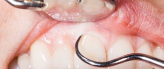
Oral abscess - symptoms and treatment
Symptoms of oral abscesses are variable and depend directly on the type and location of the abscess. In acute purulent periostitis, patients complain of pain in the area of the causative tooth or jaw segment, swelling of the soft tissues. The face of such a patient is asymmetrical.
When the causative tooth is localized in the frontal part of the upper jaw, the swelling is located in the upper lip and infraorbital region, the nasolabial fold is smoothed. If the diseased tooth is located in the frontal region of the lower jaw, swelling of the soft tissues is noted in the area of the lower lip and chin. When the causative tooth is located in the lateral part of the dentition, perifocal edema (near the infectious focus) is located in the buccal region.
Acute purulent periostitis is usually not accompanied by restrictions in mouth opening. Palpation of regional lymph nodes often reveals signs of acute lymphadenitis (enlarged lymph nodes). When examining the oral cavity, the causative tooth is identified, which usually reacts sharply to tapping (percussion). This is explained by the presence of a pathological process behind the root apex. When examining the vestibule of the oral cavity, a painful inflammatory infiltrate is determined, over which there is an edematous and hyperemic (red) mucous membrane. According to the literature, periostitis is most often located on the side of the cheek or lips, less often on the palatal and lingual side [5][8].
Often abscesses of the maxillo-lingual groove, buccal region, and pterygomaxillary space are considered as a complication of acute purulent periostitis. However, in some cases these diseases develop independently, so there is no reason not to consider them in this review.
Abscess of the maxillo-lingual groove is characterized by a more serious course. The patient complains of pain when swallowing, moving the tongue to the sides, and limited mouth opening. A visual examination reveals swelling of the submandibular area and acute lymphadenitis. Examination of the oral cavity is often difficult and is only possible after blocking the motor branches of the mandibular nerve. When examining the oral cavity, acute or aggravated periodontitis of the chewing tooth of the lower jaw or difficult eruption of the lower wisdom tooth is determined. When examining the maxillo-lingual groove, its bulging is determined; upon palpation, an inflammatory, sharply painful infiltrate can be detected.
With an abscess of the pterygomaxillary space, the patient notes an increase in body temperature, pain in the pharynx, difficulty swallowing, mouth opening is limited, in some cases almost impossible. Visually, perifocal edema is often absent. An examination of the oral cavity can be carried out only after blocking the motor branches of the mandibular nerve. In the oral cavity, difficult eruption of the lower wisdom tooth is usually detected, as well as a hyperemic and edematous pterygomandibular fold.
The clinical picture of an abscess in the buccal region largely depends on the depth of the abscess. With a superficial abscess, hyperemia (redness) of the skin, a local increase in temperature, the skin is tense and does not fold. With a medium and deep location, there is pronounced swelling of the buccal area, the skin is not externally changed, it is difficult to fold into a fold. Local hyperthermia (increased temperature) is usually not observed. When the abscess is deeply located on the mucous membrane of the cheek, marks from teeth are detected.
The condition of patients with these abscesses is usually assessed as moderate. Treatment is usually carried out in a maxillofacial surgery hospital under supervision in order to prevent the development of severe complications. Patients often exhibit symptoms of general intoxication of the body (fever, headaches and muscle pain).