Sometimes dentists jokingly say that they can diagnose any patient before he sits in the chair. “The wonders of X-ray vision” or unrivaled professional experience? I hasten to disappoint: neither one nor the other. The point is the widespread prevalence of periodontal diseases. And the diagnosis of “gingivitis” can be given in absentia to the vast majority without making a mistake. So, what is periodontium and what does it consist of?
The periodontium of a tooth is the tissue that surrounds it. More precisely, a complex of tissues. They are interconnected by their common location, development and functions. The structure of the periodontium has four main components:
- gum;
- periodontal ligament;
- tooth root cement;
- alveolar bone (alveolar part of the lower jaw and alveolar process of the upper).
Well, first things first. In this article we will look at the structure of the gums.
The gum is part of the oral mucosa that covers the alveolar bone (1) and surrounds the necks of the teeth (2).
The main task of the gum is to protect the tissues underneath it from mechanical and microbial damage.
Attached gum
From simple to complex: let's start with the attached gum. It is dense, elastic, firmly connected to the periosteum by collagen fibers. Wider in the incisor area (up to 4.5 mm in the upper jaw) than in the chewing teeth. Above, the attached gum borders on the marginal gum, below – on the mucous membrane of the alveolar bone. The border with the marginal gingiva is a small depression called the gingival groove (white arrow). And the mucous membrane of the alveolar bone originates at the mucogingival junction (black arrow).
Operations
The most common operation on the gum is gingivectomy - excision of the D. area within the patol, gingival pocket for periodontal disease. Benign tumors are removed along with the surrounding gum, often with the periosteum. When cutting out trapezoidal mucoperiosteal flaps during surgery on the alveolar process, the incision is made along the bottom of the gingival pocket; when suturing such a wound, they usually strive to restore the anatomical relationships of all elements of D. In acute odontogenic inflammation (periostitis, osteomyelitis of the jaw), an incision to open the abscess is made along the transitional fold, i.e., along the border of the gum with the mobile mucous membrane of the vestibule of the mouth.
Bibliography:
Bolotina Z. L. and Mikhailovskaya T. S. Current state of the issue of gum biochemistry, in the book: Problems, ter. Dentistry, ed. A. I. Marchenko, V. 4, p. 5, Kyiv, 1969; Vorobyov V. and Yasvoin G. Anatomy, histology, embryology of the oral cavity and teeth, M.-L., 1936; Gemonov V.V. Histochemical study of the activity of some enzymes in the mucous membrane of the human oral cavity, Dentistry, No. 1, p. 30, 1969, bibliogr.; Zhilina V.V. Glycogen content in the gingival epithelium in normal and pathological conditions, in the book: Theory and practice of dentistry, ed. L. I. Falina et al., c. 5, p. 150, M., 1961, bibliogr.; Kulazhenko V.I. Periodontal disease and its treatment using vacuum, p. 36, Odessa, 1960; Novik I. O. Periodontal disease (pathogenesis, clinical picture and treatment), Kyiv, 1964; Rybakov A. I. and Ivanov V. S. Clinic of therapeutic dentistry, M., 1973, bibliogr.; Uvarov V. M., Rusak M. K. and Kalinin V. I. Organs of the oral cavity in diseases of the blood, L., 1975, bibliogr.; Falin L. I. Histology and embryology of the oral cavity and teeth, M., 1963; Eichel V. Oxidative enzymes of gingiva, - Ann. NY Acad. Sci., v. 85, p. 479, 1960; Kraus BS, Jordan RE a. AbramsL. Dental anatomy and occlusion, a study of the masticatory system, Baltimore, 1969; Listgarten MA The infrastructure of human gingival epithelium, Amer. J. Anat., v. 114, p. 49, 1964; Plenk H. Topo-histochemische Untersuchungen an den Nerven der Gingiva, in the book: Histochem. d. Ultra-struktur, hrsg. v. H. Zimmermann, S. 439, Jena, 1971, Bibliogr.; Rateitscha k KH Prophylaxe und Frtihbehandlung der Gingivitis und Parodontitis, Soz.- u. Pravent. Med., Bd 20, S. 309. 1975, Bibliogr.; Schroeder HE a. Munzel-Pedrazzoli S. Correlated morphometric and biochemical analysis of gingival tissue, J. Microscop., v. 99, p. 301, 1973; Schultz - Haudt SD Observations on the acid mucopolysaccharides of human gingiva, Oslo, 1957, bibliogr.; Schumacher G. - H. Odontographie, Lpz., 1972, Bibliogr.; Schumacher G. - H. u. Schmidt H. Anatomic und Biochemie der Zahne, Stuttgart, 1972.
I. F. Romacheva.
Gingival sulcus
The depth of the gingival sulcus can be determined using a probe (periodontal, preferably). Normally it can be up to 3 mm. But it is worth considering that the gingival groove is not the same on different surfaces of the tooth: the approximal grooves are deeper, and they are shallower on the lingual and buccal sides. With many periodontal diseases, this depth increases (the answer to the question “why?” will be a little lower).
The gingival sulcus is lined with sulcular epithelium (or gingival sulcus epithelium). It is thin, non-keratinizing and is a kind of semi-permeable membrane for gingival fluid, on the one hand, and for microbial toxins, on the other.
Gingival fluid
Gingival fluid is transudate or exudate (more protein compared to transudate) from the connective tissue of the gum and its blood vessels. Normally there is very little of it - 0.5-2 ml; with inflammation this figure increases. The composition of this liquid is very diverse:
- Microorganisms;
- Leukocytes;
- Desquamated epithelium;
- Enzymes;
- Immunoglobulins;
- Other serum and connective tissue components.
It is logical that its composition determines the functions it performs: it cleanses the gingival sulcus, has antimicrobial properties and even improves the attachment of the epithelium to the tooth.
The gingival sulcus (2) is followed by the attachment epithelium (attachment, connective) (3).
Histological structure
In accordance with the histological characteristics, two components are distinguished in the structure of the gums. These include stratified epithelium and connective tissue.
In the case of epithelium, the basis of the basal layer are cells in the shape of a cube or cylinder, on top of which are sequentially located spiny cells, protein grains (keratohyalin), as well as the outer stratum corneum. The structure of the latter consists of flat cellular elements that do not contain nuclei and are subject to keratinization, which leads to its periodic renewal - due to deeper layers.
The absence of nerve endings or blood vessels in the epithelium is determined by the functional task of the layer. The goal is to prevent pathogenic bacteria and microbes from entering the periodontal tissue. In this case, there is a natural barrier between the basal layer and the connective tissue base, which forms in the form of a membrane septum.
The gum's own plate is two-layered:
- The upper layer is structurally a connective tissue with a loose structure, forming papillae oriented towards the surface and extending into the epithelium. The sensitivity of the gums is caused by the passage of blood vessels, nerve endings, and the central or trigeminal nerve through them.
- The deeper layer is characterized by a mesh structure and is formed from collagen fibers produced by fibroblasts. Uniting with each other, they move into the periosteum area, due to which the gums remain immobile, and the elements of the dentition are arranged in strict sequence.
The connective substance that unites and strengthens connective tissue cells is the matrix, a product of proteoglycan and glycoprotein molecules. The supply of blood is ensured by the gum branches - the location of the capillaries close to the edge of the tissue causes increased sensitivity of these areas.
Junctional epithelium
The junctional epithelium is a stratified squamous non-keratinizing epithelium (like the furrows). Formed during tooth eruption as a result of the fusion of the oral epithelium of the gum (1) + the primary enamel cuticle. Its length is very small - from 0.25 to 1.35 mm. Interestingly, at the apical end (the one closest to the enamel-cementum junction) its width can be only 1-2 cells. But, despite its far from impressive size, the attachment epithelium performs a number of important functions:
- it is a barrier against bacterial plaques;
- this is access of gingival fluid with protective components to the site of inflammation;
- this is a quick update (every 4-8 days), recovery from damage;
- and, finally, the ability to phagocytose epithelial cells (according to some scientists).
In the figure “Formation of the connective epithelium”: REE – primary enamel cuticle; OE – oral epithelium; JE – junctional epithelium
It is he who is destroyed by marginal gingivitis, and then by periodontitis. And it is precisely with the destruction of the attachment epithelium that the inevitable deepening of the gingival sulcus is associated (the promised answer to the question above).
Interdental papillae
A number of authors also make interdental papillae (interdental gum) a separate point in the anatomy of the gums. This is the gum that fills the space between the teeth below their contact point. But in terms of their structure, these are all the same parts - marginal (top and edges) and attached (central part).
The shape of the interdental papilla may vary depending on the size of the interdental space. So, in the area of the incisors it is smaller - and the interdental gum there is triangular (in the photo). In molars, its shape is more like a trapezoid.
The shape of the interdental gum may also change as a result of its recession:
Or there may be no interdental papilla at all. For example, at the site of a trema or diastema, this space is tightly connected to the alveolar bone (attached gum):
Since we have sorted out the gum anatomically, it’s time to talk about its histology. Of course, it is full of its own characteristics, but in general, like any mucous membrane, it consists of epithelium and connective tissue (the lamina propria of the mucous membrane).
A small flowchart about the gingival epithelium, so as not to get confused (checkmarks on what we have already talked about):
A few words about the oral epithelium. It covers the entire gum from the tongue, cheeks and lips. That is, the outer surface of the marginal and the entire attached gingiva. It is parakeratinized, or partially keratinized (difference from completely keratinized - in the surface, stratum corneum, there are remnants of the nucleus and organelles).
All its layers are visible in the figure:
- basal (on the basement membrane);
- spiny;
- grainy;
- horny;
- connective tissue.
Cells in its composition:
- keratinocytes (epitheliocytes) – the main ones;
- melanocytes (in figure M);
- Langerhans cells (white arrow) – macrophages;
- Merkel cells – tactile receptors;
The oral epithelium is a mechanical, chemical, water and microbial barrier. On the plus side, there are more specific protective reactions: the epithelium can respond to stimuli by changing proliferation (start dividing and regenerating faster) or cell differentiation, signaling, or even death.
Pathology
Damage to the gums
observed with fractures and dislocations of teeth, with jaw fractures, as well as during tooth extraction surgery. Damage to D. can be caused by chemicals. substances, for example, arsenic in the treatment of pulpitis; in this case, necrosis D. may occur. Thermal burns are possible.
As a rule, due to the increased regenerative ability of the oral mucosa, damage heals quickly and no special treatment is required. If there is a significant rupture of D., sutures are placed on the wound; In this case, soft, non-irritating food is recommended.
Inflammatory processes
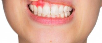
, acute and chronic, may be of local origin or be a symptom of a general disease of the body. Inflammation of the gums - gingivitis (see) - can occur as a result of the traumatic effect of an incorrectly manufactured denture, overhanging edges of a filling, or tartar; The pathological process may acquire the character of acute inflammation, hypertrophy or atrophy. Thus, often, as a reaction to chronic injury, granulations grow, which are partially or completely covered with a thin layer of epithelium, taking on the appearance of mushroom-shaped formations - gingival polyps.
D. inflammation occurs with pericoronitis, periodontitis (see), periostitis and osteomyelitis of the jaws (see Jaws); D. becomes swollen, hyperemic, and painful. D. is always involved in the process of periodontal disease (see).
Hyperemia and cyanosis of the gingival margin, as well as ulcerative gangrenous changes occur with C-vitaminosis (see Scurvy).
The gums, like the entire mucous membrane of the oral cavity, react to many pathol processes in the body, and gingivitis often occurs as a symptom of a general disease.
Often the first signs of diseases of the blood system are hemorrhagic, ulcerative-necrotic, hyperplastic processes in the D. In patients with gout. diseases due to the increased content of histamine in the blood serum, there is a persistent dilation of D.’s capillaries, which contributes to the occurrence of hron and inflammation in it. Some collagen diseases (systemic lupus erythematosus, scleroderma, etc.) are accompanied by catarrhal inflammation D. In some diseases of the cardiovascular system, radiation damage to the maxillofacial area, necrosis of the oral mucosa appears, which usually begins from the gingival margin.
Tuberculous lesion
D. is sometimes observed in active forms of pulmonary tuberculosis. At the same time, small gray-yellow tubercles appear on the D.; when they decay, painful superficial ulcerations of irregular outlines with seemingly corroded, pitted edges are formed, the gingival margin relatively soon becomes uneven, sometimes the epithelium of the bottom of the physiol. The gingival pocket loses contact with the tooth, a patol, gingival pocket is formed, and the necks of the teeth are exposed. Tuberculous lupus, when localized on the oral mucosa, according to G. A. Vasiliev and I. G. Lukomsky, also causes changes in the bone tissue of the alveoli, reminiscent of the picture of periodontal disease.
Syphilis
appears on the gums in all three periods. Hard chancre occurs more often in the area of the front teeth, sometimes in the form of a small spot of carmine-red color; in this case, an increase in regional lymph nodes is observed. Elements of the secondary period of syphilis on the oral mucosa are a spotted rash represented by papules, less often roseola. The tertiary period of syphilis is characterized by the appearance of focally located tubercles, usually in the area of two to four teeth, but a larger area may be involved in the process; the mucous membrane thickens, turns red, and becomes lumpy.
Actinomycotic process
in D. it develops as a result of the introduction of fungus through the patol, gum pocket;
There is no isolated D. lesion; actinomycosis of the soft tissues of the oral cavity usually develops. Rice.
1. Hard gingival fibroma (indicated by an arrow). Rice. 2. Fibromatous growth of the gums (indicated by an arrow). Neoplasms
. On D., a benign tumor on a stalk is more often observed - epulis (see). Fibroma D. also belongs to benign tumors (Fig. 1). Gingival fibromatosis differs from fibroma in the diffuse nature of the growth of fibrous connective tissue, the spread of the process to the gums as a whole; sometimes the disease is limited to several teeth (Fig. 2), often on both jaws. Fibromatous growths can cover a significant part of the crowns of the teeth and interfere with the closure of teeth and chewing. Unlike hypertrophic gingivitis, fibromatous gums are dense and there are no signs of inflammation.
A cancerous ulcer may be detected on the gum, usually resulting most often from a cancerous tumor of the jaw (see Jaws); Unlike an ulcer of inflammatory origin, it is painless, without a tendency to epithelialization.