Otitis externa is an inflammation of the outer ear, which includes the pinna and external auditory canal (EA).
Author:
- Filatova Evgenia Vladimirovna
otolaryngologist, rhinosurgeon
3.63 (Voted by: 
- Causes of a boil in the external auditory canal
- Causes of development of bullous otitis media
- Treatment
Otitis externa is an inflammation of the outer ear, which includes the pinna and external auditory canal (EA). Like most inflammatory diseases of the ENT organs, otitis externa can be acute or chronic.
Features of the structure of the outer ear
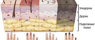
The auricle and ear canal are covered with skin that extends to the eardrum. There are two sections in the ESP: cartilaginous and bone; at the border of these sections, a physiological narrowing is determined - the isthmus, which is why the three-dimensional model of the external auditory canal resembles an hourglass. Under the skin of the auricle and the cartilaginous part of the external auditory canal there is elastic cartilage, due to which this part of the auditory canal is mobile during chewing and articulation. In the bony section, under the skin, there is periosteum and bone. The skin of the external auditory canal differs from any other skin in that its thickness contains cerumenous glands that produce earwax (cerumen). The function of cerumen is to protect the skin from constant moisture and microorganisms, and the presence of a cerumen film is an important factor in the normal functioning of the outer ear. The dimensions of the external auditory canal may vary markedly depending on individual characteristics. Often in the area of the isthmus there are osteophytes - bony protrusions that reduce the lumen of the ESP, and sometimes completely block it. Anterior, posterior and inferior to the auricle in the subcutaneous tissue there are lymph nodes that can become inflamed with otitis externa.
Acute external otitis is an inflammation of the external ear that occurs within 1 month.
Forms of external otitis:
- diffuse external otitis;
- local external otitis or NSP boil;
- acute bullous (hemorrhagic) external otitis;
- myringitis;
- dermatitis of the auricle;
- erysipelas of the auricle;
- malignant (necrotizing) external otitis;
- chondroperichondritis of the auricle.
The most common is acute diffuse external otitis, in which bacterial inflammation of the skin of the entire ear canal occurs, often spreading to the eardrum or the skin of the auricle.
Acute diffuse otitis media
Provoking factors: excessive toilet of the ears, water getting into the ear, microtrauma of the skin of the NSP, factors of decreased immunity (hypothermia, excessive tanning, ARVI, chronic diseases). Pathogens: Staphylococcus aureus, Pseudomonas aeruginosa.
Symptoms
This is severe pain in the ear, ear congestion, liquid discharge from the ear canal in a small amount, there is also an increase in the behind-the-ear or parotid lymph nodes, inflammation of the skin of the auricle, there may be an increase in temperature, and symptoms of intoxication. On examination, hyperemia and swelling of the skin of the NSP are noted, the skin is uneven, with overlays of transparent yellow or purulent discharge, and desquamated epidermis is visible. The lumen of the ESP is often sharply narrowed and the eardrum may not be visible. If it is visible, it is usually also uneven and moderately hyperemic. Enlargement and tenderness of regional lymph nodes, thickening, tenderness and hyperemia of the auricle are often detected. Hearing, while maintaining the lumen of the lumen, is slightly reduced or normal.
Diagnostics
To make a diagnosis, in addition to examination, it is necessary to exclude middle ear disease, for which tuning fork tests (a hearing test using a set of tuning forks), tympano- and impedance measurements (measurement of the mobility of the eardrum), and a study of hearing thresholds (audiometry) are performed. These studies are carried out to exclude diseases of the middle ear. Laboratory diagnostics includes a general blood test, blood sugar, microflora culture from the diseased ear to prescribe antibacterial therapy.
Treatment
The main direction of treatment is antimicrobial and anti-inflammatory therapy. Antibiotics and anti-inflammatory drugs (non-steroidal anti-inflammatory drugs, corticosteroids) are prescribed locally or systemically. For severe pain, there is a good effect from behind-the-ear blockades with corticosteroids and local anesthetics. A gentle ear toilet and rinsing is carried out to free the ear from discharge and epidermal masses. Additionally, you can use physical treatment methods: quartz tube or laser therapy.
Advantages of the EMC Clinic
EMC Aesthetic Clinic is a leading Moscow clinic in the field of cosmetology and plastic surgery. EMC has been providing medical services for more than 27 years, strictly observing international protocols and representing the highest level of medicine.
The high professional skills of EMC specialists allow us to deal with the most complex cases and show amazing results in our work. The appearance of the ears and hearing acuity do not always radically change life, but they always affect its quality. Correction, reconstructive restoration of the auricles, restoration of hearing function, EMC’s task is to help its patients in any situation at the highest level of medical capabilities.
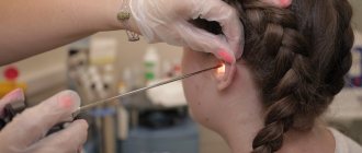
Limited (local) otitis externa or boil of the external auditory canal
A furuncle is an inflammation of the hair follicle that spreads to the subcutaneous tissue. Bristly hairs grow at the entrance to the NSP and around it, and inflammation develops in their follicles.
Causes of a boil in the external auditory canal
The most common pathogens are streptococci. Provoking factors and symptoms are similar to those of diffuse otitis media.
Diagnostics
Upon examination, a narrowing of the ESP is visible due to infiltration in the area of one of its walls. The boil goes through three stages in its development:
- Infiltration stage: at this stage, a painful infiltrate of the NSP skin is formed, local hyperemia and thickening of the skin about 0.5-1.5 cm in size appear.
- The abscess formation stage is the appearance of a purulent cavity and a necrotic core in the area of infiltration. At this stage, the presence of an infiltrate is also determined, in the center of which a yellow-green purulent core is visible, usually with a hair in the center; when pressing on the infiltrate, fluctuation (the presence of fluid in it) can be determined.
- The resolution stage is the release of pus from the cavity with gradual recovery.
Treatment
Treatment varies and depends on the stage. At the infiltration stage, systemic and local antibacterial and anti-inflammatory therapy is prescribed. In the stage of abscess formation, it is worth performing surgical treatment of the boil - opening and draining, cleaning the cavity from pus, dressings are performed within several days. Antibacterial and anti-inflammatory drugs are also prescribed. During the resolution stage, it is sufficient to use only local antibacterial drugs and ear toilet.
Acute bullous or hemorrhagic otitis externa
This is a form of inflammation in which blisters (bullas) containing bloody fluid appear on the skin and on the outer surface of the eardrum. If such a bubble is opened, then bloody discharge flows out of the ear.
Causes of development of bullous otitis media
Most often, bullous otitis occurs against the background of acute respiratory viral infection or influenza, but a bacterial infection can also be the cause.
Treatment
An ear canal toilet is required. Antibacterial and anti-inflammatory drugs are prescribed locally, and drugs that strengthen the vascular wall and reduce vascular permeability should also be prescribed systemically. Opening bullae is not recommended.
Dermatitis of the ear
It can be separated into a separate form, since inflammation of the skin of the auricle often occurs, without involving the NSP. The causes of the disease are a bacterial infection plus a factor of sensitization of the body to bacterial toxins. With this type of inflammation, redness and thickening of the skin occurs, with the formation of small serous blisters, similar to those in eczema. When the bubbles open, yellow crusts form in their place. Diagnosis is made based on examination, and consultation with a dermatologist may be required. Treatment is also antibacterial and anti-inflammatory, and antihistamines may be prescribed.
Correction of protruding ears in adults and children
Correcting protruding ears is one of the most common reasons for visiting a plastic surgeon. Prominent ears are not considered a medical pathology, so ear plastic surgery in this case is purely aesthetic.
Severe protruding ears can become the basis of psychological trauma and the cause of ridicule from others. An important feature of plastic surgery for protruding ears is the minimal level of intervention during surgery. The guaranteed safety of the operation allows otoplasty to be performed on a child.
Ear plastic surgery in children at EMC is performed from the age of 7, when the auricles are almost completely formed. The healing process goes quite quickly, the ears after otoplasty look natural and do not bother the child, after the healing process is completed the child will not even remember about the operation. Even upon closer inspection, no one will notice the small strip of cosmetic seam behind the auricle.
Erysipelas of the auricle
Acute erysipelas is an infectious disease and is caused by streptococcus pyogenes; later, other flora may join. With such inflammation there are symptoms of skin damage and a general inflammatory syndrome - weakness, fever, chills. Factors provoking the disease: decreased immunity, abrasions and burns of the skin, swimming in polluted waters, contact with a carrier of pyogenic streptococcus or a sick person. Depending on the depth of damage to the skin, three forms are distinguished:
- Erythematous, when pain occurs, redness of the skin (it has a “varnished” appearance) slightly rises above the level of healthy skin and has a clear border with it.
- The bullous form, more severe, is characterized by the same skin lesions as in the erythematous form, but with the formation of large blisters above the surface of the skin with transparent or cloudy contents.
- Necrotic form. With this form, deep damage to the skin occurs, right down to the reticular and papillary layers, and deep ulcers with purulent plaques form. Due to the addition of various microflora, it is more severe than other forms. Typically, patients with this form require surgical debridement of areas of necrosis.
Forecast
The outcome of perichondritis of the costal cartilages is generally favorable. The aseptic type of pathology is a typical inflammatory lesion, the general principles of treatment of which have been successfully developed.
The prognosis worsens with the purulent type of the disease. In this case, timely diagnosis and adequate surgical treatment will help to cope with the disease.
Patients should remember that self-medication is fraught - especially the notorious overheating, which in the case of a purulent process can lead to critical consequences and even the death of the patient.
Kovtonyuk Oksana Vladimirovna, medical observer, surgeon, consultant doctor
18,063 total views, 3 views today
( 42 votes, average: 4.50 out of 5)
Abrasions and abrasions: first aid, items from a personal first aid kit, medicinal plants
Tuberculosis of bones and joints
Related Posts
Erysipelas
This is a serious disease that often causes complications, and its treatment should take place in an infectious diseases hospital. If erysipelas is suspected, consultation with an infectious disease specialist is required. Isolated erysipelas of the auricle is rare and is usually combined with lesions of the skin of the face or neck.
Treatment
Massive antibacterial and anti-inflammatory therapy, detoxification therapy, and antistreptococcal gamma globulin are prescribed.
Differential diagnosis
Differential (distinctive) diagnosis of perichondritis of the costal cartilages is primarily carried out with such pathologies as:
chondritis – inflammatory lesion of the costal cartilage;- intercostal myositis - an inflammatory process in the intercostal muscles;
- intercostal neuralgia is a pain syndrome that develops when intercostal nerves are damaged of various types;
- mediastinitis;
- tumor of the chest wall.
Malignant otitis externa (necrotizing otitis externa)
This form of external otitis is distinguished as a separate one, but in fact it is a negative course of ordinary diffuse external otitis. During the course of the disease, inflammation spreads to the structures of the temporal bone, damage to the cranial nerves and the development of intracranial complications may occur. The name of the disease is not associated with the development of a malignant tumor, it simply reflects the course of the disease. Fortunately, this disease is rare; it affects patients with severe immune disorders, such as AIDS, conditions after radiation and chemotherapy, decompensated diabetes mellitus, and patients receiving immunosuppressive and cytostatic therapy. The leading pathogen is Pseudomonas aeruginosa, but any infection is polymicrobial. During the process of inflammation, necrosis of the skin of the ear canal occurs and inflammation passes to the periosteum and bone, followed by its destruction. Complaints in this disease include severe pain, discharge from the ear of a purulent-inflammatory nature, with a putrid odor, and hearing loss quickly occurs. Later, symptoms of damage to the cranial nerves appear: difficulty swallowing, choking when eating, numbness of the face, paralysis of the facial muscles. When the process spreads to the middle and inner ear, systemic vertigo may develop. When inflammation spreads into the cranial cavity, cerebral and meningeal symptoms develop. On examination, one notices pronounced inflammation of the skin of the NSP, skin with foci of necrosis, a large amount of purulent-necrotic discharge, the eardrum and the structures of the middle ear are often indefinable. Symptoms of damage to the cranial nerves are determined - facial asymmetry, ptosis of the lower eyelid on the affected side, asymmetry of the soft palate, impaired mobility of the epiglottis. In diagnosis, culture for bacteriological examination and computed tomography of the temporal bones are important. Treatment is always carried out in a hospital setting. Massive antibacterial and detoxification therapy is carried out. Infusions of human gamma globulin, antipseudomonas serum or hyperimmune human plasma are prescribed. Surgical treatment is also performed - necrotomy (removal of necrotic areas of skin and bone), and subsequently, upon recovery, plastic surgery of the defect is performed using one’s own tissues.
Causes
All the main causes of the development of perichondritis of the costal cartilages can be divided into groups:
- physical;
- chemical;
- infectious.
The physical reasons for the formation of this pathological process are:
- mechanical;
- thermal;
- radioactive.
Mechanical reasons that can provoke the development of perichondritis of the costal cartilages include injuries:
- non-penetrating;
- penetrating.
Non-penetrating injuries are bruises of the chest in the area of the costal cartilages. The chest wall is often injured, this explains the “leadership” of injuries among other factors that cause an inflammatory process in the perichondrium of the costal cartilages. Injury can happen:
- household;
- production;
- sports;
- criminal.
A separate mechanical cause of the occurrence of perichondritis of the costal cartilages is violation of the integrity of the periosteum during operations on the chest.
Everyday life is a “storehouse” of conditions for trauma to the chest with the subsequent development of perichondritis of the costal cartilages. Bruises can be easily caused by:
- due to inappropriate living conditions - in particular, cramped rooms filled with furniture;
- when bulky objects (mezzanines, shelves) fall;
- as a result of noisy children's games or capricious behavior of a child, who can purposefully or unintentionally hit the mother in the chest area.
Injury in industrial conditions, which can lead to the development of perichondritis of the costal cartilages, occurs in most cases when the principles of labor protection are not observed at work or when safety rules are ignored. By production we mean conditions not only in a factory or plant, but also in agriculture.
Thus, the occurrence of the described disease is quite often observed in workers who care for farm animals - a horse can hit the chest with its hoof, a cow can butt it with its horn, and so on.
Sports injury is also one of the most common causes of perichondritis of the costal cartilages. Athletes who engage in strength and team sports are mainly exposed to traumatic danger - these are:
- wrestlers;
- boxers;
- hockey players;
- basketball players;
- football players
and so on.
Also often injured:
- skiers;
- cyclists;
- jumpers.
Somewhat less frequently, perichondritis of the costal cartilages is diagnosed in people who have been subjected to physical violence (beating):
- in family;
- in a criminal environment.
Penetrating wounds of the chest, which can lead to the development of the described pathology, are puncture, cut, lacerations, chopped, and gunshot wounds.
The thermal factor that can provoke the development of perichondritis of the costal cartilages is mainly low temperatures. Banal ignorance of clothing according to the season can provoke the occurrence of this pathology. It develops especially often against the background of frostbite.
But critically elevated temperatures can also cause the formation of this disorder - for example, inflammatory processes in the perichondrium can occur during burns.
The radioactive factor leads to the development of perichondritis of the costal cartilages less often than other physical factors. Most often this happens due to:
- frequent chest X-ray examinations;
- radiation therapy prescribed for malignant tumors of the chest organs;
- contact with radioactive substances or equipment due to professional activities - often due to the same personal failure to comply with safety regulations or inadequate labor protection conditions at work.
Chemical factors that can lead to the development of perichondritis of the costal cartilages include chemicals that entered the perichondrium tissue by contact, through the blood or lymph flow, and provoked the occurrence of an inflammatory process in it.
Such toxic compounds are:
- exogenous;
- endogenous.
Exogenous provoking toxins include chemicals that are used in everyday life, in production and in agriculture.
Endogenous toxins are those produced in the human body. Most often their synthesis occurs:
- directly in human tissues due to a pathological process - most often suppuration and necrosis (death);
- during the development of an infectious process - such toxic substances are toxins of microorganisms that are released during their life activity or are formed as decay products of dead microbial bodies.
In the latter case, damage to the perichondrium develops against the background of a general infectious disease:
- malaria;
- flu
Less commonly, perichondritis of the costal cartilages is formed due to a local infectious lesion.
As for the specifics of the pathogen, the disease described is mainly caused by damage to a nonspecific infectious agent - this is:
- staphylococci;
- streptococci;
- Proteus;
- coli
and others.
Very rarely, specific tuberculous and syphilitic perichondritis of the costal cartilages are formed, but the possibility of their development should also be remembered.
Purulent perichondritis of the ribs often appears due to reasons such as:
- open chest injury accompanied by damage to the costal cartilages;
- contact spread of infection.
The latter can occur with diseases such as:
- mediastinitis – inflammation of the mediastinum (organs located between the lungs);
- pleural empyema – diffuse purulent lesion of the pleura;
- osteomyelitis of the sternum and ribs (for example, post-traumatic) - purulent melting of their bone tissue, which leads to the formation of fistulas (pathological tracts).
Chondroperichondritis of the auricle
This disease is an inflammation of the perichondrium and cartilage of the auricle caused by bacterial flora. Chondroperichondritis can be caused by trauma to the auricle with damage to the skin, suppuration of an auricular hematoma, deep burns of the auricle, a boil or suppurating atheroma of the ear, and erysipelas. There are infiltrative and purulent forms. A few days after the described traumatic factor, infiltration and hyperemia of the skin of the auricle develops, accompanied by pain, with or without fever. An important diagnostic criterion is that the earlobe remains unaffected, since there is no cartilage in its thickness. Treatment: antibacterial and anti-inflammatory therapy. If there are purulent cavities, an opening with removal of pus and gentle necrotomy of the destroyed cartilage is performed. The disease is difficult to treat and can last for weeks or months. Often the consequence of chondroperichondritis is cicatricial deformation of the auricle; sometimes there may be complete destruction of the cartilage.
Symptoms of rib perichondritis
The clinical picture of perichondritis of the costal cartilages largely depends on its shape.
Important
With aseptic perichondritis, symptoms, having arisen, will increase, then a gradual regression of symptoms is noted. At the same time, the symptoms of the purulent form of the described pathology can only increase over time - this state of affairs is explained by the destruction (destruction) of cartilage and the formation of fistulas.
The clinical picture of aseptic perichondritis is manifested by local signs - these are:
- pain;
- breathing disorder.
Characteristics of pain syndrome:
- by localization - in the area of the affected costal cartilages;
- in terms of distribution - there is no irradiation as such, but in some cases pain can spread to the bone fragment of the affected rib;
- by nature - the “range” of pain sensations is different, it can be stabbing, aching, tugging pain;
- by severity - intensity depends on the degree of damage, as well as the pain threshold of the patients. Often, with minor perichondritis of the costal cartilages, patients complain that pain prevents them from working, sleeping and generally living as usual;
- by occurrence - they appear almost with the beginning of the development of the pathological process in the perichondrium, and as it progresses they intensify. Also, increased pain is observed with movements and deep breathing.
With the aseptic form of the described pathology, the general condition often does not worsen.
The purulent form of perichondritis of the costal cartilages manifests itself:
- local signs;
- violation of the general condition of the body.
Local symptoms are most indicative. When visiting a doctor, the patient complains of typical signs of suppuration, which successively follow each other. First, an infiltrate forms in the affected area - tissue compaction. After a certain period of time (on average it can last from 3-4 to 7 days), the dense lesion softens. In severe cases, inflammation can spread to the entire lower chest and upper part of the anterior abdominal wall. The formed abscess is opened - often through the skin (in this case the patient complains of purulent discharge from the “hole”), less often through the posterior perichondrium (pain in the chest will bother you). In the first case, a fistula is formed, in the second - purulent leaks in the soft tissues.
The characteristics of pain with purulent perichondritis of the costal cartilages are as follows:
- by localization - in the area of the affected costal cartilages;
- by distribution - along the ribs on the affected side;
- by nature - at first vague aching, then twitching, with a large abscess - pulsating;
- in terms of intensity - in the early stages the disease is insignificant, as the abscess matures it increases and can be very strong;
- by occurrence - appear when the first inflammatory changes appear in the perichondrium, intensify with any activity in the chest area on the affected side.
Signs of a general condition disorder are:
- hyperthermia – increased body temperature. Can reach 38.5-39.0 degrees Celsius;
- general weakness caused by the products of the purulent process entering the bloodstream;
- malaise;
- loss of appetite;
- decreased performance – both mental and physical.