Article for the “bio/mol/text” competition: “The boy who was left without skin.” This is not the title of another horror film. This is a documentary film directed by Patrick Collerton, who captured the last months of the life of 36-year-old Johnny Kennedy, who suffered from a rare genetic disease - dystrophic epidermolysis bullosa. The film reached five million viewers across the UK and helped raise £500,000 for charity DEBRA, an international association dedicated to the research and treatment of epidermolysis bullosa. The life of people with this diagnosis and their relatives is a path that not everyone can overcome: it is difficult both physically and mentally, and requires courage and an incredible will to live. Children suffering from this disease are called butterfly children, comparing the fragility of their skin to the fragility of a butterfly's wing. What is epidermolysis bullosa and what are the prognoses for the future?
Note!
This work was published in the “best review article” category of the “bio/mol/text” competition 2015.
The sponsor of the nomination “Best article on the mechanisms of aging and longevity” is the Science for Life Extension Foundation. The audience award was sponsored by Helicon.
Competition sponsors: Biotechnology Research Laboratory 3D Bioprinting Solutions and Scientific Graphics, Animation and Modeling Studio Visual Science.
I know only one way to be in harmony with your own conscience: this way is not to shy away from suffering.
Antoine de Saint-Exupery
General information
Epidermolysis bullosa (abbr. EB) is a concept that unites a whole group of rare hereditary diseases that have genetic and clinical heterogeneity. Most often, pathological processes occur in keratinocytes and lead to the formation of blisters with a benign course, the appearance of erosive areas of the skin and mucous membranes, increased vulnerability and sensitivity of the skin to any minor mechanical trauma, and the development of “ mechanobullous disease ,” also called Butterfly syndrome . The occurrence of different types of pemphigus is only 1 case per 30-100 thousand people.
The concept of epidermolysis bullosa was introduced by the German dermatologist Heinrich Koebner back in 1886, although similar cases of skin diseases had been encountered earlier.
Life expectancy with epidermolysis bullosa
Despite the rapid development of global medicine, the life expectancy of patients with congenital epidermolysis – “butterfly children” – is most often not high. It all depends on the subtype of the disease, the depth and extent of the pathological lesions. With proper care and complex therapy, simple emidermolysis bullosa is easier, the number of blisters is minimized with age, and the prognosis for such patients is favorable. Other types of genodermatous disease, especially with the addition of infections, most often staphylococcal or streptococcal, with the development of systemic complications and sepsis most often lead to death in the early years of children's lives.
Treatment
Unfortunately, there is currently no cure for EB. The main therapy is aimed at preventing the formation of new blisters and erosions, treating wounds and preventing their infection. The main objectives of treatment are to protect patients’ fragile skin from mechanical stress (using special dressings) and treating existing wounds (using antiseptics, applying bandages).
But science does not stand still, and scientists around the world are desperately looking for a cure for this terrible disease. Three promising types of therapy for BE have been most developed: protein, cellular and gene therapy. Let's look at these approaches in more detail.
Protein therapy
With this approach, a sufficient amount of normal protein is introduced into the patient's body. Studies have shown that intradermal injection of purified human collagen VII into mice with recessive dystrophic EB leads to the formation of normal collagen fibrils at the junction of the epidermis and dermis. When purified protein is administered intravenously, systemic deposition of collagen VII in the skin is observed. It has also been shown that such therapy not only improves the functional state of the skin, but also promotes wound healing. Based on the results of preclinical studies, Lotus Tissue Repair, which later became part of Shire Pharmaceuticals, began conducting extensive preclinical studies of intradermal and intravenous injections of collagen VII in a canine model of recessive dystrophic EB, as well as the first stage of clinical studies [3, 5].
Cell therapy
With this method of treatment, the necessary and sufficient number of cells containing a normal gene encoding the desired protein are introduced into the patient’s body.
One of the promising types of cell therapy is intradermal injection of allogeneic (obtained from healthy donors) fibroblasts. Thus, it was shown that such an approach restores the synthesis of collagen VII and stabilizes the junction of the epidermis with the dermis in a mouse model of recessive dystrophic EB. The next step towards the implementation of this type of therapy has already been taken: several patients with recessive dystrophic EB were injected with allogeneic fibroblasts. Although the injected cells were not detectable two weeks after the injection, collagen production levels remained elevated both after two weeks and three months. Scientists believe that the main therapeutic effect of allogeneic fibroblast injection is to increase the production of the epidermal growth factor HB-EGF, as well as to increase the expression of the collagen VII gene in the recipient's keratinocytes and fibroblasts. However, the mechanism of this phenomenon has not yet been established [3, 6].
Allogeneic bone marrow transplantation may also be a promising treatment for BE. Thus, in a mouse model of recessive dystrophic EB, it was shown that bone marrow transplantation leads to relief of the symptoms of the disease. Following encouraging results, clinical trials of a similar therapy method were started. However, the results so far are disappointing: five out of twenty patients died from disease progression or complications after transplantation [3, 6].
Gene therapy
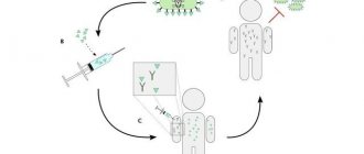
Finally, the last and perhaps most promising approach is gene therapy (Fig. 6). In this case, patients are transplanted with autologous (made from their own cells) transplants, in the cells of which the defective gene is replaced with a normal one using genetic editing methods. And although such a procedure does not lead to complete healing, it can temporarily eliminate the symptoms of EB, and is also relatively simple in technical terms.
Figure 6. Editing your own DNA in the treatment of EB. a — The principle of gene therapy. At the first stage, epidermal stem cells are cultured from skin biopsies of a patient with EB. The stem cells are then subjected to a genetic editing procedure, which replaces the defective gene with a normal one. Once the genetically edited keratinocyte layer is tested for safety, it is transplanted into patients. b — Transplants formed from “correct” keratinocytes of a patient with recessive dystrophic EB. Figure from [7].
The first successes of gene therapy were achieved in Italy in 2006, when the normal LAMB3 gene, encoding one of the subunits of the laminin-332 protein, was delivered into the epidermal stem cells of a patient with borderline EB using a retroviral construct. Epidermal grafts were created from the "corrected" cells and then transplanted onto the patient's legs. As a result, they formed functionally complete skin. The study showed that the survival of even a small number of stem cells in such grafts leads to the successful restoration of normal skin functions. Eight years later, production of the target LAMB3 protein was still maintained in the patients' skin. There were no blisters or signs of inflammation, tumor growth or any immune response in the graft area. In July 2014, a second similar operation was carried out in Austria. Professor Alfred Lane from Stanford University (California) and his collaborators began clinical trials of a similar technology aimed at restoring the synthesis of collagen VII in epidermal stem cells of a patient with recessive dystrophic EB. 30 days after surgery, no abnormalities were noted in the transplanted areas, and the level of collagen VII production was much higher than baseline [7].
Of course, in the case of gene therapy, there are many difficulties. Thus, gene replacement using viral constructs and non-viral DNA editing systems (ZFN, TALEN and CRISPR/Cas9)* is associated with the risk of inserting the gene into a location other than the target one in the genome. It is quite difficult to assess the consequences of such embedding. Therefore, this approach requires great caution.
* — The ZFN, TALEN and CRISPR/Cas9 methods are based on the site-specific action of nucleases in vivo. These systems usually consist of two modules, one of which recognizes the target oligonucleotide sequence, and the other cuts the DNA strands: “Shouldn’t we try to... change the genome?” [8], “CRISPR systems: immunization of prokaryotes” [9], “Mutagenic chain reaction: genome editing on the verge of science fiction” [10]. - Ed.
Another problem with such therapy is the possible risk of developing an immune response to the new protein being expressed. The risk is especially great for those patients whose cells carried mutations that completely eliminate the synthesis of a certain protein. At the same time, it is known that some patients with EB have areas of skin that do not form blisters and produce increased levels of protein. This phenomenon is called reverse mosaicism, or natural gene therapy, and can prevent the development of an immune response to the protein synthesized as a result of gene therapy. Moreover, such “self-healing” cells can be used for cell therapy, and then the need for gene correction will disappear. However, so far attempts to create transplants based on such cells in patients with borderline EB have not been successful due to the small number of such cells in the transplanted material [11].
Pathogenesis
The pathology is detected from birth - on the skin there are blistering formations with serological contents, which, when opened after about 2-3 days, leave long-term non-healing erosions, most often leading to the development of atrophic scar tissue, hyper- or hypokeratosis , and re-development of blisters. There are known cases of the disease manifesting itself at a later age - in infancy, early childhood and even adolescence.
Skin damage - the occurrence of ulcers and blisters is caused by any mechanical impact, so the areas with the most frequent friction - the armpits and various folds, the places where clothes and straps fit - are the most affected.
The pathology is based on mutations in genes encoding proteins of various layers of the skin - basement membranes, dermis. In this case, an imbalance occurs in the system of enzymes and inhibitors, proteins become the target of attack by the body’s defense systems - enzymatic cytolysis, causing swelling of the cytoplasm , rupture of cell membranes and, as a result, disruption of intercellular connections, the structure of the epidermis with the formation of intra- or subepidermal bullae and vesicles.
New skin for the butterfly
In November, Nature
published an article, the authors of which, biologists and doctors from Italy and Germany under the leadership of Michele De Luca, spoke about the successful experience of transplanting transgenic skin to a patient suffering from a severe genetic disease - epidermolysis bullosa. Because the new skin was grown from the patient's own skin, it took root and, most importantly, allowed the patient - currently a nine-year-old boy - to feel healthy. This event is a real breakthrough in the field of treatment of so-called “butterfly children”. We talked with the staff of the Butterfly Children charity foundation about its significance and clinical prospects, as well as what epidermolysis bullosa is in general, what its types and features are, how many patients suffer from it in Russia and what kind of help they receive.
Epidermolysis bullosa (EB) is a rare genetic disease in which blisters and erosions occur on the skin and mucous membranes. It can affect certain areas of the skin (most often the feet and hands), or it can affect its entire surface. The disease is sometimes very difficult - the skin of those affected is sensitive to even minor mechanical influences, and in some forms of EB it is very easily damaged.
Patients with epidermolysis bullosa require ongoing care and treatment aimed at preventing injury and eliminating itching, pain and other complications. The disease affects many internal systems of the body. Therefore, patients need a multidisciplinary treatment approach: active dynamic observation and treatment not only from dermatologists, but also from other specialists, such as surgeons, dentists, gastroenterologists, pediatricians, oncologists, ophthalmologists, hematologists, psychologists, nutritionists.
What happens to the skin
EB manifests itself in early childhood and can significantly affect quality of life and also be a cause of premature death. Children suffering from this disease are often called “butterflies” - by analogy with fragile insects whose wings pollen cannot withstand even the lightest touch.
Recall that human skin consists of epidermis, dermis and subcutaneous fat. The epidermis, in turn, consists of five layers, the lowest of which is located on the basement membrane. The basement membrane on the upper side is called the light lamina of the skin (lamina lucida), and on the dermis side it is called the dense lamina (lamina densa). Basement membrane proteins provide “anchoring” of the epidermis to the dermis.
EB occurs due to mutations in genes encoding proteins in various layers of the skin. Depending on which layer bubbles form, BE is divided into four types. Have a simple form
the targets are proteins of the upper layers of the epidermis, keratins, plakophilin-1, desmoplactin and others.
The borderline form
affects the level of the light plate of the skin - namely, laminin proteins, type XVII collagens and integrins.
In dystrophic EB,
type VII collagens from the papillary layer of the dermis, below the dense lamina of the skin, are damaged.
Finally, if the blisters form at different levels, it is called Kindler syndrome
and is associated with mutations in the Kindlin-1 protein gene.
Different proteins perform different functions, but in general they are all structural proteins that ensure the integrity of the layers of skin and their connection with each other. When these proteins are missing or their structure is damaged, the skin becomes very vulnerable to damage, and in addition, blisters similar to burns constantly form in it. One of the most common and unpleasant symptoms of the disease is frequent pain.
Charitable Foundation "Butterfly Children"
Share
Where does the disease come from?
“Broken” genes are inherited from parents in an autosomal dominant or autosomal recessive manner. “Autosomal” inheritance means that damage occurs in genes that are not located on the sex chromosomes (X or Y) - that is, this disease is not directly related to gender. In the case of “dominant” inheritance, it manifests itself even when only one of the parental genes is broken, and in the case of “recessive” (more common) inheritance, it is necessary that the child receive mutations from both parents.
In borderline EB, mutations are most often found in the LAMB3 gene. Typically, borderline EB is inherited in an autosomal recessive manner, but, for example, cases of somatic mosaicism are known, when the defect manifests itself in some cells, but not in others, and then the skin begins to resemble a mosaic, that is, it becomes heterogeneous in terms of diseases.
How to diagnose and treat
Doctors make the diagnosis of epidermolysis bullosa without difficulty; most often, the specific form of the disease is immediately clear. More detailed diagnosis can be carried out by examining skin samples taken during biopsy using transmission electron microscopy or immunohistochemistry (indirect immunofluorescence). In this case, the absence or decrease in the level of various structural proteins of epidermal cells (keratinocytes) and the basement membrane is determined. In this way, EB is classified - the structural protein whose gene is broken in the patient is identified, and a clinical prognosis is obtained. Genetic tests are also carried out to reliably confirm the diagnosis.
Until today, there were no treatment methods for any of the forms of EB, with the exception of symptomatic ones - dressing and wound treatment. The patient needs to constantly make efforts to ensure that his skin is injured as little as possible - in addition to severe discomfort and pain, the skin with EB is susceptible to infections, and erosions (destroyed areas of skin that appear at the site of damaged blisters) significantly increase the risk of developing cancer.
Transgenic skin
Recently, a major breakthrough occurred in the scientific and medical communities - Italian and German scientists, led by Professor Michele De Luca from the Stefano Ferrari Center for Regenerative Medicine, managed to create transgenic skin from the own cells of a seven-year-old boy with borderline EB and replace 80 percent of his skin with it.
Using genetic engineering methods, a working copy of the LAMB3 gene was introduced into the cells, as a result of which the symptoms of the disease in the transgenic tissue disappeared. Due to the fact that a working copy is introduced into stem cells, such tissue renews itself. Two years have passed since the transplantation, and the boy is alive, his skin is healthy, he is studying at school and playing sports, although before that he was in a hospital ward, needed morphine daily and was fed only through a tube. You can read more about this here.
In their work, scientists also talk about their previous patient, a boy who had leg skin transplanted in 2006. Eleven years have passed since then, and the transgenic areas still maintain their normal functions. This, in turn, allows us to hope for successful long-term results of such operations.
Charitable Foundation "Butterfly Children"
Share
Charitable Foundation "Butterfly Children"
Share
With questions about epidermolysis bullosa, how doctors assess Michele de Luca’s achievement, and about the prospects for treating EB in Russia, we turned to dermatovenerologist Natalia Mikhailovna Marycheva, an employee of the Butterfly Children charity foundation and the Moscow Scientific and Practical Center for Dermatovenereology and cosmetology.
N+1: How common is this disease? How many people have it in Russia?
Natalia Marycheva:
Currently, there are no accurate statistics on people suffering from epidermolysis bullosa in our country. About three hundred children are registered in our fund - these are patients under the age of 18, but this is not even all the patients who come to us - some do not register with the fund because they do not want to advertise the child’s illness, and we, of course, do not care for them either We refuse help: we advise, we make appointments. It is clear that in reality there are even more sick people in the country.
In the scientific community, it is believed that approximately one person in 30–100 thousand suffers from various forms of EB; These figures are probably no different in Russia.
What is the Butterfly Children Foundation?
The Butterfly Children Charitable Foundation was established in 2011 to provide comprehensive support to children diagnosed with epidermolysis bullosa.
The foundation is supported by private donations and government grants, and is also financed by corporate support, partnership projects with companies and shares. The main goal of the foundation is to create a system in Russia for providing qualified medical care to patients with EB. To do this, first of all, it is necessary to train specialists. Currently, the medical community in Russia has too little knowledge about this disease. A timely diagnosis and proper care from birth using special dressings and creams open the door to a normal life for the “butterfly child” - with some reservations. Therefore, the educational program is one of the most important areas of the foundation’s activities. As part of the program, we send doctors for training to the best medical centers and clinics in Europe that specialize in the treatment of patients with EB. Also, as part of the program, our doctors and nurses train regional doctors.
Three years ago, on the initiative of our foundation, the first and so far only medical department for “butterfly children” was opened on the basis of the country’s leading pediatric institution in Moscow - the National Medical Research Center for Children’s Health of the Russian Ministry of Health. Here, the foundation's beneficiaries from all over Russia undergo a comprehensive examination and the necessary symptomatic treatment.
We work in cooperation with the National Medical Research Center for Children's Health using a well-functioning system. We determine the order of children who undergo treatment. Thanks to patronage, we know what condition our children are in. In emergency cases, we hospitalize newborns as a matter of urgency.
For our patients, treatment at the National Medical Research Center for Children's Health is free. We hospitalize children from all over Russia and pay for travel for the child and parent, dressings for the duration of treatment and procedures, operations and examinations that are not included in compulsory medical insurance. We fully support patients during hospitalization: collecting documents, necessary certificates, tests, calling to the federal center, taxi picking them up from the airport or train station, taking them to the hospital and also back.
At the same time, on average, four of our wards are at the National Medical Research Center for Children's Health, but sometimes there are eight or nine.
Polina Reutova Director of Public Relations of the Butterfly Children Foundation
Share
What are the prognosis for patients' life expectancy?
It depends on the form of the disease. We don't really like to announce numbers, in particular because of the patients themselves. People sometimes come and immediately say that they know about the poor prospects for their child, because they have heard about the likelihood of cancer.
I can say that with very severe forms we can talk about, for example, several years of life or even shorter periods. However, the prognosis is not always so bad; a lot depends on the form of the disease, on its individual manifestations in the patient and on care. Yesterday, for example, a boy with a dystrophic form, our regular patient, came to see me. He takes excellent care. On the street, if you look at him when he is next to his friends, it is impossible to understand which of them is sick. Up close, upon closer examination, of course, you can see that he has wounds on his skin, no nails, mucous membranes are affected - but he runs, plays, studies.
The psychological moment is very important; we usually tell parents not to think of their child as sick, but to think of him as a child with skin characteristics. This is also very important for him. Patients with not the most severe manifestations of the disease also successfully grow into adulthood and have children of their own.
What is the current treatment for this disease?
This mainly includes skin care, application of special dressings, treatment with antiseptics, pain relief, elimination of itching. When they call us and inform us that a child has been born or has been found with EB, we immediately go out to examine the child, assess the condition of his skin and health, and educate the mother. how to care for your skin, what dressings, creams, sprays to use.
How does the Butterfly Children Foundation work?
When a butterfly child is born, parents find themselves face to face with the disease and many questions.
What medications should I use? How to apply bandages? How to bathe a child? Where to look for doctors? Our foundation operates a medical patronage service. The foundation's doctors and nurses, who are today leading experts on EB in Russia, visit the “butterfly children” at home. To examine, assess the state of health and development, and prescribe treatment. And also during primary patronage - to train parents and medical staff on how to care for the child’s skin and how to live with the diagnosis. Doctors go immediately to newborn “butterflies”, new patients and in emergency cases. The foundation’s medical team is always in touch with the parents of the wards. We learn about newborn “butterflies” from doctors or from parents who contact the foundation with a request to come, train, and help. Sometimes our clients report that a “butterfly child” has appeared in their city. Then we look for the child - through doctors, the local Ministry of Health. It is important to find the child in the first days of life in order to preserve his skin, teach the parents, explain to them that it is possible to live with this disease, so that they do not abandon the child while under extreme stress. It is important at this moment to support both the child and the mother. Almost all dressing materials that “butterflies” require are imported and are very expensive. On average, caring for a butterfly child requires from 50 to 150 thousand rubles per month, depending on the age and severity of the disease. Families are unable to cope with such financial burden on their own. They all need help. Mothers are forced to stop working to care for a sick child. For the 300 children who are currently registered with our fund, 375-400 million rubles per year are required for dressings alone. Of course, this is an unaffordable amount for us. And we cannot provide all our patients with medicines in full. At the same time, it is important to understand that in Russia, according to unconfirmed data, there are approximately 2.5 thousand patients with EB.
It is important that the state undertakes to provide the “butterflies” with medicines. We are doing a lot of work in this direction. We establish relationships and cooperate with regional ministries and health departments. On our initiative, “butterfly children” are currently provided with medicines from the local budget in three regions. Starting from 2021, provision will begin in three more regions. This is a lot of work that we have been doing for several years. Our doctors describe the quantities and names of medications for each child. After all, in the case of BE, everything is individual: one child is suitable for one dressing, another - for another.
Polina Reutova Director of Public Relations of the Butterfly Children Foundation
Share
And there are no other methods of treatment?
Treatment at the moment is only symptomatic. But this news about transgenic skin, which has just appeared, has caused a lot of noise. So now we can rely on this technique.
I consulted with my colleagues, and we understand that the procedure with this boy, who had almost all his skin transplanted, was a step taken by people who had no other chance. But the results are very impressive, and this was far from the mildest form of EB. This means there is hope for other patients.
In addition, from the borderline form to the dystrophic, the most severe, is also, in principle, not so far from the point of view of transplantation techniques.
Considering that EB is a genetic disease, is it possible to predict it with a reasonable degree of probability?
Yes, and this is done in Russia. The disease can be detected at an early stage of pregnancy. Not long ago, we helped a young woman with dystrophic EB give birth to a healthy child. Recently, this practice has become increasingly popular in families with cases of EB.
Varieties of the disease, as far as I understand, are monogenic, that is,
caused by mutations in one gene. Therefore, genetic tests are apparently not very complicated?
Yes, but different mutations make different contributions to the disruption of each gene. Therefore, the analysis of genetic tests is not as straightforward and simple a process as it seems. However, much can be predicted.
That is, it is possible to test parents and understand how likely their children are to get sick?
Yes. Most often, such tests are done when there are already cases of epidermolysis bullosa in the family. Healthy people, at least in our country, do not undergo such tests now. In addition, genetic tests are performed on patients to clarify the diagnosis, as well as to collect data for statistics and study of the disease. Well, sometimes to understand something about sisters and brothers.
Does it happen that the disease does not manifest itself immediately?
Very rarely. Usually, a newborn baby will immediately notice damage to the skin - blisters and erosion. Previously, doctors in our country had a lot of problems with the diagnosis, because it’s easy to think that it’s just some kind of skin infection if you don’t know about such a phenomenon as EB. After all, this is a fairly rare disease, and many dermatologists simply could not recognize it. Over the past few years, the situation has changed, largely thanks to the work of our foundation. For example, parents or doctors contact us, and we help, among other things, make a diagnosis.
Do you think transgenic skin transplantation is a real solution to the problem?
No, because there are many different forms of BE and their nuances. Even with the same form of epidermolysis in different children, it can be expressed in completely different symptoms. It would probably be correct to say that this treatment is definitely suitable for some forms of epidermolysis.
In addition, epidermolysis bullosa often affects, for example, the mucous membranes of the respiratory and digestive tracts, including in the mouth, and this also needs to be treated. De Luca's study does not say anything about this; the treatment is focused specifically on the epidermis. But this is already very, very much.
On the one hand, we can say that this is still a symptomatic treatment, because the child’s genetic disease itself has not gone away after a skin transplant. But on the other hand, scientists say that now he does not require any bandages, any constant treatment and daily skin treatment, which other children with such a diagnosis need not only to go outside, but also to just sit up in bed.
There is a lot of talk about the risk of transgenic stem cells degenerating into cancer cells. How do you assess these prospects? How dangerous is this really and is it worth talking about here?
If a person has epidermolysis bullosa, the risk of cancer is very high. When the skin is constantly damaged, erosion occurs and cancerous growths very often appear in these areas. So this disease itself is dangerous from an oncological point of view.
But in the case of transgenic skin, scientists say that the insertion of new genes occurs in predominantly non-coding regions of the genome, so the risks are not as great as they could be. In addition, these are incomparable things - a severe form of the disease, in which a person needs to be saved as soon as possible, or a small potential risk of developing cancer in the future.
So far we are talking about only two patients, while for a complete picture much more observations are needed, but in general these are completely different risk categories.
What does it mean to live with a diagnosis of EB?
Epidermolysis bullosa began to be dealt with in Russia in 2011, when our foundation was formed.
And the disease was added to the orphan list only in 2011, on our initiative. Families with butterfly children lived in a kind of parallel world: there were no medicines, no government support, no specialists who could help, but there were patients. They survived on their own as best they could. Therefore, the statistics of adult “butterflies” with severe EB are poor, there are very few of them. But in other countries where the disease has been studied for much longer, for example in England, the USA (there are many more patients with EB than in Russia), the statistics are good. Recently, together with the EKSMO publishing house, we published an art book “My Life in His Paws”, it is sold in bookstores. This is the true story of a woman in England named Wendy Hilling who has EB. Wendy is now 68 years old. She gave birth to and raised two healthy children. Married, educated, worked. Of course, it also describes very difficult moments associated specifically with EB. Wendy has serious problems with the mucous membrane of her respiratory tract; she cannot cry or laugh too much, otherwise she may suffocate. But it's an inspiring story. In the spring of 2021, one of our adult “butterflies” gave birth to a healthy daughter. During pregnancy, we carried out prenatal diagnostics, specifically for BE. The analysis showed that the disease was transmitted to the fetus. Then the mother decided to terminate the pregnancy. But on the second attempt, the examination showed that the fetus was healthy. And she, calm and happy, went through the entire pregnancy, knowing that the child would be healthy. My daughter is now one and a half years old.
Those children we look after from birth are now much better in terms of skin and health than those we find when they are five, seven or more years old.
Polina Reutova Director of Public Relations of the Butterfly Children Foundation
Share
When can a new technique become widely used in medicine?
It's hard to say for sure because there are still many clinical trials ahead. Probably five to ten years, more likely even five. Another question is when this technique will come to Russia.
Now many children are sent abroad for treatment, in particular, with money from charitable foundations. Can “butterfly children” even fly somewhere?
Of course, a child with EB can fly abroad to receive treatment. And five to seven years ago, they had to do this a lot, even for the simplest medical procedures, like vaccinations, fillings, and so on. Now the situation has changed, the necessary equipment and doctors with clinical experience have appeared, and all these routine operations are now performed in Russia. Even balloon dilatation, examination of the mucous membrane of the digestive system, dental treatment, including under anesthesia, and surgery to separate the fingers are carried out. Now, in order to expand the esophagus, patients with EB no longer have to fly abroad.
But in the case of transgenic skin transplantation, since we are talking about a completely unprecedented technique, it is difficult to say exactly how everything will work.
Our foundation is a member of the DEBRA International association. This is an international community dealing with the problem of EB, there are doctors, clinicians, scientists who conduct research, participate in conferences and constantly monitor what is happening. As soon as the method is fully justified, and the treatment is safe and tested, we will find out about it: where to go, how it will all go and how much it will cost. Our children, along with children from other DEBRA member countries, will have the opportunity to receive this treatment. Now it is necessary to conduct a molecular study on our charges in order to determine in each one the gene in which the error occurred. And we plan to do this. Material prepared by Anna Kaznadzei
Classification
Studying at the ultrastructural level the morphology of the skin (the number, types of skin structures such as keratin , desmosomes, hemidesmosomes, anchor threads, anchor fibers, etc.) and the localization of vesicular formations in the layers of the epidermis. There are three main types of epidermolysis mechanobullosa, which are divided into more than 30 subtypes depending on the phenotypic and genetic characteristics of the patients, as well as the type of inheritance:
- The simple type ( epidermolytic according to the Pearson classification of 1962) is the most common, occurs in 75% of cases and is characterized by the formation of blisters in the upper layers of the epidermis on the skin of the feet, hands, and in severe cases, the whole body. Divided into 2 main subtypes : suprabasal , in which the target proteins are plakophilin-1 , desmoplactin , etc., as well as the basal subtype, which causes changes in the structure of alpha-6/beta-4 integrin , and skin disorders can be localized, generalized (blisters occur in groups) and in the form of patchy pigmentation. The most common clinical forms are lethal acantholytic, superficial, with muscular dystrophy, with pyloric atresia, Ognassian, migratory annular , etc.
- The borderline type ( lucidolytic ) leads to the formation of blisters in the area of the light plate of the basement membrane and occurs in the form of the Herlitz subtype , which disrupts the structure of laminins 332 and 5 , as well as other subtypes involving not only laminins, but also collagen type XVII and α alpha-6 /beta-4 –integrin. In addition, there is a clinical form of inversion , with a late onset, in the form of laryngo-onycho-cutaneous syndrome .
- Dystrophic type ( dermolytic ) - affects the upper part of the papillary layer of the dermis, which comes in dominant and recessive subtypes and develops due to disruption of the structure of type 4 collagen. It can be generalized, peripheral, pretibial, centripetal, pruriginous , affecting only the nails, inverse .
- Kindler syndrome is considered a separate form, the rarest and least studied , since blisters can form in different layers of the epidermis and this is caused by a violation of the structure of proteins - kindlin-1 . They are detected immediately at the birth of a child, most often on the skin of the hands and feet. As a consequence, the occurrence of dystrophic changes in the nails, the development of caries , periodontitis , various diseases of the oral cavity, gastrointestinal tract, eye membranes, and genitourinary system may occur. With age, the number of newly formed blisters decreases, but the skin still remains thin, easily vulnerable, sensitive, the capillary network is located too close to the surface of the epidermis.
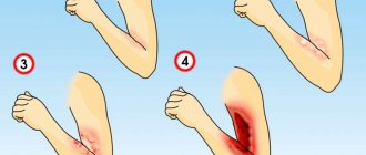
Clinical manifestations of BE
Epidermolysis bullosa manifests itself already during childbirth: the baby’s skin is injured when he passes through the birth canal. The nose, chin and heels are usually affected. In rare cases, the disease makes itself felt at 1-6 months of life. With age, depending on the type of EB, new symptoms of the disease may appear.
The main clinical manifestations of epidermolysis bullosa are blisters on the skin that appear at sites of friction, bruise, pressure, with increased body temperature, the environment, or spontaneously. Blistering can also occur on the mucous membranes of any organ. Most often the mucous membrane of the oral cavity, esophagus, intestines, genitourinary system, and eye mucosa are affected.
Causes of epidermolysis bullosa
The main causes of the pathology are mutations in more than 10 genes responsible for encoding proteins of various layers of the skin, most often in 75% - these are the genes KRT5 and 14, LAMC2, LAMA3, LAMB3, COL17A1. As the online resource Wikipedia indicates, for almost each of the established subtypes of EB, it was possible to identify mutations in certain genes, among them the most common are:
- missense point mutations;
- nonsense point mutations;
- deletions and insertions – losses and, accordingly, insertions of chromosome sections;
- frameshift mutations;
- splicing.
The type of inheritance of epidermolysis bullosa occurs as autosomal recessive (most often), autosomal dominant, and uniparental disomy, and somatic mosaicism .
Factors provoking congenital epidermolysis bullosa
Congenital Butterfly syndrome can develop even in healthy parents who do not carry mutated genes. Spontaneous mutations occur in utero and are caused by:
- bad habits of a pregnant woman - smoking, alcohol abuse;
- chaotic use of medications and other teratogenic factors.
Literature
- Boeira V., Souza E., Rocha B., Oliveira P., Oliveira M., Rêgo V., Follador V. (2013). Inherited epidermolysis bullosa: clinical and therapeutic aspects. An. Bras. Dermatol. 88 (2), 185–198;
- Fine J. D. (2010). Inherited epidermolysis bullosa. Orphanet. J. Rare Dis. 5, 12;
- Shinkuma S. (2015). Dystrophic epidermolysis bullosa: a review. Clin. Cosmet. Investig. Dermatol. 8, 275–284;
- Albanova V.I., Chikin V.V., Epishev R.V. (2014). On the issue of diagnosing congenital epidermolysis bullosa. Bulletin of dermatology and venereology. 3, 53–59;
- Bruckner-Tuderman L., McGrath J. A., Robinson E. C., Uitto J. (2013). Progress in epidermolysis bullosa research: summary of DEBRA International Research Conference 2012. J. Invest. Dermatol. 133, 2121–2126;
- Soro L., Bartus C., Purcell S. (2015). Recessive dystrophic epidermolysis bullosa. A Review of disease pathogenesis and update on future therapies. J. Clin. Aesthet. Dermatol. 8 (5), 41–46;
- Murauer E.M., Koller U., Pellegrini G., De Luca M., Bauer J.W. (2015). Advances in gene/cell therapy in epidermolysis bullosa. Keio J. Med. 64 (2), 21–25;
- Shouldn't we try to… change the genome?;
- CRISPR systems: immunization of prokaryotes;
- Mutagenic chain reaction: genome editing on the verge of science fiction;
- Kiritsi D., Garcia M., Brander R., Has C., Meijer R., Jose Escámez M. et al. (2014). Mechanisms of natural gene therapy in dystrophic epidermolysis bullosa. J. Invest. Dermatol. 134 (8), 2097–2104..
Symptoms
Symptoms of epidermolysis bullosa are associated with a violation of the structure of keratinocytes and manifest as:
- burning, pain in the affected areas and relief when emptying the blisters;
- itching during the healing of opened blisters - peeling off of the dried cap of the vesicle, which can subsequently cause peeling and pigmentation;
- development of skin areas with erosion;
- increasing the sensitivity and vulnerability of the skin to any mechanical damage;
- spontaneous occurrence of tense blisters containing clear, colorless liquid or hemorrhagic contents.
The pathology is usually detected immediately at birth, because thin and “brittle” skin is easily injured even from passing through the birth canal. Moreover, skin lesions have varying degrees of severity and prevalence, even within a family. Multiple widespread blisters can cause death in a newborn, especially if a secondary infection occurs. Exacerbations usually occur in the summer - warm season; with age, the prevalence of blisters is minimized.
Additional symptoms of simple EB
Simple epidermolytic lesions usually occur on the hands and feet or may cover the entire body. In addition, you may experience:
- development of palmoplantar hyperkeratosis of a widespread or confluent type;
- dystrophic changes in the nail plates;
- laryngeal stenosis;
- milia;
- hyper- or hypopigmentation ;
- growth retardation.
Healing of blisters most often occurs without scarring, sometimes even reminiscent of herpes simplex, but the likelihood of relapse is very high.
Epidermolysis bullosa simplex
Features of the course of borderline EB
This type is characterized by the development of a pathognomonic symptom - the appearance of areas of abundant granulation tissue on various parts of the body (as in the photo of borderline epidermolysis bullosa), for example, localized symmetrically around the mouth, in the middle part of the face, around the nose, on the upper back, in the armpits or nail folds. The pathology can lead to onychodystrophy and even complete loss of nail plates, a large number of milia , serious scars on the body, including cicatricial alopecia , crusts on the skin, oral ulcers, enamel hypoplasia and severe dental caries. Possible systemic complications usually include polyetiological anemia , growth retardation, erosion, vesicular skin rashes and detachment, and strictures in the gastrointestinal tract. The risk of their development increases depending on the area, degree of destructiveness and depth of skin lesions.
Borderline epidermolysis bullosa
Borderline epidermolysis bullosa is characterized by extremely high mortality, mainly in the first years of life, caused by cessation of weight gain, sepsis, pneumonia , or obstruction of the trachea and larynx.
Extracutaneous manifestations of dystrophic EB
In addition to generalized skin lesions - recurrent formation of blisters, erosions, milia, atrophic scarring and loss of nails, “Butterfly syndrome” causes:
- disorders of the gastrointestinal tract;
- damage to the genitourinary tract and outer eye membranes;
- chronic anemia;
- osteoporosis;
- growth retardation;
- compaction, degeneration and even loss of nail plates;
- contractures of various joints of the limbs - elbow, knee, wrist, metatarsal-tarsal, metatarsophalangeal, etc.;
- increased risk of developing neoplasms, for example, aggressive squamous cell carcinoma .
Dystrophic epidermolysis bullosa
What kind of disease is this
Congenital epidermolysis bullosa includes a whole group of diseases (more than 30 forms), which are united by one name. A common symptom of these diseases is the appearance of blisters on the skin at the slightest mechanical injury. This pathology is also called butterfly disease, but not because sick children grow wings, but because their characteristic excessive fragility and vulnerability of the skin (it is easy for a butterfly to damage its wings). The mechanism behind the formation of blisters is a shift between the upper and lower layers of the skin. The German dermatologist Heinrich Koebner suggested calling the disease this term. The group of these diseases is genetically determined and can be inherited in an autosomal recessive or autosomal dominant manner; it has been proven that mutations affect more than 10 genes.
Epidemiology
The disease epidermolysis bullosa is widespread in 70 (total 85) constituent entities of the Russian Federation. Its frequency is 0 – 19.73 cases per 1,000,000 population. In Russia, the incidence of epidermolysis bullosa reaches 1:50 thousand - 1:300 thousand of the population.
Pathology researchers have found that in many countries around the world, epidermolysis bullosa simplex predominates in the structure of the disease, and in some countries - dystrophic epidermolysis bullosa. Sex differences are not typical for this pathology. It was noted that children and adolescents predominate among the registered patients, which is associated with the high mortality rate of such patients.
Tests and diagnostics
The greatest importance in making a diagnosis is played by a biopsy - the examination of skin samples using transmission electron microscopes, which allow visualization and semi-quantitative analysis of various structures of the epidermis. Due to the availability of monoclonal and polyclonal antibodies against proteins of different layers of the epidermis involved in the pathogenesis of epidermolysis bullosa, immunohistological methods are gaining increasing popularity today.
The immunohistochemical research method and the method of indirect immunofluorescence make it possible to determine the state of expression of structural proteins with hereditary defects in skin cells - keratinocytes and basement membranes (the basis of the epithelium, which performs a barrier and trophic function), as well as the distribution pattern of proteins in previously formed or artificially provoked blisters, including number - their localization depth.
Thanks to modern DNA diagnostic methods, it is possible to quickly classify the type of pathology, identify structural proteins that have undergone mutation, and make a clinical prognosis. An innovative method of genetic analysis - direct sequencing - makes it possible to identify mutations, their type and location, and reliably confirm the diagnosis.
In addition, an important role is played by collecting a family history and medical history of the patient, a comprehensive examination of the whole body, and conducting laboratory tests.
Discussion
The diagnosis of VBE in Russia is mainly established on the basis of anamnesis and clinical picture, as in the cases described by us. The disease is included in the orphan list, and patients need to be provided with dressings, medications, medical nutrition products and the creation of special conditions at home, which requires high material costs. However, the implementation of these measures, due to the lack of statistical records of patients with VBE, complicates the provision of targeted assistance. At the same time, auxiliary laboratory diagnostic methods - transmission electron microscopy and immunofluorescence antigen mapping are absent in most regions, which makes it difficult to identify the type of VEB and predict the life expectancy of an individual and the future medical care he will need from specialists in various fields. To provide them with regular and complete palliative care, it is advisable to train mothers in caring for such patients during inpatient treatment. A full-scale registry and clinical surveillance, as well as an algorithm for their routing in the region, will improve the quality of life of these patients. These issues need to be addressed.
The authors declare no conflict of interest.
In children
Children diagnosed with epidermolysis bullosa are called butterflies because their skin is so vulnerable, delicate and unprotected like the wings of these insects.
The main way to protect your future child from this diagnosis is responsible family planning and genetic testing. Do not forget about the importance of screening, which can identify abnormalities in fetal development. For example, it is important to undergo prenatal diagnosis of pemphigus simplex, which can be detected in the second trimester of pregnancy by the high content of fetoprotein in the blood serum.
Life expectancy of patients
How long do butterfly children live? It is difficult to answer this question unambiguously. The life expectancy of patients with epidermolysis bullosa is directly proportional to the form of the disease, the degree of damage to the structure of genes, the depth of skin damage and the general condition of the child. Unfortunately, such patients rarely survive to adulthood, and this is primarily due to caring for them, which requires a lot of effort, patience and courage. Children with severe generalized forms of the disease live only a few years and die in preschool age, for example, Görlitz or Allopo-Siemens syndrome. Mild forms, especially simple EB, can go into stable remission over time, and the formation of blisters is associated only with trauma to the skin.
Diet for epidermolysis bullosa
Diet 15 table
- Efficacy: therapeutic effect after 2 weeks
- Timing: constantly
- Cost of food: 1600-1800 rubles per week
Regardless of the patient’s age and type of pathology, the therapeutic diet must fully compensate for the loss of proteins, salts, and fluids that are caused by pathological changes in the external integument and internal systems. When it comes to infants, the most recommended is breast milk, supplemented with protein foods - at least 20%. Nutrition should be fractional, mechanically, thermally and chemically sparing, and should be eaten in small portions. With age, the diet should include:
- non-acidic fruit juices and purees;
- add unrefined vegetable oil to breakfast to facilitate bowel movements and provide healthy fats;
- vegetables rich in plant fiber, for example, cabbage, zucchini, dried fruits, beets, which are best consumed mashed and boiled.
Caramel and candies, sweets, cookies, alcoholic drinks, hot and spicy foods - any food that can provoke the formation of bullous lesions in the esophagus and other parts of the gastrointestinal tract are strictly prohibited.