What is this?
Tularemia is a bacterial infection that can be contracted through contact with wild animals or ticks.
It is not as well known as rabies or tick-borne encephalitis, although it causes a lot of problems for humans. The disease causes inflammation and suppuration of the lymph nodes, which sometimes have to be removed. The clinical form of tularemia depends on the method of penetration of the pathogen into the human body. Most often, after infection with tularemia, damage to the lymphatic system develops (the lymph nodes become inflamed); the disease affects the skin and mucous membranes of the eyes, pharynx and lungs. An acute disease is accompanied by high fever and signs of intoxication.
The source of tularemia infection is the bacterium Francisella tularensis. This is a very tenacious microbe that can survive for a long time in water, soil, grain and straw. It is stable in the environment, able to survive at very low temperatures. Surprisingly, F. tularensis remains viable even after being deep frozen for a long time. It can be stored in ice for more than 10 months, and in frozen meat for up to 3 months. At the same time, the bacterium is sensitive to ultraviolet radiation, heat and the effects of disinfectants. For example, upon contact with a Lysol solution, the causative agent of tularemia dies within 3–5 minutes.
How does infection occur? Vectors of infection and routes of transmission
Various types of rodents, birds, dogs, sheep and goats make a major contribution to the spread of infection. Despite the fact that the natural reservoir of tularemia is mainly mammals, the main carrier of the disease is insects that feed on the blood of infected animals, including ticks of the genus Ixodes. In addition to a tick bite, the cause of illness in humans can be:
- direct contact with an infected animal;
- eating raw foods and unboiled water;
- inhalation of dust in industries where plant materials are processed or livestock is slaughtered (Fig. 1).
The disease is not transmitted from person to person.
Important! Tularemia is extremely contagious and, when it enters the human body, causes disease in almost 100% of cases.
Figure 1. How a person becomes infected with tularemia. Source: CC0 Public Domain
Foci of tularemia appear every now and then in: Russia, Kazakhstan, Turkmenistan, countries of Eastern Europe. To a lesser extent, tularemia is common in Western Europe, the USA, Canada, China and Japan.
In Russia, the European part of the country and Siberia are considered dangerous from the point of view of tularemia infection. The spread of the infection is being carefully monitored, despite the relatively low incidence - in 2021, only 71 cases of the disease were registered in our country.
The seasonality of the infection (increased incidence usually occurs in summer and autumn) is associated with the period of activity of ticks and other blood-sucking insects. Previously, most human cases were recorded in rural areas, where people are more likely to come into contact with animals and be in nature. However, in recent years, about ⅔ of all cases are city residents. The reason for this is the frequent trips of city residents to the forest or to their dachas, as well as the consumption of raw meat.
Who is at risk?
The risk group for tularemia includes people who frequently come into contact with animals or agricultural raw materials:
- hunters,
- fishermen,
- veterinarians,
- shepherds,
- animal shelter staff,
- livestock workers,
- cooks,
- agricultural workers, etc.
What is Tularemia
Tularemia is an acute infectious natural focal disease affecting the lymph nodes, skin, sometimes eyes, pharynx and lungs and is accompanied by severe intoxication.
What provokes / Causes of Tularemia:
The causative agent is non-motile gram-negative aerobic encapsulated bacteria F. tularensis of the Francisella genus of the Brucellaceae family. Exhibit pronounced polymorphism; most often have the form of small coccobacilli.
Bacteria have three subspecies:
- Nearctic (African);
- Central Asian;
- Holarctic (European-Asian).
The latter includes three biological variants: Japanese biovar, erythromycin-sensitive and erythromycin-resistant. Intraspecific differentiation of the causative agent of tularemia is based on differences in subspecies and biovars according to a number of phenotypic characteristics: biochemical activity, composition of higher fatty acids, degree of pathogenicity for humans and animals, sensitivity to certain antibiotics, as well as environmental characteristics and habitat of the pathogen. O- and Vi-antigens have been found in bacteria. The bacteria grow on yolk or agar media supplemented with rabbit blood or other nutrients. Among laboratory animals, white mice and guinea pigs are susceptible to infection. Outside the host's body, the pathogen persists for a long time. Thus, in water at 4 °C it remains viable for 1 month, on straw and grain at temperatures below 0 °C - up to 6 months, at 20-30 °C - up to 20 days, in the skins of animals that died from tularemia, at 8 -12 °C - more than 1 month. Bacteria are not resistant to high temperatures and disinfectants. For disinfection, use a 5% phenol solution, a mercuric solution 1:1000 (kills bacteria within 2-5 minutes), 1-2% formaldehyde solution (kills bacteria within 2 hours), 70° ethyl alcohol, etc. For complete disinfection of infected corpses animals should be kept for at least 1 day in a disinfectant solution, after which they should be autoclaved and incinerated.
Epidemiology
The reservoir and source of infection are numerous species of wild rodents, lagomorphs, birds, dogs, etc. Bacteria were isolated from 82 species of wild animals, as well as from domestic animals (sheep, dogs, artiodactyls). The main role in maintaining infection in nature belongs to rodents (water rat, common vole, muskrat, etc.). A sick person is not dangerous to others.
The transmission mechanism is multiple, most often transmission. The pathogen persists in nature in the tick-animal cycle and is transmitted to farm animals and birds by ticks and blood-sucking insects. Specific vectors of tularemia are ixodid ticks. A person becomes infected with tularemia as a result of direct contact with animals (skinning, collecting dead rodents, etc.), as well as through the nutritional route through food and water infected with rodents. Often infection occurs through blood-sucking carriers (ticks, mosquitoes, fleas, horseflies and other arthropods). Infection through the respiratory route is also possible (by inhaling infected dust from grain, straw, and vegetables). Cases of human diseases have been registered in industries associated with the processing of natural raw materials (sugar, starch-molasses, alcohol, hemp factories, elevators, etc.), in meat processing plants, during the slaughter of sheep and cattle, which had infected ticks, in outskirts of cities located near natural centers. There are known cases of infection being imported during the transportation of products and raw materials from areas unfavorable for tularemia.
The natural receptivity of people is high (almost 100%).
Basic epidemiological signs. Tularemia is a common natural focal disease, found mainly in the landscapes of the temperate climate zone of the Northern Hemisphere. The wide distribution of the pathogen in nature, the involvement of a large number of warm-blooded animals and arthropods in its circulation, and the contamination of various environmental objects (water, food products) also determine the characteristics of the epidemic process. There are various types of foci (forest, steppe, meadow-field, name-swamp, in river valleys, etc.). Each type of outbreak corresponds to its own species of animals and blood-sucking arthropods that take part in the transmission of the pathogen. Adults predominate among the cases; often the incidence is associated with the profession (hunters, fishermen, agricultural workers, etc.). Men get sick 2-3 times more often than women. Anthropurgic foci of tularemia arise during the migration of infected rodents from their habitats to populated areas, where they come into contact with synanthropic rodents. Tularemia remains a disease of rural areas, but currently there is a steady increase in the incidence of the urban population. Tularemia is recorded throughout the year, but more than 80% of cases occur in summer and autumn. In recent years, the incidence has been sporadic. In some years, local vector-borne, commercial, agricultural, and waterborne outbreaks are noted, and, less frequently, outbreaks of other types. Vector-borne outbreaks are caused by the transmission of the infectious agent by blood-sucking dipterans and occur in foci of epizootic tularemia among rodents. Vector-borne outbreaks typically begin in July or June, peak in August, and end in September–October; Haymaking and harvesting contribute to the rise in morbidity.
The industrial type of outbreak is usually associated with the capture of water rats and muskrats. Fishing outbreaks occur in spring or early summer during the flood period, and their duration depends on the harvesting period. Infection occurs through contact with animals or skins; The pathogen penetrates through lesions in the skin, which is why axillary buboes often appear, often without ulcers at the site of penetration.
Water outbreaks are determined by the entry of pathogens into open water bodies. The main water pollutant is water voles that live along the banks. Diseases usually occur in the summer with an onset in July. Diseases are associated with field work and the use of drinking water from random reservoirs, wells, etc. In 1989-1999. the proportion of tularemia pathogen isolates from water samples reached 46% or more, which indicates the important epidemiological significance of water bodies as long-term reservoirs of infection.
Agricultural outbreaks occur when airborne dust aerosol is inhaled when working with straw, hay, grain, and feed contaminated with the urine of sick rodents. The pulmonary, less frequently abdominal and anginal-bubonic forms predominate. The household type of outbreaks is characterized by infection in the home (at home, on the estate). Infection is also possible during sweeping the floor, sorting and drying agricultural products, distributing food to pets, and eating contaminated foods.
Pathogenesis (what happens?) during Tularemia:
Bacteria enter the human body through the skin (even intact), mucous membranes of the eyes, respiratory tract and gastrointestinal tract. In the area of the entrance gate, the localization of which largely determines the clinical form of the disease, a primary affect often develops in the form of successive spots, papules, vesicles, pustules and ulcers. Subsequently, tularemia bacilli enter the regional lymph nodes, where they multiply and develop the inflammatory process with the formation of the so-called primary bubo (inflamed lymph node). When Francisella dies, a lipopolysaccharide complex (endotoxin) is released, which enhances the local inflammatory process and, when released into the blood, causes the development of intoxication. Bacteremia does not always occur during the disease. In the case of hematogenous dissemination, generalized forms of infection develop with toxic-allergic reactions, the appearance of secondary buboes, and damage to various organs and systems (primarily the lungs, liver and spleen). In the lymph nodes and affected internal organs, specific granulomas are formed with central areas of necrosis, accumulation of granulocytes, epithelial and lymphoid elements. The formation of granulomas is facilitated by the incompleteness of phagocytosis, due to the properties of the pathogen (the presence of factors that prevent intracellular killing). The formation of granulomas in primary buboes often leads to their suppuration and spontaneous opening, followed by prolonged healing of the ulcer. Secondary buboes, as a rule, do not suppurate. In the case of replacement of necrotic areas in the lymph nodes with connective tissue, suppuration does not occur, the buboes resolve or become sclerotic.
Symptoms of Tularemia:
In accordance with the clinical classification, the following forms of tularemia are distinguished:
- by localization of the local process: bubonic, ulcerative-bubonic, oculo-bubonic, anginal-bubonic, pulmonary, abdominal, generalized;
- by duration of course: acute, protracted, recurrent;
- by severity: mild, moderate, severe.
Incubation period. Lasts from 1 to 30 days, most often it is 3-7 days.
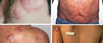
Signs of the disease, common to all clinical forms, are expressed in an increase in body temperature to 38-40 ° C with the development of other symptoms of intoxication - chills, headache, muscle pain, general weakness, anorexia. Fever can be remitting (most often), constant, intermittent, wave-like (in the form of two or three waves). The duration of fever varies, from 1 week to 2-3 months, most often it lasts 2-3 weeks. When examining patients, hyperemia and pastiness of the face, as well as the mucous membrane of the mouth and nasopharynx, injection of the sclera, and hyperemia of the conjunctiva are noted. In some cases, exanthema of various types appears: erythematous, maculopapular, roseolous, vesicular or petechial. The pulse is slow (relative bradycardia), blood pressure is reduced. A few days after the onset of the disease, hepatolienal syndrome develops.
The development of various clinical forms of the disease is associated with the mechanism of infection and the entry gates of infection, which determine the localization of the local process. After the pathogen penetrates the skin, the bubonic form develops in the form of lymphadenitis (bubo), regional in relation to the gate of infection. Isolated or combined damage to various groups of lymph nodes - axillary, inguinal, femoral - is possible. In addition, during hematogenous dissemination of pathogens, secondary buboes can form. Soreness occurs, and then the lymph nodes become enlarged to the size of a hazelnut or a small chicken egg. In this case, pain reactions gradually decrease and disappear. The contours of the bubo remain distinct, the phenomena of periadenitis are insignificant. In the dynamics of the disease, the buboes slowly (sometimes over several months) dissolve, suppurate with the formation of a fistula and the release of creamy pus, or become sclerotic.
Forms of the disease
Ulcerative bubonic form . It often develops during transmissible infection. At the site of introduction of the microorganism, a spot, papule, vesicle, pustule, and then a shallow ulcer with raised edges successively replace each other over the course of several days. The bottom of the ulcer is covered with a dark crust in the shape of a “cockade”. At the same time, regional lymphadenitis (bubo) develops. Subsequently, scarring of the ulcer occurs slowly.
In cases of pathogen penetration through the conjunctiva, the oculobubonic form of tularemia occurs. In this case, damage to the mucous membranes of the eyes occurs in the form of conjunctivitis, papular, and then erosive-ulcerative formations with the separation of yellowish pus. Corneal lesions are rarely observed. These clinical manifestations are accompanied by severe swelling of the eyelids and regional lymphadenitis. The course of the disease is usually quite severe and long-lasting.
Anginal-bubonic form . Develops after penetration of the pathogen with contaminated food or water. Patients complain of moderate sore throat and difficulty swallowing. On examination, the tonsils are hyperemic, enlarged and swollen, fused with the surrounding tissue. On their surface, usually on one side, grayish-white necrotic deposits form, which are difficult to remove. Swelling of the palatine arches and uvula is pronounced. Subsequently, the tonsil tissue is destroyed with the formation of deep, slowly healing ulcers, followed by the formation of a scar. Tularemia buboes occur in the submandibular, cervical and parotid areas, most often on the side of the affected tonsil.
Abdominal form . Develops due to damage to the mesenteric lymph nodes. Clinically manifested by severe abdominal pain, nausea, occasionally vomiting, and anorexia. Sometimes diarrhea develops. On palpation, pain around the navel is noted, and positive symptoms of peritoneal irritation are possible. As a rule, hepatolienal syndrome is formed. It is rarely possible to palpate the mesenteric lymph nodes; their enlargement is determined using ultrasound.
Pulmonary form . It occurs in the form of bronchitis or pneumonia.
- The bronchitis variant is caused by damage to the bronchial, mediastinal, paratracheal lymph nodes. Against the background of moderate intoxication, a dry cough, chest pain appears, and dry wheezing is heard in the lungs. Usually this option is easy and ends with recovery in 10-12 days.
- The pneumonic variant is characterized by an acute onset, a sluggish, debilitating course with high, prolonged fever. Pathology in the lungs is clinically manifested by focal pneumonia. Pneumonia is distinguished by a rather severe and acyclic course, a tendency to develop complications (segmental, lobular or disseminated pneumonia, accompanied by an increase in the above groups of lymph nodes, bronchiectasis, abscesses, pleurisy, cavities, gangrene of the lungs).
Generalized form . Clinically reminiscent of typhoid-paratyphoid infections or severe sepsis. High fever becomes irregularly remitting and persists for a long time. Symptoms of intoxication are pronounced: headache, chills, myalgia, weakness. Confusion, delirium, and hallucinations are possible. The pulse is labile, heart sounds are muffled, blood pressure is low. In most cases, hepatolienal syndrome develops from the first days of the disease. In the future, persistent exanthema of a roseolous and petechial nature may appear with localization of rash elements on symmetrical areas of the body - forearms and hands, legs and feet, neck and face. With this form, the development of secondary buboes, caused by hematogenous dissemination of pathogens, and metastatic specific pneumonia is possible.
Complications
In most cases, they develop in a generalized form. The most common are secondary tularemia pneumonias. Infectious-toxic shock is possible. In rare cases, meningitis and meningoencephalitis, myocarditis, polyarthritis, etc. are observed.
Prevention of Tularemia:
Epizootological and epidemiological surveillance
Includes constant monitoring of the incidence of people and animals in natural foci of tularemia, circulation of the pathogen among animals and blood-sucking arthropods, monitoring the state of immunity in people. Its results form the basis for planning and implementing a set of preventive and anti-epidemic measures. Epidemiological surveillance involves epizootological and epidemiological examination of natural foci of tularemia, generalization and analysis of the data obtained, which determines epidemic manifestations in natural foci of tularemia in the form of sporadic, group and outbreak morbidity in people.
Preventive actions
The basis for the prevention of tularemia consists of measures to neutralize sources of the infectious agent, neutralize transmission factors and carriers of the pathogen, as well as vaccination of endangered populations. Elimination of conditions for infection of people (general sanitary and hygienic measures, including sanitary educational work) has its own characteristics for various types of morbidity. In case of vector-borne infections through blood-sucking insects, repellents and protective clothing are used, and access of the unvaccinated population to unfavorable areas is limited. The fight against rodents and arthropods (deratization and disinfestation measures) is of great importance. To prevent nutritional contamination, you should avoid swimming in open water, and only boiled water should be used for household and drinking purposes. When hunting, it is necessary to disinfect your hands after skinning and gutting hares, muskrats, moles and water rats. Vaccination is carried out as planned (among the population living in natural foci of tularemia and populations at risk of infection) and according to epidemiological indications (unscheduled) when the epidemiological and epizootological situation worsens and there is a threat of infection of certain groups of the population. For immunoprophylaxis, a live attenuated vaccine is used. Vaccination ensures the formation of stable and long-term immunity in vaccinated people (5-7 years or more). Revaccination is carried out after 5 years for contingents subject to routine vaccination.
Activities in the epidemic outbreak
Each case of human tularemia requires a detailed epizo-otological-epidemiological examination of the outbreak with clarification of the route of infection. The issue of hospitalization of a patient with tularemia and the timing of discharge from the hospital is decided by the attending physician purely individually. Patients with abdominal, pulmonary, oculo-bubonic and anginal-bubonic, as well as moderate or severe cases of ulcerative-bubonic and bubonic forms must be hospitalized according to clinical indications. Patients are discharged from the hospital after clinical recovery. Long-term non-absorbable and sclerotic buboes are not a contraindication for discharge. Dispensary observation of the patient is carried out for 6-12 months in the presence of residual effects. The separation of other persons in the outbreak is not carried out. As an emergency preventive measure, antibiotic prophylaxis can be carried out by prescribing rifampicin 0.3 g 2 times a day, doxycycline 0.2 g 1 time a day, tetracycline 0.5 g 3 times a day. The patient's home is disinfected. Only things contaminated with secretions from patients are subject to disinfection.
Compiled based on Internet materials
Forms of the disease
After entering the body, the tularemia bacillus attacks the lymph nodes, as well as internal organs - the liver, spleen, and less often - the lungs. Depending on how a person became infected, the disease manifests itself differently (Table 1).
| Table 1. Clinical forms (classification) of the disease | |||
| Form of the disease | Type of infection | Which organs are affected? | Symptoms |
| Bubonic | Through the skin | The lymph nodes | Lymph nodes enlarge significantly, sometimes reaching the size of a chicken egg. The contours of the lymph nodes (buboes) are clear. They hurt when pressed. As the disease progresses, the buboes may dissolve or fester. Possible formation of abscesses and fistulas |
| Ulcerative bubonic | After being bitten by a tick or other insect | Bite site | A small ulcer with a dark bottom appears at the site of the bite, which heals very slowly |
| Oculo-bubonic | In case of contact with eyes with dust or dirty hands | Eyes, lymph nodes | Conjunctivitis: inflammation of the eyes with redness, swelling, tenderness and stinging. Papular formations, which then turn into wounds and purulent ulcers |
| Anginal-bubonic | After drinking contaminated food or water | Pharyngeal mucosa, lymph nodes | High temperature, enlarged lymph nodes. Sore throat, problems swallowing, redness and swelling of the tonsils. The tonsils are enlarged and covered with a gray coating that is difficult to remove. Later, long-healing ulcers form on the tonsils, and then scars |
| Abdominal | After drinking contaminated food or water | Lymphatic vessels in the intestines | Abdominal pain, nausea, vomiting, weight loss |
| Pulmonary | After inhalation of contaminated dust | Bronchial lymph nodes, lungs | Fever, cough, chest pain |
| Generalized | Does not depend on the type of infection, but most often develops when the pathogen is inhaled | Spread of infection through blood, sepsis | Fever, weakness, confusion, headache, muscle pain |
The incubation period of tularemia, when there are no symptoms yet, usually does not exceed 3-7 days. The first manifestation of the disease is a high temperature, which rises to 38-40 °C. After this, the patient begins to suffer from muscle pain and headaches, feels constant weakness and dizziness. The fever lasts a long time and subsides only after 2-3 weeks.
During the examination of the patient, the doctor observes redness and swelling of the face, a noticeable increase in the vascular network of the eyes or “red eye” syndrome, and dark red spots – hemorrhages – are visible on the mucous membrane of the mouth. The tongue is covered with a white or gray coating. The main characteristic sign of infection is greatly enlarged lymph nodes (Fig. 2).
Figure 2. Lymphadenitis due to tularemia. The disease often affects the cervical lymph nodes. Source: Coronation Dental Specialty Group
In the later stages of the disease, blood pressure decreases and the pulse becomes rarer. On the fifth day from the moment the first symptoms appear, a severe dry cough develops. Most patients have an enlarged liver and spleen.
The causative agent of tularemia
The disease received its name “Tularemia” in honor of Tulare Lake (California), where a disease similar in clinical picture to plague was discovered in ground squirrels. The bacterium Francisella tularensis is named after the researcher E. Francis, who established the fact of transmission of the disease to humans.
Francisella tularensis is a gram-negative rod (Gram stained pink), which means the bacterium has a capsule. The causative agent of tularemia is an aerobe. Does not create a dispute.
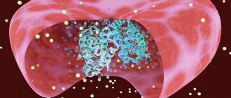
Rice. 2. The photo shows the bacteria Francisella tularensis under a microscope (left, Gram stain) and computer visualization of pathogens (right). The causative agent of tularemia has the form of a coccobacilli, but may have the appearance of filaments.
Tularemia bacteria have the following abilities that determine their pathogenicity:
- adhesion (sticking to cells);
- invasion (penetration into tissue);
- intracellular reproduction in phagocytes with subsequent suppression of their killer effect;
- the presence in bacteria of receptors for the Fc fragments of IgG (class G immunoglobulins), which leads to disruption of the activity of the complement system;
- When microbes are destroyed, endotoxins are released. They play a leading role in the pathogenesis of the disease and determine its clinical manifestations;
- toxins and components of microbial cells have strong allergenic properties, which contributes to even greater tissue damage.
Antigenic structure of bacteria
The O and Vi antigens were found in virulent forms of tularemia bacteria.
- Vi-antigen (envelope). The virulence of bacteria and immunogenicity depend on it.
- O-antigen (somatic). In tularemia bacteria, the somatic antigen is endotoxin.
Resistance of bacteria in the external environment
The causative agents of tularemia exhibit high resistance in the external environment:
- they remain viable in water and moist soil at a temperature of 4°C for up to 4 months, and for up to 2 months at a temperature of 20–30°C;
- in straw and grain crops, bacteria persist for up to 6 months at a temperature of 0°C;
- bacteria remain in the skins of killed animals for up to 20 days, and in their excrement for up to 120 days;
- Bacteria can be stored in frozen meat for up to 6 months, and in milk for up to 8 days.
When boiled, bacteria die instantly; when exposed to sunlight, they die after 30 minutes. Solutions of sublimate, chloramine and 50% alcohol have a detrimental effect on bacteria.
Rice. 3. The photo shows a colony of tularemia pathogens.
When growing on solid nutrient media, they are white with a bluish tint.
Diagnostics
Considering the strong seasonality, locality, occupation characteristic of a high risk of infection, and pronounced symptoms, the clinical diagnosis of tularemia is not very difficult.
Laboratory diagnosis is confirmed by identifying antibody titers or x-ray (for the pulmonary form). Bacteriological and PCR methods make it possible to detect the presence of a pathogen in biological samples taken from a patient.
Which doctor should I contact?
Having felt the symptoms and arrived at the clinic, the patient goes to the local therapist. He, in turn, gives a referral to an infectious disease specialist, who makes a decision on hospitalization.
An infectious disease specialist diagnoses and treats tularemia.
Tests for tularemia
There are several methods for laboratory diagnosis of tularemia.
Allergological methods
At an early stage of the disease, starting from the third day after the onset of symptoms, an allergy test is used for diagnosis. The test involves injecting the patient subcutaneously with tularemia antigen. In case of infection, the diameter of the swelling at the injection site is larger than usual in healthy people - at least 0.5 cm. The test results are assessed 24, 48 and 72 hours after the injection.
Serological methods
Serological diagnostic methods (agglutination reactions and passive hemagglutination) involve examining patient blood samples for the presence of specific antibodies. Depending on the chosen method, the disease can be diagnosed already on the 7-15th day of its course.
Bacteriological diagnostic methods
The bacteriological method consists of isolating the infectious agent from blood samples or the contents of the patient’s ulcers and buboes on days 7-10 after infection. The bacteria are then grown in a special nutrient medium. This approach to diagnosing tularemia is used very rarely because of its labor intensity, and also because not all laboratories have permission to work with F. tularensis.
Polymerase chain reaction
Polymerase chain reaction (PCR) is a diagnostic method that allows you to detect pathogen DNA in a biological sample. The sensitivity and specificity of PCR are not inferior to agglutination and passive hemagglutination reactions, and the method can be used already on the 10th day of illness.
“Tularemia in Russia in the recent past,” reports A.A. Nafeev, a sanitary doctor at the Center for Hygiene and Epidemiology in the Ulyanovsk Region, was one of the most common natural focal infections. Despite the successes achieved in the fight against this infection, cases of the disease are still registered annually. This dictates the need to improve sanitary and epidemiological surveillance, as well as the constant vigilance of outpatient doctors in relation to the first symptoms of tularemia.”
Treatment
The treatment of tularemia is based on antibiotic therapy. To defeat a disease, it is necessary to destroy its source - the bacterium.
Drug therapy aimed at combating the infectious agent
The generally accepted standard of treatment for tularemia is antibiotics from the aminoglycoside and tetracycline groups. Third-generation cephalosporins and fluoroquinolones are used as second-line drugs.
The course duration is usually two weeks. In the event of a relapse of the disease, a drug that was not used during the first wave of the disease is chosen to continue treatment, since the bacteria could have acquired resistance to the previously used antibiotics.
Symptomatic treatment
Patients with symptoms are recommended bed rest and a balanced diet rich in vitamins. The ward is disinfected daily.
As symptomatic treatment, drugs are used to reduce the harmful effects of intoxication, allergic reactions and inflammation. Vitamins and medications may be prescribed to support the cardiovascular system. In case of eye damage, they are washed and instilled with a 20-30% solution of sodium sulfacyl 2-3 times a day. For tularemic sore throat, gargling is prescribed.
Treatment of ulcers
To treat ulcers and speed up the resorption of buboes, compresses and bandages with ointment are used. Physiotherapy procedures may be indicated: exposure to heat, blue light, laser irradiation, etc.
If the bubo has festered, it is opened and cleaned.
Possible complications
The vast majority of patients with tularemia can be cured without consequences. But in approximately 1-2% of cases of the disease, complications are observed, mainly related to the generalized form of tularemia.
Common complications include:
- meningitis and meningoencephalitis,
- secondary pneumonia,
- infectious psychosis after tularemia,
- chronic joint damage - polyarthritis,
- progressive heart disease - myocardial dystrophy,
- chronic course with frequent relapses.
Mortality with tularemia is less than 3%, with a generalized form - 30-60%.
Prevention
To protect yourself from infection, you should remember to take precautions while being in nature, when contacting animals, and eating meat and animal products. Vaccination remains the most effective method of prevention.
Is there a vaccine? Is it effective?
The most reliable way to protect yourself from infection is to get vaccinated. The effectiveness of vaccination against tularemia exceeds 90%; in Russia it is recommended for adults and children over 7 years old living in dangerous regions.
To protect against infection, a live vaccine containing weakened bacteria that cannot cause disease is used. On days 5–7 and 12–15 after vaccination, the strength of immunity is assessed. If immunity is not formed, re-vaccination is carried out. The state of immunity of vaccinated people is checked 5 years after vaccination and subsequently every 2 years.
Emergency unscheduled preventive vaccination is carried out in the presence of epidemiological indications:
- in case of illness of people in this territory,
- with intensive reproduction of rodents,
- with widespread carriage of tularemia among one or more animal species over a large territory.
Precautions in nature
Measures to reduce morbidity include controlling the spread of infection, carrying out deratization measures and disinsection of hazardous areas.
In case of a water outbreak, it is forbidden to drink unboiled water, as well as to swim. To reduce the risk of infection from tick and mosquito bites, it is recommended to use repellents, wear closed clothing, and limit the entry of unvaccinated people into disadvantaged areas.
To prevent commercial infections, it is advisable to use gloves when removing skins from killed rodents and to disinfect your hands.
It is necessary to carry out sanitary education work among the population of areas disadvantaged by tularemia. Persons who have been in contact with the patient are not isolated, since the sick are not contagious. The patient's home is disinfected.
Epidemiology of tularemia
In the Russian Federation, 50 to 380 cases of human tularemia are registered annually. These are mainly small or isolated outbreaks of the disease in the summer-autumn periods, caused by tick attacks, handling of muskrat and hare carcasses, and consumption of infected food and water. The mechanization of agriculture has minimized cases of mass accumulation of small rodents and mice in agricultural fields. Persons with summer cottages and garden plots, hunters and fishermen, geologists and agricultural workers are at risk.
Places where rodents actively breed are particularly dangerous for tularemia.
Rice. 4. The photo shows carriers of tularemia pathogens.
Reservoir of infection
- In nature in the Russian Federation, tularemia bacteria most often affect hares, rabbits, hamsters, water rats and voles. Their disease progresses rapidly and always ends in death. Black rats, gophers and ferrets also suffer from tularemia. The second place for tularemia is in cattle, pigs and sheep.
- Contaminated food products can become a source of infection.
- Water can become a source of infection. Voles living along the banks of rivers, lakes and ponds pollute the water. The source of infection can be water from random abandoned wells. The causative agents of tularemia make water bodies long-term reservoirs of infection.
- Infected dust particles that are formed during grain threshing, dust from straw and mixed feed can also become a source of tularemia pathogens. In this case, the respiratory organs are most often affected.
A sick person does not pose a danger to others.
Tularemia vectors
The infection is carried by mosquitoes, horseflies and ixodid and gamas ticks.
Rice. 5. In the photo there is a male ixodes taiga tick (Ixodes persulcatus) on the left and a gamasid tick on the right.
Routes of transmission
- Contact (involves contact with sick animals and their biological material).
- Nutritional (consumption of contaminated food and water).
- Transmissible (bites from infected bloodsuckers).
- Aerogenic (inhalation of infected dust).
Rice. 6. Contact with the skins of killed infected animals and bites of blood-sucking animals are the main routes of transmission of infection.
Mechanism of transmission of infection
Tularemia has multiple routes of transmission:
- through damaged skin,
- through the mucous membrane of the oropharynx and tonsils,
- through the mucous membrane of the eyes,
- through the respiratory tract,
- through the digestive tract.
One microbial cell is enough to become infected with tularemia.


