Cytomegalovirus infection in pregnant women and newborns
What are the difficulties in diagnosing CMV? What is the mechanism of CMV infection?
In recent years, there has been a tendency towards an increase in the frequency of viral infections among pregnant women and their ability, under certain conditions, to spread epidemically has increased.
The problem of cytomegalovirus infection (CMV) during pregnancy is of particular importance, due to the fact that cytomegalovirus (CMV) can cause obstetric pathology, embryo- and fetopathies, intrauterine and perinatal infections [1].
The presence of latent forms of CMV with an atypical course, inapparent manifestations and damage to organ tissue presents great difficulties for the timely recognition of infection and is the cause of diagnostic errors.
Among those born alive, 0.2-2.2% of children are infected with cytomegalovirus in utero. Intrauterine CMV infection, regardless of the form of the disease, can be dangerous due to damage to the central nervous system and lead to psychomotor disorders and mental retardation in children even several years after birth [2].
Pregnant women account for a significant percentage of the epidemiology of CMV, which is found in them twice as often as rubella.
CMV infection can contribute to the development of complications during pregnancy, childbirth and the postpartum period. Obstetric pathology most often manifests itself in the form of spontaneous miscarriages, stillbirths, and the birth of nonviable children [3, 4, 11].
The problem of studying cytomegalovirus infection and its influence on the course and outcome of pregnancy, developing the foundations of comprehensive prevention and treatment are extremely relevant and especially important for practical healthcare.
Cytomegalovirus (CMV) belongs to the family of herpetic viruses, numbering about 40 members. Eight types of herpes viruses are characteristic of humans: herpes simplex (types 1 and 2), cytomegalovirus, chickenpox-zoster, Epstein-Barr virus, and herpes virus types 6, 7 and 8. They cause exacerbations of chronic infection and demonstrate the ability to persist in the body [10].
Cytomegalovirus is a large DNA gene with relatively low virulence and a special ability to sharply suppress cellular immunity.
The effects of exo- and endogenous factors lead to a decrease in immunity and, accordingly, to the activation of chronic cytomegalovirus infection.
Sources of infection can be CMV carriers, their biological fluids and secretions: blood, urine, tears, saliva, breast milk, cerebrospinal fluid, amniotic fluid, vaginal discharge, mucus from the nasopharynx, semen, feces, etc.
CMV exhibits special tropism for the salivary glands. Hence the real possibility of transmitting CMV from mother to child through kissing. There is a direct correlation between infection with cytomegalovirus and sexual activity of partners.
Taking into account the age and intimacy of contacts between people, two waves of CMV attack are identified: the first is achieved by the age of three, the second by the period of puberty.
The entry gates for the virus are the respiratory tract, digestive tract, and mucous membranes. CMV, penetrating into the blood, reproduces in leukocytes and in the system of mononuclear phagocytes.
In most cases, there is an asymptomatic virus carriage or a subclinical, inapparent, chronic infection, which, outside of immunosuppression, does not cause any subjective disorders or objective clinical manifestations. Long-term (often lifelong) latency is facilitated by the intracellular preservation of CMV in lymphocytes, where it is reliably protected from the action of specific antibodies and interferon.
Morphological changes manifest themselves in the form of complexes of cytomegalic cells (CMCs) and interstitial lymphohistiocytic infiltration [3].
A specific transformation of cytomegalic cells occurs, acquiring the characteristic appearance of typical giant cells (“owl’s eye”) with a diameter of 35 to 65 microns with inclusions in the nucleus and a light perinuclear zone.
Cytomegalic changes are most often observed in the salivary glands, lungs, kidneys and brain.
Local cytomegalic damage to the eyes (choreoretinitis), intestines (colitis), skin (dermatitis), adrenal glands, lungs, kidneys, and brain have been described [3].
The question of the influence of gestational age on the transmission of infection from mother to fetus remains open. It is believed that the greatest threat to the fetus is the development of cytomegalovirus infection in the first half of pregnancy.
Reactivation of latent infection leads to transmission of the virus from mother to fetus in 0.15%–0.36% of cases [5].
CMV infection and immunosuppression are the most important features of the pathogenesis of cytomegaly. This is due to the fact that CMV is characterized by reactivation under conditions of immunodeficiency, in particular during the so-called “physiological immunosuppression” that accompanies pregnancy.
There are several possible ways of transmitting infection from mother to fetus:
- transplacental;
- through infected discharge in the birth canal;
- feeding with infected breast milk.
Transplacental transmission of CMV most often leads to infection of the fetus [7].
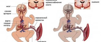
Cytomegalovirus from the mother can penetrate the placenta at any stage of pregnancy and cause damage to the fetus.
Cytomegalovirus infection in pregnant women can be primary or recurrent. Primary maternal CMV infection occurring during pregnancy is best diagnosed by studying the seroconversion of antibodies to CMV. The presence of IgG and IgM antibodies can be considered evidence of a primary maternal infection. Recurrent CMV infection during pregnancy includes both reactivation of one's own CMV strain and possible reinfection with a new strain of the virus [9].
In most cases, in populations with a high prevalence of antibodies to CMV, the development of CMV during pregnancy is more likely to occur as a result of reinfection.
Congenital CMV infection occurs either after a primary infection or as a result of an exacerbation of a chronic one. More severe consequences are observed during pregnancy with primary infection.
During pregnancy in conditions of immunodeficiency, nonspecific depression of the immune system occurs. With CMV, cellular immunity is most affected. It is the nature of the changes in the cellular immune system that largely determines the course and outcome of cytomegaly.
In response to the introduction of the virus, antibodies are produced. With cytomegaly, complement-binding antibodies persist throughout life, which makes it possible to identify cases of past disease. It is clear that specific antibodies are important in protection against cytomegalovirus.
IgG antibodies indicate that the woman has previously had an infection. Pregnant women have a more intense and prolonged IgG response. High levels of antibody titers indicate activation of the chronic process.
The absence of IgG in umbilical cord or neonatal serum excludes congenital CMV infection, while their presence may indicate passive transfer from mother to fetus.
A significantly higher level of IgG in the newborn than in the mother may indicate a congenital infection.
When examined in the first six months of a child’s life, congenital CMV infection is excluded if the IgG level gradually decreases. The presence of IgM in the blood serum characterizes the primary infection. IgM antibodies remain in the blood from 12 to 18 weeks from the onset of primary infection.
IgM antibodies to CMV are a marker of “fresh” primary infection.
Antibodies of the IgM class do not cross the placenta; they are synthesized by the fetus and newborn in response to antigenic stimulation.
The mechanism of transmission of CMV involves close contact between a pregnant woman (parturient woman) and the fetus (newborn). After pregnancy and childbirth, CMV can be found in almost all body fluids, especially in cervicovaginal secretions and breast milk, which can be a source of peri- and postnatal transmission of CMV from mother to newborn.
Transmission of infection can occur through blood during blood transfusions and parenteral procedures, as well as through damaged skin and macerated mucous membranes. It has been established that intranatal or early postnatal transmission of CMV infection occurs 10 times more often than transplacental transmission.
In the postpartum period, the epidemiological danger to the newborn remains in cases of a woman’s violation of hygiene standards, as well as due to transmission of the virus through breast milk (20% of seropositive mothers have CMV in breast milk and 30% of them can excrete the virus into milk within a year after birth) [ 8].
Perinatal and postnatal manifestations of CMV infection in children are almost always mild and asymptomatic.
The variety of variants of the course of CMV infection in pregnant women is associated in most cases with asymptomatic latency and polymorphism of the clinical manifestations of the disease.
Variants of the course of CMV and the wide range of their manifestations are due to the complexity of the relationship between CMV and the host cell.
Often clinical symptoms manifest themselves in the form of low-grade fever, nasopharyngitis, cough, and the development of mononucleosis-like syndrome.
The range of clinical variants varies from mild sialadenitis and favorable current mononucleosis-like disease (lymphocytosis, atypical mononuclear cells) to severe damage to the liver, lungs and brain.
In pregnant women, symptomatic CMV usually manifests itself in the form of mononucleosis syndrome, which is confirmed by the presence of lymphocytosis, the presence of CMV IgG and IgM antibodies in the serum and CMV cultures in the blood, urine, saliva, and cervical secretions.
Mononucleosis-like syndrome is characterized by prolonged fever, chills, fatigue, muscle pain, headaches, and is more common in young people [6].
A pregnant woman most often experiences an exacerbation of chronic CMV infection, which, as a rule, does not have obvious clinical manifestations. However, this category of women often has a history of recurrent miscarriage or non-developing pregnancy, stillbirth, the birth of non-viable children, as well as disabled children with congenital malformations [11].
Infection is possible in any trimester, however, the most severe consequences occur with a primary infection that occurs in the mother during the first 20 weeks of pregnancy. However, even with the development of primary CMV during pregnancy, 90-95% of women have a chance to give birth to a healthy child [12].
Intrauterine cytomegalovirus infection can lead to the birth of children with severe neurological disorders, damage to the optic and auditory nerves.
The extent of fetal damage depends on the duration of infection. Infection in early pregnancy often leads to fetal death and spontaneous miscarriages.
Children with CMV infection (IgM positive) from seropositive mothers have lower birth weight and height than children from seronegative mothers.
CMV can infect the placenta, fetus, and amniotic fluid. Viral culture and characteristic cytomegalic cells can be detected in fetuses from women with active primary CMV infection. However, the presence of signs or symptoms of CMV in the fetus does not always predict serious disease at birth or in the postnatal period.
If symptoms of the disease are detected during the first three weeks of a child’s life, CMV infection can be considered congenital; the occurrence of the disease at a later date more often characterizes the acquired form of CMV infection (postnatal infection).
Congenital CMV is characterized, as a rule, by generalized damage to the fetal organs and a whole complex of clinical symptoms.
The acquired form of CMV is characterized by a latent course, mainly with local damage to the salivary glands.
In 90% of newborns infected in utero, there are no symptoms at birth; 5-17% of them develop complications in a later period [6].
The development of symptoms in a newborn with CMV correlates with the symptoms present at birth.
The remaining children with congenital CMV are completely asymptomatic and appear healthy at birth.
Clinical forms of CMV infection can have an acute or chronic course.
According to the severity of the CMV disease, the following are identified:
- mild (including erased and subclinical forms of infection), in which damage to internal organs is insignificant and is not accompanied by functional disorders (compensation);
- moderate, in which there is damage to internal organs, accompanied by functional disorders of varying degrees (subcompensation);
- severe, in which intoxication is pronounced and damage to internal organs is accompanied by severe functional impairment (decompensation);
In severe cases of congenital CMV infection, the death of a child often occurs in the first days and weeks of life; in mild cases, the disease takes on a wave-like course.
It should be noted that children with congenital or perinatal active cytomegaly are extremely predisposed to the development of bacterial superinfections, which is primarily due to immune deficiency.
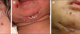
Symptoms of congenital CMV infection include signs of intrauterine growth retardation, intrauterine malnutrition, conjugation jaundice, hepatosplenomegaly, interstitial pneumonia, and cerebrovascular accidents. Complications from the central nervous system are also very common and manifest in newborns with increased drowsiness, poor sucking reflex, macro- and microcephaly, hydrocephalus, porencephaly, chorioretinitis, and sensorineural deafness [4, 11].
Congenital CMV infection may also have unusual manifestations such as ventriculomegaly, periventriculitis or cyst formation, hemolytic anemia, petechiae, hemorrhagic purpura, thrombocytopenia, chronic hepatitis and ascites [3, 8].
However, despite the very large number of symptoms and complications associated with congenital CMV infection, it is difficult to predict the physical and intellectual development of a child based on the symptoms present at birth.
Each of the listed clinical manifestations of CMV disease deserves separate consideration.
Hepatomegaly, together with splenomegaly and petechiae, is the most convincing evidence of the disease in children with congenital CMV. Usually there are symptoms of hepatitis with impaired liver function (jaundice, hyperbilirubinemia).
Liver function usually returns to normal in the first weeks of life.
Splenomegaly can often be the only indicator of congenital CMV infection at birth.
Petechiae are usually transient and observed within two to three days after birth. With congenital CMV, a combination of petechiae with hepatosplenomegaly and thrombocytopenia is more common.
A blood test reveals a picture of hypochromic anemia, erythroblastosis, and thrombocytopenia.
Congenital CMVI leads to visual impairment in 20% of children. The most common condition is chorioretinitis (unilateral or bilateral), which can lead to blindness. However, congenital CMV infection does not usually cause microophthalmia or catarcts.
Microcephaly may be partly the cause of delayed development of the newborn and be combined with intracranial calcifications, which are localized in the periventricular structure and cerebrospinal fluid.
Computed tomography is the most sensitive method for their determination. Ultrasound and skull radiography are less sensitive methods.
Many children with microcephaly and intracranial calcifications have neurological manifestations.
Up to 10% of children with asymptomatic infection may experience complications, even at the age of five to seven years, most often in the form of sensory hearing loss, neurological disorders or mental retardation. Half of children with congenital CMV have concomitant hearing loss. Deafness is bilateral sensorineural in nature. Congenital cardiovascular, gastrointestinal, and musculoskeletal anomalies of organ development are also often diagnosed [7].
Thus, for pregnant women with cytomegalovirus infection, a set of diagnostic, preventive and therapeutic measures should be provided, aimed at reducing the incidence of congenital cytomegalovirus infection, which should be a prerequisite for creating an effective system for the prevention and protection of maternal and child health.
Literature
- Kozlova V.I., Puchner A.F. Viral, chlamydial and mycoplasma diseases of the genitals // M., 1995. 307 p.
- Samokhin P. A. Cytomegalovirus infection in children. M., 1987. 161 p.
- Serov V.N., Muzykantova V.S., Kalashnikov V.G. Congenital cytomegaly - clinical and anatomical characteristics // Obstetrics and Gynecology. 1992. No. 3-7. pp. 33-36.
- Serov V.N., Kuzmin V.N. Cytomegalovirus infection in pregnant women // A&G Inform. 1998. No. 1. P. 32-33.
- Farber N. A., Savitsky G. I., Martynova V. N. Clinical and immunological features of cytomegalovirus infection in pregnant women // Obstetrics and Gynecology. 1990. No. 8. P.73-75.
- Alford CA, Stagno S., Pass RF, Britt WJ Congenital perinatal cytomegalovirus infections // Rev. Infect. Dis., 1991, 12S7, 745-753.
- Boppana SB, Pass RF, Britt WJ Symptomatic congenital cytomegalovirus infections: Neonatal morbility and mortality // Pediatr. Infect. Dis., 1992, 11, 93-99.
- Demmler GJ Cytomegalovirus // Viral Diseases in Pregnancy, edited by Bernard Gonic. New York, 1994, p. 35-68.
- Donner C., Liesnard C., Content J. Prenatal diagnosis at risk for congenital cytomegalovirus infections // Obstet. Gynecol., 1993, 418-486.
- Ho M. History of cytomegalovirus // Ho M. ed. Cytomegalovirus: Biology and Infection. New York, 1991. 1-6,7-10, 57-60, 189-203.
- Kuzmin VN Influence of cytomegalovirus infection on development anomaly and antenatal death of fetus // XV FIGO World Congress of Gynecology and Obstetrics, Copenhagen, 1997, p. 77.
- Pass RF Commentary: Is there a role for prenatal diagnosis of congenital cytomegalovirus infections // Pediatr. Infect. Dis., 1992, 11, 608-609.
Note!
- Pregnant women account for a significant percentage in the epidemiology of CMV
- Depending on the age and intimacy of contacts between people, two waves of CMV attacks are detected: the first is achieved by the age of three, the second by puberty.
- In most cases of CMV infection, asymptomatic virus carriage or inapparent chronic infection occurs
- With CMV, changes are most often observed in the salivary glands, lungs, kidneys and brain
- Clinical symptoms of CMV often manifest as low-grade fever, nasopharyngitis, cough, and mononucleosis syndrome.
- Intrauterine CMV infection can lead to the birth of children with severe neurological disorders, damage to the optic and auditory nerves
The danger of cytomegalovirus during pregnancy
Infection with the virus in the first trimester of pregnancy is very dangerous. Cytomegalovirus can penetrate the placenta into the fetus. Infection can cause intrauterine death.
If infection occurs later, the following situation is possible: the pregnancy will continue, but the infection will affect the internal organs of the child. A baby may be born with congenital deformities, various diseases (edema of the brain, microcephaly, jaundice, inguinal hernia, heart disease, hepatitis).
Dire consequences can be avoided if the virus is detected in time, so it is very important to plan your pregnancy and get tested for any infections before conception, as well as visit your doctor regularly throughout your pregnancy. With proper treatment, the baby can be born healthy, being only a passive carrier of cytomegalovirus.
Intrauterine infection
A child may become ill at the moment of conception if there is a pathogen in the ejaculate. But in most cases, infection occurs during the birth process itself, when the child passes through the mother’s reproductive tract. As noted above, the virus is also transmitted through breast milk. Therefore, sick mothers are not recommended to breastfeed their babies naturally.
Infection during birth poses less danger to the child than intrauterine infection. If a pregnant woman gets the virus before the 12th week of gestation, this threatens:
- spontaneous abortion
- miscarriage
- stillbirth
In case of infection from the 12th week of gestation until the last day of pregnancy, the baby is almost always diagnosed with a congenital form of cytomegaly. The disease sometimes manifests itself immediately, and sometimes after a certain period.
Cytomegalovirus during pregnancy can manifest itself with the following symptoms:
- weakness in the body
- headache
- general malaise
- high temperature, etc.
The congenital form of CVM infection is found in children in the first 2-3 months. Possible variant of virus carriage. In 17 cases out of 100 with a congenital disease, manifestations of the disease begin on the first day of birth, and rarely - only at 2-5 years. If the child is sick, tests performed 30 days apart show a 4-fold increase in the titer of IgG antibodies.
Diagnostic methods
Laboratory diagnosis of CMV infection is based on the detection of viral DNA in biological material (blood, urine) and secretions of various organs (saliva, vaginal secretions, cervical mucus, urethral scrapings, semen). The virus is detected by cytological (owl eye phenomenon), virological (infection of fibroblast or thyroid cell culture with biomaterial), molecular genetic (using polymerase chain reaction) methods.
To identify the activity of the process and the level of viral load in the blood serum, the total concentration of antiviral antibodies and their individual fractions (immunoglobulins of classes M and G) is determined. This study is carried out using various serological methods (enzyme immunoassay, neutralization reactions, complement fixation or indirect hemagglutination).
IgG antibody avidity
The time when infection occurred can also be determined by the avidity of IgG antibodies. This test is less time-sensitive than confirmatory immunoblot testing.
Low-avidity antibodies occur in acute, recent infection
High-avidity antibodies – if the encounter with the infection took place a long time ago.
Determining the avidity of antibodies helps the doctor assess the infection situation and decide on further actions.
Who needs a full screening for infections?
- women during preparation for pregnancy
- pregnant women
- newborns for diagnosing congenital infection
Additional information: if tests were taken during pregnancy
| Toxoplasma, rubella | CMV | |
| IgG in high titers, IgM negative | There is immunity, no further research is needed | There is immunity. But during pregnancy, activation of the infection is also important, so observation is required (IgM retesting during pregnancy). |
| IgG in low titers or “gray zone”, IgM negative | Testing: - IgG by immunoblot method - avidity of IgG antibodies | Testing: - IgG by immunoblot method - avidity of IgG antibodies |
| IgG positive, IgM positive | There is immunity, no further research is needed | Testing: - IgM using immunoblot method |
| IgG negative, IgM negative | Monitoring is required (IgM retest during pregnancy). | Monitoring is required (IgM retest during pregnancy). |
| IgG negative, high titer IgM or “gray zone” | Testing: - IgM using immunoblot method | Testing: - IgM using immunoblot method |
| In mid-pregnancy (first antibody test) IgG positive, IgM positive | Testing: - IgG by immunoblot method - avidity of IgG antibodies - IgM by immunoblot method | Testing: - IgG by immunoblot method - avidity of IgG antibodies - IgM by immunoblot method |
Indications for examination
Only a large-scale examination for CMV can detect and treat cytomegalovirus in a timely manner.
Tests are recommended for women who:
- planning pregnancy;
- have a burdened obstetric history (miscarriage, frozen pregnancy, stillbirth, birth of children with intrauterine infection or developmental defects);
- are pregnant and, according to ultrasound results, an intrauterine infection has been detected in the fetus;
- are pregnant and have immunodeficiency.
Along with these women, children previously born to them and the women’s sexual partners are also subject to a comprehensive examination for CMV infection.
Treatment with neocytotectoma
The drug is relevant for the treatment of newborns and pregnant women. When the disease enters an acute phase, symptoms resembling influenza or acute respiratory infections are very likely. The disease lasts 2-6 weeks, eventually the person recovers. Manifestations of cytomegalovirus:
- rhinitis
- heat
- enlarged lymph nodes
- fast fatiguability
- weakness in the body
- headache
- swollen pharynx
In men, slightly different symptoms are possible, but in this article we are talking about women, so it is worth noting that in female patients, under the influence of cytomegalovirus, inflammation may occur in the ovaries, vagina, and cervix. Cervical erosion is also possible, mainly through sexual transmission. A whitish-blue substance may be discharged from the vagina, and pain in the lower abdomen is likely.
Briefly about cytomegaly
Due to the lack of immunity of many modern people, the statistics of cytomegaly are growing every year. The CVM virus is transmitted from person to person. Moreover, the patient can transmit the infection if he has a latent or acute form of the disease. Many people do not know what cytomegaly is, because there are no typical manifestations of this disease, and there are many cases of a hidden (latent) form. It is very dangerous to catch cytomegalovirus if you have HIV or are expecting a child.
People infected with the CVM virus are doomed to live in the body for life, but in a dormant state. Transmission occurs with physiological fluids:
- breast milk
- ejaculate
- feces (hard stool)
- urine
- saliva
- blood
The maximum risk of disease is in those who have weak body defenses. Pregnant women are known to have a weakened immune system, so they are also at risk. There is a very high chance that the virus will be transmitted to a child from a pregnant woman with cytomegaly.