Neuroma (also known as schwannoma, schwannoglioma, perineural lymphoblastoma, lemmoblastoma, neurilemmoma) is a benign tumor that develops from nerve cells and, accordingly, grows on the nerve. Acoustic neuroma usually develops on the vestibular branch of the auditory nerve, less often on the cochlear branch.
This disease accounts for 11-12% of all cases of intracranial tumors. It most often develops between the ages of 30 and 50 and primarily affects women. Acoustic neuroma does not occur in children before puberty, or such cases are exceptional.
Symptoms of acoustic neuroma
Schwannoma of the auditory nerve has three stages of development. Symptoms vary depending on the stage (i.e., size of the tumor).
Stage 1
The size of the tumor is 2-2.5 cm in diameter. The patient has
- decreased hearing acuity,
- persistent noise or ringing in the ears (tinnitus),
- inflammation of the auditory nerve,
- numbness of the face from the side of the tumor,
- facial nerve paresis,
- impaired swallowing and articulation,
- taste changes or loss are possible.
Damage to the vestibular apparatus leads to impaired coordination of movements. Sometimes a patient may completely lose hearing in the “affected” ear already at the first stage.
Stage 2
The size of the tumor reaches a walnut. The symptoms of the first stage increase. In most cases, the patient loses hearing, and coordination problems significantly interfere with life. Compression of the brain stem by a tumor leads to disruption of the formation of reflexes, the functioning of the oculomotor nerves (which is expressed in nystagmus) and even changes in the patient’s behavior.
Stage 3
The tumor reaches the size of a chicken egg. All of the above symptoms of neuroma intensify. Severe compression of the brain stem leads to hydrocephalus with corresponding symptoms (for example, severe headaches, frequent nausea and vomiting, constant weakness, disturbances of consciousness). Also, due to hydrocephalus, compression of the occipital lobe and the visual analyzer located in it can begin, which provokes visual impairment (first of all, diplopia develops). Often third degree acoustic neuroma provokes mental disorders (psychosis). If the tumor compresses the areas where the respiratory and vasomotor centers are located, this can lead to the death of the patient.
Acoustic neuroma is a slow-growing tumor, and therefore the first symptoms that a person will notice may appear only several years after the tumor begins to grow. Also, the manifestation of symptoms very much depends on the location of the neuroma on the auditory nerve - the closer to the brain stem the tumor is located, the brighter and faster it will manifest itself. In addition, schwannoma on the cochlear part of the nerve primarily manifests itself in hearing impairment, and on the vestibular part - in impaired coordination of movements.
In some cases, an accidentally discovered neuroma is so small that it does not cause discomfort to the patient, and therefore its treatment is not insisted upon, but requires periodic monitoring.
Thus, today, the risk of major functional disorders during VS radiosurgery is many times lower than as a result of surgery
Myrseth E. et al: Vestibular Schwannoma: Surgery or Gamma Knife Radiosurgery? A Prospective, Nonrandomized Study. Neurosurgery. 64(4):654-663, April 2009
Is there a risk of developing a radiation-induced tumor after radiosurgery?
A tumor is considered radioinduced if it meets the following three criteria:
1. Irradiation was carried out more than 3 years ago
2. The tumor develops inside or along the border of the irradiation zone
3. The tumor differs in its histology from that previously irradiated
The work of American neurosurgeons (2003), devoted to the use of Gamma Knife in pregnant patients, indicates that the level of radiation measured in experiment and in practice at the level of critical organs, incl. at the fetal level – extremely low. Therefore, if necessary, it is possible to use Gamma Knife even in pregnant women, in the II-III trimester of pregnancy.
English authors (2007) analyzed data from 5,000 patients after stereotactic radiosurgery, with a total follow-up of 30,000 patient-years, of which more than 1,200 patients were followed for more than 10 years. In this group of patients, only one newly emerging astrocytic tumor was identified, with the expected average probability of occurrence of similar tumors in the general population being almost 2.5. This result once again demonstrates the extremely negligible probability of developing a radiation-induced tumor after stereotactic radiosurgery.
According to the most recent studies (2019), the likelihood of developing a radiation-induced tumor after radiosurgery corresponds to the same risk in the general population in the United States and some European countries with a cumulative incidence of 0.00045% over more than 10 years of follow-up.
At the same time, mortality after surgical removal of vestibular schwannomas averages 0.8-1%.
Thus, the hypothetical risk of developing a malignant tumor due to radiosurgery within 3 years of follow-up after treatment is approximately 2000 times less than the risk of patient death in the immediate postoperative period after surgery.
Yu C. et al. Fetal radiation doses for model C gamma knife radiosurgery/ Neurosurgery. Mar 2003; 52(3):687-93; discussion 693
Rowe J et al: Risk of malignancy after gamma knife stereotactic radiosurgery/Neurosurgery. 2007 Jan; 60(1):60-5; discussion 65-6.
Wolf A et al: Risk of radiation-associated intracranial malignancy after stereotactic radiosurgery: a retrospective, multicentre, cohort study. //Lancet Oncol. Jan;20(1):159-164, 2019
ACOUSTIC NEUROMA: a comprehensive guide to symptoms, treatment, research and support //updated: Jan 2021. Medifocus.com, Inc
Diagnosis of acoustic neuroma
Typically, complaints about hearing impairment or coordination of movements appear in the patient only after the tumor reaches a fairly large size. By chance, a neuroma can be detected during an MRI of the brain, prescribed for the diagnosis of some other disease. It is impossible to detect schwannoma using radiography only if the tumor has also affected bone structures. For targeted diagnosis of neuroma, the following is prescribed:
- audiogram
- MRI or computed tomography (will register a tumor from 1.5 cm)
- Ultrasound of the ear
- auditory brainstem response test
- electronystagmography
- tumor biopsy
List of sources
- Rezakova N.V., Gudkova A.A., Pavlova L.V., Luzin R.V., Gaskin V.V. Clinical observation of acoustic neuroma // General Medicine. — 2021. No. 3. pp. 97-99.
- Skobskaya O. E., Kiseleva I.G., Gudkov V.V. Current state of the problem of early diagnosis of vestibular schwannoma // Ukrainian Neurological Journal. 2012.- No. 3. pp. 4-6.
- Gaidar B.V., Khilko V.A., Parfenov V.E., Shcherbuk Yu.A., Martynov B.V., Trufanov G.E., Asaturyan M.A. Tumors of the posterior cranial fossa // Practical neurosurgery / Ed. B.V. Gaidar. - St. Petersburg: Hippocrates, 2002. 648 p.
- Stupak V.V., Pendyurin I.V. Results of surgical treatment of large and giant acoustic neuromas // Modern problems of science and education. – 2021. – No. 5.
- Tastanbekov, M.M. Recurrent vestibular schwannomas: treatment results, features of surgical tactics and technique / M.M. Tastanbekov, T.N. Fadeeva, S.V. Pustovoy et al. // Polenov readings: Materials of the XI All-Russian. scientific-practical conf. - St. Petersburg, 2012. - P. 277.
Treatment of acoustic neuroma
In medicine, there are three tactics for treating neuroma: watchful waiting, radiation therapy and surgical treatment. A patient with a neuroma is treated by an otolaryngologist and a surgeon.
Tumor surveillance
It is usually used in cases where the neuroma was discovered by chance and does not yet bother the patient. This condition can last for many years. The task of the ENT doctor and the patient is to regularly monitor the level of hearing and urgently respond to its decrease, the appearance of ear pain or loss of coordination of movements.
Radiation therapy
Local irradiation of the tumor to stop its growth. This method is more convenient than surgical intervention, as it allows you to “euthanize” tumors located in hard-to-reach places and not amenable to conventional surgery. Side effects include nausea, pain in the neck, in the place where the stereotaxic frame was installed during gamma knife treatment.
Surgery
To remove the tumor, craniotomy is required, so the recovery period can take from six months to a year. There is also a risk of bleeding, infection or effects of general anesthesia, as with any surgery.
Consequences and complications
The postoperative period may be accompanied by complications such as liquorrhea , hemorrhage in the surgical site, hemiparesis ; / brainstem edema , meningitis , wound infection, paresis of the 8th cranial and other cranial nerves. Long-term consequences can be manifested by headache , lack of function of the facial nerve, cerebellar disorders, severe disorders of the function of the facial muscles, and bulbar symptoms. And such postoperative complications as oculomotor disorders, swallowing disorders, and paralysis of the facial nerve significantly increase the patient’s risk of disability.
Consequences, prognosis, prevention
The general prognosis for neuroma is positive. Due to the fact that the tumor grows extremely slowly, during the first years you can adhere to a wait-and-see approach. The operation to remove a schwannoma is also not too complicated, and if the tumor is located in a place convenient for surgical intervention, then it can be completely removed without any negative consequences for the body. In some rare cases, both after surgery and after irradiation, paresis of the facial nerve and hearing loss may develop. At the same time, untreated acoustic neuroma can lead to dangerous conditions, including mental illness and even death. Therefore, if you experience any pain in the ears or complaints about dysfunction of the vestibular apparatus, contact an otolaryngologist or neurologist.
Classification
The classification of neuroma is based on the size/position of the tumor relative to the internal auditory canal and brain stem, according to which there are several classifications.
W. Koos classification (stages of tumor growth):
- Stage I - the neuroma does not extend beyond the internal auditory canal, the size of the tumor does not exceed 10 mm;
- Stage II - neuroma extends into the cerebellopontine angle, tumor size up to 20 mm;
- Stage III - the neuroma reaches the brain stem, but does not compress it, the tumor size is up to 30 mm;
- Stage IV - the neuroma compresses the brain stem, the size of the tumor exceeds 30 mm.
Classification according to M. Samii (neurinoma stages):
- T1—neurinoma is located directly in the internal auditory canal;
- T2 - neuroma grows from the internal auditory canal;
- T3a - neuroma fills the cistern of the cerebellopontine angle;
- T3b - neuroma reaches the brain stem;
- T4a - neuroma compresses the brain stem;
- T4b - neuroma grossly deforms the 4th ventricle/brain stem.
There are also unilateral and bilateral acoustic neuromas.
Morphology of the disease
The neoplasm macroscopically looks like a dense round or irregularly shaped node. Its surface is lumpy and has a connective tissue capsule on the outside. Diffuse or local cavities filled with brownish liquid may be observed inside the tumor. Depending on the blood supply, the neuroma has a pale pink, bluish or brownish color on the cut.
Microscopic examination reveals that acoustic schwannoma consists of cells with rod-shaped nuclei. The cells form a palisade-like structure, separated by fibrous areas. As the neuroma grows, it becomes fibrotic and deposits of hemosiderin are formed.
General information about the disease
The auditory or vestibulocochlear nerve is the VIII pair of cranial nerves. It consists of an auditory and vestibular part. The first part delivers information from the auditory receptors of the cochlea to the cerebral centers. The vestibular part, in turn, transmits data from the vestibular receptors. Neuroma most often develops in the vestibular nerve. The symptomatic picture characteristic of damage to the auditory department is caused by its compression. Since the trigeminal, vagus, glossopharyngeal nerves and the trunk of the facial nerve pass near the vestibulocochlear nerve, as the tumor grows, signs of compression and damage to adjacent structures of the brain stem may appear.
Neuromas grow from Schwann cells that surround the axons of nerve fibers. Accordingly, it is often called acoustic or vestibular schwannoma. Prevalence of the pathology: one patient per 100,000 people. This tumor accounts for about 13% of all cerebral neoplasms. At risk are people aged 30-40 years. Not a single case of the disease has been reported in pre-pubescent children.
Clinical picture
Due to the slow growth of the tumor, there is a gradual development of the symptomatic picture, which is preceded by an asymptomatic period. In 95% of cases, the first sign is a gradual or sudden deterioration in hearing. In 60% - the appearance of ringing and noise in the ears. If compression of the auditory nerve is unilateral, patients may not notice the development of hearing loss for a long time. In 2/3 of cases, patients also have vestibular disorders. They are characterized by:
- Dizziness;
- Instability;
- Nystagmus;
- Vestibular crises accompanied by nausea and vomiting.
Schwannoma at stage I needs to be differentiated from Meniere's disease, cochlear neuritis, labrinthitis, and otosclerosis.
Over time, the progressive growth of the tumor causes complete deafness of the ear on the side where the neuroma is located. Symptoms characteristic of damage to nearby structures are also noted. But the severity of the symptomatic picture is not always comparable to the size of the neuroma. Its localization and direction of growth are also of great importance.
When the facial nerve is compressed, a schwannoma can provoke paresthesia and facial pain that is dull and aching in nature. At first they occur as paroxysms, and then take a constant course. They can be confused with trigeminal neuralgia or toothache. Patients may note that they experience:
- Paresis of facial muscles;
- Facial asymmetry;
- Impaired salivation;
- Atrophy of taste on the anterior two-thirds of the tongue.
If the manifestation of symptoms of compression of the facial nerve occurs at an early stage of the disease, neuritis should be excluded.
As the tumor progresses, it affects the glossopharyngeal and vagus nerves. This leads to impaired taste loss in the posterior third of the tongue, phonation, extinction of the pharyngeal reflex and dysphagia. In this case, paresis almost does not occur, and motor and sensory conduction disturbances are almost not expressed.
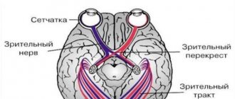
Intracranial hypertension manifests itself at stage III of acoustic schwannoma. It is accompanied by vomiting, cephalalgia in the back of the head and frontal part of the head. During ophthalmoscopy, congested optic discs are noticeable. Perimetry reveals hemianopia or isolated scotomas due to compression of the optic tracts and chiasm.
Treatment methods
If you suspect a brain tumor, you should seek medical help as soon as possible. After the collected medical history and examination, the doctor will prescribe a series of diagnostic examinations:
- audiometry;
- electronystagmography;
- radiography of the temporal bones;
- biopsy;
- MRI or CT.
The research results will help the doctor determine the location and size of the formation, make the correct diagnosis, and prescribe the necessary treatment.
Treatment for vestibular schwannoma on the right or left depends on the size of the tumor. At the initial stage, treatment can be carried out using conservative therapy, which will include taking several groups of medications to reduce the clinical signs of the disease and slow or stop tumor growth. Basically, doctors prescribe drugs from the following pharmacological groups:
- diuretics;
- anti-inflammatory;
- glucocorticosteroids;
- painkillers;
- cytostatics.
The choice of a specific medication, dose and duration of use is determined by the doctor for each patient.
It is strictly forbidden to self-medicate or use traditional methods, since they will not only not bring the desired result.
Treatment of neuroma of the cerebellopontine angle or auditory nerve with a small formation is carried out using radiation therapy, which also gives good results. If the tumor does not respond to conservative methods or is large enough, the only way to get rid of the disease will be surgical treatment of acoustic neuroma.
Radical methods include surgical removal of the acoustic neuroma. The procedure can be carried out in three ways:
- Translabyrinthine - carried out through the outer wall of the labyrinthine part of the middle ear. During the operation, a small segment of the middle ear and the tumor itself are removed. The patient completely loses the ability to hear in one ear, and there is also a high risk of dysfunction of the vestibular nerve.
- Suboccipital - access to the tumor is carried out by opening the skull under the back of the head. Used for large formations. It is possible to preserve hearing after removal of an acoustic neuroma using this method in 45% of cases.
If the size of the formation does not exceed 2 cm in diameter, it can be removed through the middle cranial fossa. The prognosis for such surgery is positive.
All operations are performed under general anesthesia with craniotomy. The duration of surgery depends on many factors and can take from 2 to 6 hours. The recovery period after surgery is long. The patient remains in the hospital for a long time, then is transferred to outpatient treatment.