Some numbers
There are more than 95 thousand kilometers of blood vessels in the body of a healthy adult. More than seven thousand liters of blood are pumped through them every day. The size of the blood vessels varies from 25 mm
(aortic diameter)
to eight microns
(capillary diameter).
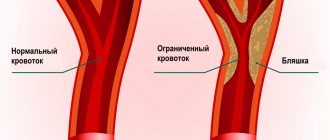
Down with atherosclerosis!
Atherosclerosis is one of the most common vascular diseases. It is characterized by the formation of fatty plaques on the walls of the arteries. Find out how to prevent atherosclerosis.
Blood vessel diseases
- The most dangerous vascular diseases that pose a threat to life: aneurysm of the abdominal and thoracic aorta, arterial hypertension, coronary artery disease, stroke, renal vascular diseases, atherosclerosis of the carotid arteries.
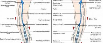
- Vascular diseases of the legs are a group of diseases that lead to impaired blood circulation in the vessels, pathologies of the vein valves, and blood clotting disorders.
- Atherosclerosis of the lower extremities - a pathological process affects large and medium-sized vessels (aorta, iliac, popliteal, femoral arteries), causing them to narrow. As a result, the blood supply to the extremities is disrupted, severe pain appears, and the patient’s performance is impaired.
- Varicose veins are a disease that results in dilation and lengthening of the veins of the upper and lower extremities, thinning of their walls, and the formation of varicose nodes. The changes that occur in the vessels are usually persistent and irreversible. Varicose veins are more common in women - in 30% of women after 40 and only 10% of men of the same age. (
What types of vessels are there?
All vessels in the human body can be divided into arteries, veins and capillaries
.
Despite the difference in size, all vessels are constructed approximately the same. The inside of their walls are lined with flat cells - endothelium. With the exception of capillaries, all vessels contain tough and elastic collagen fibers and smooth muscle fibers that can contract and dilate in response to chemical or nerve stimuli. Arteries
carry oxygen-rich blood from the heart to tissues and organs.
This blood is bright red
, which is why all the arteries appear red.
Blood moves through the arteries with great force, which is why their walls are thick and elastic. They are composed of a large amount of collagen, which allows them to withstand blood pressure. The presence of muscle fibers helps turn the intermittent blood supply from the heart into a continuous flow to the tissues. As they move away from the heart, the arteries begin to branch, and their lumen becomes thinner and thinner. The thinnest vessels that deliver blood to every corner of the body are capillaries
.
Unlike arteries, their walls are very thin, so oxygen and nutrients can pass through them into the cells of the body. This same mechanism allows waste products and carbon dioxide to move from cells into the bloodstream. The capillaries through which oxygen-poor blood flows are collected into thicker vessels - veins
.
Due to the lack of oxygen, venous blood is darker
than arterial blood, and the veins themselves appear bluish. Through them, blood flows to the heart and from there to the lungs to be enriched with oxygen. Vein walls are thinner than arterial walls because venous blood does not create as much pressure as arterial blood.
What are blood vessels?
Vessels are tube-like formations that extend throughout the human body and through which blood moves. The pressure in the circulatory system is very high because the system is closed. Through this system, blood circulates quite quickly.
After many years, obstacles to the movement of blood - plaques - form on the vessels. These are formations on the inside of blood vessels. Thus, the heart must pump blood more intensively in order to overcome obstacles in the blood vessels, which disrupts the functioning of the heart. At this point, the heart can no longer deliver blood to the organs of the body and cannot cope with its work. But at this stage it is still possible to recover. The vessels are cleansed of salts and cholesterol deposits.
When the blood vessels are cleansed, their elasticity and flexibility returns. Many diseases associated with blood vessels go away. These include sclerosis, headaches, a tendency to heart attack, and paralysis. Hearing and vision are restored, varicose veins are reduced. The condition of the nasopharynx returns to normal.
What is blood pressure?
Blood pressure is the force with which blood presses against the walls of the arteries. It increases when the heart contracts and pumps out blood, and decreases when the heart muscle relaxes. Blood pressure is stronger in the arteries and weaker in the veins. Blood pressure is measured with a special device - tonometer
.
Pressure readings are usually recorded in two numbers. Thus, normal blood pressure for an adult is considered to be 120/80
.
The first number, systolic pressure
, is a measure of the pressure during a heartbeat.
The second is diastolic pressure
- the pressure during relaxation of the heart.
Such dangerous excess weight
Professor Sergei Boytsov, chief specialist in preventive medicine at the Russian Ministry of Health and Social Development, talks about how extra pounds affect the heart and blood vessels.
Pressure is measured in the arteries and expressed in millimeters of mercury. In the capillaries, the pulsation of the heart becomes invisible and the pressure in them drops to approximately 30 mm Hg. Art. A blood pressure reading can tell your doctor how your heart is working. If one or both numbers are higher than normal, this indicates high blood pressure. If it’s lower, it means it’s reduced. High blood pressure indicates that the heart is working too hard: it requires more effort to push blood through the vessels. It also indicates that a person has an increased risk of heart disease.
Functional groups of blood vessels
The entire circulatory system is divided into six different groups of vessels according to functional load. Thus, in human anatomy one can distinguish shock-absorbing, exchange, resistive, capacitive, shunting and sphincteric vessels.
Shock absorbing vessels
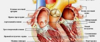
This group mainly includes arteries in which the layer of elastin and collagen fibers is well represented. It includes the largest vessels - the aorta and pulmonary artery, as well as areas adjacent to these arteries. The elasticity and resilience of their walls provides the necessary shock-absorbing properties, due to which the systolic waves that occur during heart contractions are smoothed out.
The shock absorption effect in question is also called the Windkessel effect, which in German means “compression chamber effect”.
To clearly demonstrate this effect, the following experiment is used. Two tubes are connected to a container filled with water, one made of elastic material (rubber) and the other made of glass. From a hard glass tube, water splashes out in sharp intermittent bursts, while from a soft rubber tube it flows out evenly and constantly. This effect is explained by the physical properties of the tube materials. The walls of the elastic tube are stretched under the influence of liquid pressure, which leads to the generation of so-called elastic tension energy. Thus, the kinetic energy resulting from pressure is converted into potential energy, which increases voltage.
The kinetic energy of cardiac contraction acts on the walls of the aorta and large vessels that extend from it, causing them to stretch. These vessels form a compression chamber: blood entering them under the pressure of heart systole stretches their walls, kinetic energy is converted into elastic tension energy, which contributes to the uniform movement of blood through the vessels during diastole.
Arteries located further from the heart are of the muscular type, their elastic layer is less pronounced, and they have more muscle fibers. The transition from one type of vessel to another occurs gradually. Further blood flow is ensured by contraction of the smooth muscles of the muscular arteries. At the same time, the smooth muscle layer of large elastic arteries has virtually no effect on the diameter of the vessel, which ensures the stability of hydrodynamic properties.
Resistive vessels
Resistive properties are found in arterioles and terminal arteries. The same properties, but to a lesser extent, are characteristic of venules and capillaries. The resistance of blood vessels depends on their cross-sectional area, and the terminal arteries have a well-developed muscular layer that regulates the lumen of the vessels. Vessels with a small lumen and thick, strong walls provide mechanical resistance to blood flow. The developed smooth muscles of resistive vessels provide regulation of blood volumetric velocity, controls the blood supply to organs and systems due to cardiac output.
Sphincter vessels
Sphincters are located at the end sections of the precapillaries; when they narrow or expand, the number of working capillaries that provide tissue trophism changes. When the sphincter expands, the capillary enters a functioning state; in non-functioning capillaries, the sphincters are narrowed.
Exchange vessels
Capillaries are vessels that perform an exchange function, carrying out diffusion, filtration and trophism of tissues. Capillaries cannot independently regulate their diameter; changes in the lumen of blood vessels occur in response to changes in the sphincters of the precapillaries. The processes of diffusion and filtration occur not only in capillaries, but also in venules, so this group of vessels also belongs to the exchange vessels.
Capacitive vessels
Vessels that act as reservoirs for large volumes of blood. Most often, capacitive vessels include veins - their structural features allow them to hold more than 1000 ml of blood and eject it as needed, ensuring stability of blood circulation, uniform blood flow and complete blood supply to organs and tissues.
Humans, unlike most other warm-blooded animals, do not have special reservoirs for storing blood from which it could be released as needed (in dogs, for example, this function is performed by the spleen). Veins can accumulate blood to regulate the redistribution of its volume throughout the body, which is facilitated by their shape. Flattened veins accommodate large volumes of blood, without stretching, but acquiring an oval lumen shape.
Capacitive vessels include large veins in the abdominal area, veins in the subpapillary plexus of the skin, and veins of the liver. The function of depositing large volumes of blood can also be performed by the pulmonary veins.
Shunt vessels
- Shunt vessels are an anastomosis of arteries and veins; when they are open, blood circulation in the capillaries is significantly reduced. Shunt vessels are divided into several groups according to their function and structural features:
- Pericardial vessels - these include elastic arteries, vena cava, pulmonary arterial trunk and pulmonary vein. They begin and end the systemic and pulmonary circulation.
- Great vessels are large and medium-sized vessels, veins and arteries of the muscular type, located outside the organs. With their help, blood is distributed to all parts of the body.
- Organ vessels - intraorgan arteries, veins, capillaries, providing trophism to the tissues of internal organs.
Circulation circles
The blood flowing through the cardiovascular system can be compared to an athlete running different distances. When it passes through the pulmonary circulation, it is a sprint. And a big circle is already a marathon. The Englishman William Harvey described these circles back in 1628. During a large circle, blood is distributed throughout the body, not forgetting to provide it with oxygen and take away carbon dioxide. During this “race”, arterial blood becomes venous.
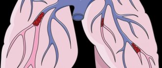
The pulmonary circulation is responsible for the flow of blood into the lungs, where the blood releases carbon dioxide and is enriched with oxygen. Blood from the pulmonary circulation returns to the left atrium. The systemic circulation, starting in the left ventricle, transports blood throughout the body. Oxygenated blood is pumped by the left ventricle into the aorta and its many branches - the various arteries. Then it enters the capillary vessels of organs and tissues, where oxygen from the blood is exchanged for carbon dioxide. The systemic circulation ends in small veins that merge into two large veins (vena cava) and return blood to the right atrium. The superior vena cava drains blood from the head, neck and upper extremities, and the inferior vena cava drains blood from the torso and lower extremities.