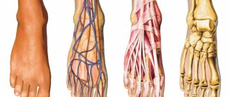
Anatomy of the veins of the lower extremities
The veins of the lower extremities are divided into superficial and deep.
Superficial veins of the lower limb
The superficial venous system of the lower extremities begins from the venous plexuses of the toes, forming the venous network of the dorsum of the foot and the cutaneous dorsal arch of the foot. From it originate the medial and lateral marginal veins, which pass into the greater and lesser saphenous veins, respectively. The plantar venous network anastomoses with the deep veins of the fingers, metatarsals and the dorsal venous arch of the foot. Also, a large number of anastomoses are located in the area of the medial malleolus.
The great saphenous vein is the longest vein in the body, contains from 5 to 10 pairs of valves, and its normal diameter is 3-5 mm. It originates in front of the medial epicondyle and rises in the subcutaneous tissue behind the medial border of the tibia, bends around the medial femoral condyle behind and passes to the anteromedial surface of the thigh, parallel to the medial border of the sartorius muscle. In the area of the oval window, the great saphenous vein pierces the ethmoidal fascia and flows into the femoral vein. Sometimes the great saphenous vein on the thigh and leg can be represented by two or even three trunks. From 1 to 8 large tributaries flow into the proximal portion of the great saphenous vein, the most constant of which are: the external genital, superficial epigastric, posteromedial, anterolateral veins and the superficial vein surrounding the ilium. Typically, tributaries flow into the main trunk in the area of the fossa ovale or somewhat distally. In addition, muscle veins can flow into the great saphenous vein. The small saphenous vein begins behind the lateral malleolus, then it rises in the subcutaneous tissue, first along the lateral edge of the Achilles tendon, then along the middle of the back surface of the leg. Starting from the middle of the leg, the small saphenous vein is located between the layers of the fascia of the leg (N.I. Pirogov’s canal) accompanied by the medial cutaneous nerve of the calf. That is why varicose veins of the small saphenous vein are much less common than the large saphenous vein. In 25% of cases, the vein in the popliteal fossa pierces the fascia and flows into the popliteal vein. In other cases, the small saphenous vein can rise above the popliteal fossa and flow into the femoral, large saphenous vein, or into the deep vein of the thigh. Therefore, before the operation, the surgeon must know exactly where the small saphenous vein flows into the deep one in order to make a targeted incision directly above the anastomosis. The constant estuarine tributary of the small saphenous vein is the fenopopliteal vein (vein of Giacomini), which flows into the greater saphenous vein. Many cutaneous and saphenous veins flow into the small saphenous vein, most in the lower third of the leg. It is believed that the small saphenous vein drains blood from the lateral and posterior surface of the leg.
Deep veins of the lower limb
The deep veins begin as the plantar digital veins, which become the plantar metatarsal veins, which then drain into the deep plantar arch. From it, blood flows through the lateral and medial plantar veins into the posterior tibial veins. The deep veins of the dorsum of the foot begin with the dorsal metatarsal veins of the foot, which drain into the dorsal venous arch of the foot, from where blood flows into the anterior tibial veins. At the level of the upper third of the leg, the anterior and posterior tibial veins merge to form the popliteal vein, which is located lateral and somewhat posterior to the artery of the same name. In the area of the popliteal fossa, the small saphenous vein and the veins of the knee joint flow into the popliteal vein. Then it rises in the femoral-popliteal canal, now called the femoral vein. The femoral vein is divided into the superficial vein, located distal to the deep vein of the thigh, and the common vein, which is located proximal to it. The deep vein of the thigh usually flows into the femoral vein 6-8 cm below the inguinal fold. As you know, the femoral vein is located medial and posterior to the artery of the same name. Both vessels have a single fascial sheath, while doubling of the trunk of the femoral vein is sometimes observed. In addition, the medial and lateral veins surrounding the femur, as well as muscular branches, flow into the femoral vein. The branches of the femoral vein widely anastomose with each other, with the superficial, pelvic, and obturator veins. Above the inguinal ligament, this vessel receives the epigastric vein, the deep vein surrounding the ilium and passes into the external iliac vein, which merges with the internal iliac vein at the sacroiliac joint. This section of the vein contains valves, in rare cases, folds and even septa, which causes thrombosis to be frequently localized in this area. The external iliac vein does not have many tributaries and collects blood mainly from the lower limb. Numerous parietal and visceral tributaries flow into the internal iliac vein, carrying blood from the pelvic organs and pelvic walls. The paired common iliac vein begins after the confluence of the external and internal iliac veins. The right common iliac vein, somewhat shorter than the left, runs obliquely along the anterior surface of the 5th lumbar vertebra and has no tributaries. The left common iliac vein is slightly longer than the right and often receives the median sacral vein. The ascending lumbar veins flow into both common iliac veins. At the level of the intervertebral disc between the 4th and 5th lumbar vertebrae, the right and left common iliac veins merge to form the inferior vena cava. It is a large vessel without valves, 19-20 cm long and 0.2-0.4 cm in diameter. In the abdominal cavity, the inferior vena cava is located retroperitoneally, to the right of the aorta. The inferior vena cava has parietal and visceral branches, which supply blood from the lower extremities, lower torso, abdominal organs, and pelvis. Perforating (communicating) veins connect the deep veins with the superficial ones. Most of them have valves located suprafascially and thanks to which blood moves from the superficial veins to the deep ones. About 50% of the communicating veins of the foot do not have valves, so blood from the foot can flow from deep veins to superficial ones, and vice versa, depending on the functional load and physiological conditions of outflow. There are direct and indirect perforating veins. Direct ones directly connect the deep and superficial venous networks, indirect ones connect indirectly, that is, they first flow into the muscular vein, which then flows into the deep vein. The vast majority of perforating veins arise from tributaries rather than from the trunk of the great saphenous vein. In 90% of patients, there is incompetence of the perforating veins of the medial surface of the lower third of the leg. On the lower leg, incompetence of the perforating veins of Cockett, which connects the posterior branch of the great saphenous vein (vein of Leonardo) with the deep veins, is most often observed. In the middle and lower thirds of the thigh there are usually 2-4 most permanent perforating veins (Dodd, Gunter), directly connecting the trunk of the great saphenous vein with the femoral vein. With varicose transformation of the small saphenous vein, incompetent communicating veins of the middle, lower third of the leg and in the area of the lateral malleolus are most often observed. In the lateral form of varicose veins, the localization of perforating veins is very diverse.
Venous networks
To carry out a correct diagnosis of the disease and make a correct diagnosis in the field of venous diseases, you need to clearly understand the system of blood vessels of the lower extremities.
There is a distinction between the deep and superficial network of veins. The deep one consists of paired vessels passing next to the arteries on the fingers, foot and lower leg. The tibial veins converge in the femoropopliteal canal and create the azygos popliteal vein, which passes into the femoral vein. Before passing into the ileum, up to 8 peripheral vessels join it. In addition to them, the deep vein also adds, carrying blood cells from the back of the thigh. The superficial circulatory network is located directly under the skin. It consists of the great and small saphenous veins, respectively.
Functions of the leg veins
The veins of the legs have a difficult task - without contractility, they must deliver a mass of blood from the most distant parts of the body to the heart.
This is what predetermined the structure of the network, divided into superficial and deep vessels, connected by a network of perforating ducts. Their walls consist of three layers:
- Intima is the inner layer of endothelium, separated from the middle layer by a thin membrane.
- The medial layer is the middle “layer” of the tube, represented by elastic fibers and a small proportion of muscle fibers. It is this layer that gives them strength and stretchability.
- The outer layer, consisting of connective tissue bordering the membrane that separates the blood tubes from the muscle tissue.
Despite the fact that in the lower extremities the drainage network is represented by tubes of different diameters (from 1.5 to 11 mm), the anatomy of the veins is almost the same. The only difference is the thickness of each layer and the number of valves. For example, the veins of the lower leg have more valves, but their diameter is 2 times smaller than that of the great saphenous vein.
In addition to blood pressure, superficial vessels experience significant stress due to external influences, so the thickness of their middle layer is much greater than that of deep-lying tubes. For example, the walls of the great saphenous vein are 1.3 times thicker and stronger than those of the deep vein.
The main functions of the VNK are:
- Ensuring uninterrupted outflow of blood, in which carbon dioxide and waste products of tissues located within their reach are dissolved.
- Delivery to tissues of hormones, organic compounds (enzymes, amino acids, proteins), vitamins and microelements coming from the intestines.
- Regulation of general blood pressure.
It is the variety of tasks assigned to the VNK that has led to close attention to the condition of the blood vessels. Any deviation in their functionality can cause irreparable harm to health.
Why is sural vein thrombosis dangerous?
Chronic venous insufficiency of the lower extremities is the most common complication of sural vein thrombosis. This condition leads to severe edema and impaired nutrition of the tissues of the lower extremities. As a result, the patient may develop eczema on his legs and trophic ulcers that do not heal for a long time. In addition, the person will constantly experience heaviness in his legs, and cramps will begin to bother him at night. The skin will lose its elasticity, become dry, and become covered with pigmented rashes. This will lead to the patient having difficulty withstanding both physical and mental stress.
An even more serious complication of sural vein thrombosis is pulmonary embolism. In this case, pieces of the blood clot break off, which rise higher and higher in the bloodstream, reaching the pulmonary artery and blocking its lumen. As a result, the patient develops pulmonary and heart failure, which leads to death. If a particle of a blood clot clogs a small branch of the pulmonary artery, then a person develops a serious condition such as a pulmonary infarction.
Causes of sural vein thrombosis
In order for a person to develop thrombosis of the sural veins, a combination of several pathogenetic factors is necessary:
- Damage to the inner layer of the vein. Such injuries can be caused by high blood pressure, penetration of bacterial endotoxins into the blood, exposure to radiation, metabolic disorders, toxic effects of cigarette smoke, etc.
- Impaired blood flow. Blood stasis is one of the most significant reasons that lead to the formation of blood clots in the veins. Blood turbulence, previous myocardial infarction, rheumatic mitral valve stenosis, polycythemia vera, and sickle cell anemia have a negative effect on the veins.
- Hypercoagulation of blood (thrombophilia). Causes of blood hypercoagulation: high levels of fibrinogen in the body, prothrombin mutations, fibrinolysis disorders, etc.
There is a high probability of developing thrombosis in people who are forced to adhere to bed rest for a long period of time. This is especially true for hospital patients. However, even a long plane flight can take its toll. It’s not for nothing that doctors call deep vein thrombosis an “economic class disease.” Therefore, any prolonged time spent sitting with your legs down can result in thrombosis of the sural veins.
Other factors that cause a high risk of sural vein thrombosis:
- Previous myocardial infarction;
- Atrial fibrillation;
- Damage to tissues of the lower extremities, including deep burns, leg fractures, surgical interventions on veins and soft tissues;
- The presence of a malignant tumor in the body;
- Having a prosthetic heart valve;
- Cardiomyopathy;
- Nephrotic syndrome;
- Increased levels of estrogen in the blood during pregnancy and in the early period after childbirth;
- Burger's disease;
- Obesity and diabetes;
- Frequent infectious diseases;
- Excessive physical activity and physical inactivity;
- Taking hormonal medications to prevent unwanted pregnancy;
- Smoking;
- Elderly age.
Material and methods
Since the beginning of 2015, MSCT venography was performed on 121 people. Initially, 30 lower extremities were examined in individuals without signs of CVD (control group). The study group included 91 patients (52 women and 39 men aged 32 to 65 years) with CVD with clinical class C0-C6. Class C0—C1 occurred in 15 (16.5%) patients, C2—C3 — in 45 (49.5%). 31 (34%) patients had various trophic skin disorders (C4-C6).
All studies were performed on a 128-slice Philips Ingenuity CT multislice computed tomograph with the Intell Space Portal image processing software package, followed by reconstruction of a volumetric image in 3D mode.
The scanning was carried out in an automatic program mode, which implied sequential non-stop administration of a bolus of contrast agent and saline solution.
Scan mode: collimation 64×0.625; pitch 0.923; kilovoltage 100 kV; mAS 188; reconstruction parameters: axial + cranial orientation.
MSCT venography was performed according to the method we developed. In a clean dressing room or in a study room, the vein of the dorsum of the foot was catheterized using an intravenous catheter G22-G24. The patient was placed on the table on his back. One of two infusion syringes (A) was filled with 50 ml of non-ionic contrast agent (Ultravist). An isotonic sodium chloride solution was drawn into the second infusion syringe (B) at the rate of 1 ml of 0.9% saline per 1 cm of height of the subject. Both infusion syringes were inserted into the auto-injector. Using an infusion line, the injector was connected to an intravenous catheter and the infusion mode was switched on from A to B, with an injection rate of the radiopaque mixture of 4 ml/s.
Computer marking of the scanned limb was carried out, including the pelvis and foot. After preliminary scanning, the scanning area was finally set (the entire lower limb and pelvic area) with a direction from the pelvis to the foot. The scanning time parameters were entered into the program.
A pneumatic cuff was placed over the ankles, the pressure in which was raised to 60 mm Hg, and the administration of a radiopaque mixture began, which, depending on the calculated volume, lasted about 40 s. After completing the administration of the entire volume of contrast and isotonic sodium chloride solution, the pressure in the second cuff placed at the mid-thigh was raised to 60 mmHg, and the patient took a deep breath, held his breath and tensed the muscles of the anterior abdominal wall. From this moment, the first main scan began, the total duration of which was 12-15 s. After this, the patient exhaled and performed five dorsiflexion movements of the foot. After completing the test, the patient returned to the starting position. After 40 s, the second main scan began, after which the study was completed and a three-dimensional image of the limb and veins was reconstructed using automatic Intell Space Portal data processing protocols embedded in the computer.