What is thrombophlebitis
Thrombophlebitis
(acute thrombophlebitis, superficial vein thrombosis, TPV) is an inflammation of the venous walls with further formation of blood clots. The thrombus is tightly attached to the inflamed vessel. If treatment is started in time, the blood clot will resolve, the inflammation will go away, and the venous lumen will be restored and will not affect blood flow.
The incidence of thrombophlebitis is determined by many factors, one of which is age. Thus, superficial vein thrombosis debuts annually in 0.3-0.6 per 1000 people under 30 years of age and in 0.7-1.8 per 1000 elderly patients. There are also gender differences. In men under 30 years of age, acute thrombophlebitis occurs in 0.05 cases per 1000 patients annually. For women, these figures are significantly higher. Thus, in the first 30 years of life, the onset of TPV is detected in 0.31 per 1000 women, but with age the detection frequency increases to 2.2 per 1000 patients i Bogachev V.Yu. Thrombophlebitis (thrombosis of superficial veins): modern standards of diagnosis and treatment / V.Yu. Bogachev [et al.] // Outpatient surgery. — 2021. — No. 3-4 (63-64). — P. 16-23. .
In 3-11% of patients, thrombophlebitis is localized on the lower extremities. The system of the great saphenous vein is affected in 60-80% of cases, the small saphenous vein - in 10-20%, and bilateral TPV occurs in 5-10% of cases i Bogachev V.Yu. Thrombophlebitis (thrombosis of superficial veins): modern standards of diagnosis and treatment / V.Yu. Bogachev [et al.] // Outpatient surgery. — 2021. — No. 3-4 (63-64). — P. 16-23. .
Against the background of varicose veins, thrombophlebitis occurs in 4-62% of patients i Bogachev V.Yu. Thrombophlebitis (thrombosis of superficial veins): modern standards of diagnosis and treatment / V.Yu. Bogachev [et al.] // Outpatient surgery. — 2021. — No. 3-4 (63-64). — P. 16-23. .
Types (classification) of disease
Thrombophlebitis is divided into several types, taking into account the origin and origin of the disease, the localization of the lesion, and the extent of the lesion.
Venous thrombophlebitis is divided according to the affected area into:
- Migratory
. A blood clot appears in one place. Over time, it disappears and appears in another area. The condition is dangerous and requires prompt medical attention. - Local
. A blood clot forms in a specific area. It doesn't have to be the shins. For example, thrombophlebitis of the hand is diagnosed.
Depending on the presence or absence of pathogens, subcutaneous thrombophlebitis occurs:
- Septic
. The infection spreads quickly through the bloodstream, so there is a risk of developing sepsis. Diagnosed after childbirth, surgery and erysipelas. Treated with antibiotics. - Aseptic
. There is no infection, the disease occurs after injury. Most often, varicose thrombophlebitis is diagnosed due to improper or absent treatment.
Depending on the size of the blood clot, the disease can be:
- Occlusive
. The lumen of the vein is completely blocked, so the outflow of blood is impaired. There is a risk of necrosis (tissue death), so immediate surgery is required. - Non-occlusive
. An attached blood clot partially blocks the venous lumen. Blood circulates.
Based on the extent of the pathological process, the following is revealed:
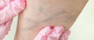
- Thrombophlebitis of the superficial veins
. Symptoms appear immediately. The patient complains of pain along the vein. The surrounding skin turns red and thickens. Over time, swelling forms, which is difficult to eliminate. The temperature is kept within 39 °C. Treated with medication. - Deep vein thrombosis
. This form is detected less frequently and is more severe. The symptoms are the same, but their severity is stronger. The limbs become very swollen and turn blue. If there is a high risk of developing pulmonary embolism, surgery is performed.
First, thrombophlebitis of the superficial veins of the lower extremities develops. If left untreated, the disease develops into deep vein thrombosis, thrombophlebitis of the great saphenous vein.
The main causes of phlebitis
The main reason for the development of phlebitis of the lower extremities is varicose veins. This is one of the most common causes of phlebitis. Also, this disease can occur as a result of infection (the causative agents of which are streptococci), mechanical damage to venous vessels (intravenous injection or infusion (infusion), blood sampling), chemical burns after injections. Other causes of phlebitis include: excessive weight of the patient; prolonged immobility or tightness of the limb; bruises and injuries; complications after childbirth, etc.
Stages
The sequence of development of the disease is as follows:
- Acute stage
. Clinical signs of thrombophlebitis are bright and appear unexpectedly. The temperature rises, the skin around the affected area turns red, and the patient shudders. If treatment is started within two weeks, the development of the disease can be avoided. The blood clot will dissolve. The duration of exacerbation is up to one month. - Subacute stage
. Lasts up to two months. Signs of the disease subside, but do not fade away. The skin remains red and the swollen veins turn blue. The swelling goes away, but the seals remain. - Chronic stage
. Diagnosed two to three months after the disease is detected. Symptoms are either absent or worsened. During remission, the limbs remain swollen, quickly become tired and itchy, but there is no pain. Discomfort occurs in the lower extremities when walking for a long time.
How is thrombophlebitis different from varicose veins?
Varicose veins are increased pressure in thinned and inelastic veins, and thrombophlebitis is an inflammation of the venous walls with subsequent formation of blood clots. Most often, the diagnosis of thrombophlebitis is a consequence of varicose veins.
Thrombophlebitis differs from varicose veins in the following symptoms:
| Sign | Varicose veins | Thrombophlebitis |
| Pain | pulling | bursting and stabbing |
| Convulsions | Yes | No |
| Changing the appearance of your legs | noticeable tortuosity and bulging of veins | compactions and lumps when palpated |
| Change in skin color | pale skin, blue veins | red skin |
| Burning feeling | Yes | No |
Both pathologies develop as a result of impaired hemostasis, but inflammation of the venous walls is more often associated with the entry of an infectious agent. The difference is the absence of local inflammation. With varicose veins, the veins do not become inflamed. In this case, only the blood circulation is disrupted.
Causes and risk factors for the development of thrombophlebitis
Thrombophlebitis of the lower extremities rarely begins spontaneously. Most often, the disease becomes a complication of leg diseases. The development mechanism is associated with improper outflow of lymph and blood through the veins and vessels. After injury to the inner layer of the vein, bleeding begins. In response to this, a blood clot forms. It prevents blood loss, but over time it grows and completely blocks the lumen of the vessel, preventing blood from circulating.
The following factors precede this process:
- Damage to the vascular wall. This group includes mechanical injury to veins due to trauma and compression of the limbs. Venous walls are damaged when injections are performed incorrectly, frequent and long IVs, or operations.
- Deterioration of blood supply to certain areas of the body. The blood flows poorly due to wearing a cast for a long time, bed rest and physical inactivity. Less commonly, slowing of blood flow occurs as a result of heart failure.
- Improper flow of blood in the veins due to venous insufficiency. The complication develops against the background of pregnancy and diseases of the pelvic organs.
- Increased blood clotting. The viscosity and homogeneity of blood and plasma change with long-term use of hormonal drugs, discrepancies in the levels of estrogen and progesterone, and infections.
Other causes of thrombophlebitis may be:
- obesity;
- allergic reactions and sensitization;
- a history of venous thromboembolic complications (VTEC);
- taking a number of medications (diazepam, amiodarone, vancomycin, chemotherapy drugs, heroin addiction).
Acute thrombophlebitis often develops against the background of some autoimmune diseases, such as systemic lupus erythematosus, vasculitis, Behcet's and Buerger's diseases. In particular, in Behçet’s disease, TPV is detected in 53.3%, and DVT in 29.8% of patients i Bogachev V.Yu. Thrombophlebitis (thrombosis of superficial veins): modern standards of diagnosis and treatment / V.Yu. Bogachev [et al.] // Outpatient surgery. — 2021. — No. 3-4 (63-64). — P. 16-23. .
The risk group includes:
- Patients with heart disease - chronic heart failure, ischemic stroke. Limbs swell, motor activity decreases, and after a stroke there may be paralysis, which impedes the flow of blood through the veins.
- Patients with concomitant phlebological diseases - varicose veins, venous thrombosis. The speed of blood movement in the veins decreases, the blood stagnates, forming a blood clot.
- Bedridden patients, people with complex fractures. Their mobility is impaired and the likelihood of blood clot formation increases.
- People with a hereditary predisposition. Hemostasis disorders are transmitted genetically. The likelihood of the disease is increased if blood flow pathologies are detected in first-degree relatives.
How to treat phlebitis?
First of all, the patient is provided with bed rest. The affected limb is fixed in an elevated position. Anticoagulants and anti-inflammatory drugs are prescribed. After relief of inflammation - physiotherapy.
Surgical treatment is carried out in case of complications, for example. thrombosis
To prevent the development of phlebitis, it is important to promptly treat infectious and inflammatory diseases, as well as follow the rules for performing intravenous injections.
Symptoms
Signs of thrombophlebitis appear immediately. The disease can be suspected based on the following clinical picture:
- the limb swells;
- the skin over the inflamed vein turns red, and the vessel itself darkens and is clearly visible under the skin;
- a compaction is felt to the touch along the course of the vessel;
- there is a feeling of heaviness in the legs and bloating of the veins;
- in a supine position, the patient cannot lift his leg and rotate his foot;
- The leg is cold to the touch.
At the initial stage, the symptoms are local, that is, the general condition does not change. As the disease progresses, mobility becomes impaired. The patient notes weakness in the legs and cannot stand for a long time. When bending the foot, he feels tension in the lower leg and thigh. Temperatures range from 37°C to 39°C.
Distinctive signs of thrombophlebitis of the lower leg are that the calf muscle turns blue and the veins on it swell. The pain is so severe that the patient cannot step on his foot.
Thrombosis of the iliofemoral vein is manifested by pain in the lumbosacral spine, pain in the groin and the lower third of the abdomen on one side.
Femoral vein thrombosis is characterized by swelling of the veins in the upper thigh and discomfort in the groin area.
Symptoms of phlebitis of the veins of the upper and lower extremities
Symptoms of phlebitis are quite varied and depend on the form and type of disease.
The following symptoms are characteristic of acute and chronic phlebitis of the superficial veins:
- the vein becomes tense and very painful;
- there is redness and thickening of the skin, local hyperthermia (increased temperature of a given area of the body);
- the appearance of red stripes along the location of the venous vessel.
Acute and chronic phlebitis of the deep veins are accompanied by:
- pain and swelling in the area of inflammation;
- increased temperature of the whole body (general hyperthermia);
- the skin takes on a milky white hue.
With cerebral phlebitis the following occurs:
- headache;
- blood pressure increases;
- neurological symptoms.
A characteristic symptom of pylephlebitis
is severe purulent intoxication.
Diagnosis of thrombophlebitis
Diagnosis of the condition of the lower extremities includes:
- Vascular ultrasound in Doppler mode
. The blood flow of the superficial and deep veins is analyzed. Areas of narrowing of the vascular lumen and slowing down of blood flow are identified. - CT venography
. The transition of the femoral vein to the iliac vein is poorly visualized on ultrasound, so computed tomography shows a splash of blood after slow passage through a certain area. It is carried out with contrast enhancement, that is, a dye is injected into the vein. - D-dimer assay
. When a blood clot forms, the amount of fibrin breakdown products in the blood increases. The analysis is performed after an ultrasound. - Blood analysis
. The diagnosis is confirmed by high coagulogram (clotting test) values. The presence of C-reactive protein indicates inflammation. Leukocytes also grow, ESR increases, and the number of rods in the blood increases.
Treatment of thrombophlebitis
Therapeutic actions are aimed at suppressing inflammation, resolving blood clots and thinning the blood.
Drug treatment
venous thrombophlebitis involves taking:
- anticoagulants to thin and prevent blood clotting, in the acute phase are prescribed by injection;
- antibiotics for the infectious nature of the disease;
- angioprotectors to increase the tone of the veins, reduce venous stagnation;
- disaggregants for breaking down cells that form a blood clot;
- phlebotonics to strengthen and increase the elasticity of the walls of blood vessels;
- non-steroidal analgesics to relieve acute pain.
Compression therapy
using bandages, elastic stockings or class 2 sleeves accelerates blood flow in the superficial and deep veins, thereby preventing the growth of a blood clot. In addition, compression has an analgesic effect.
Physiotherapy
– an effective method of treating venous thrombophlebitis. After acute symptoms have been relieved, a course of magnetic therapy, infrared radiation, ultraviolet radiation, or darsonvalization of your choice is prescribed. The procedures reduce inflammation, increase blood circulation, and accelerate the regeneration of the internal walls of veins.
Surgical intervention
is prescribed if the blood clot grows and the threat of blocking the lumen of the vessel increases. There are several types of operations for thrombophlebitis:
- crossectomy – access to the saphenous vein through the groin and an incision in the thigh;
- thrombectomy – access to the thrombus through one incision;
- invagination stripping - with the help of a probe, the vein is everted, the blood clot is removed;
- stenting – the vein is expanded mechanically by installing a special balloon.
Treatment of deep vein thrombosis
In almost all cases, deep vein thrombosis is treated in a hospital. An exception may be deep vein thrombosis of the leg, provided there is no threat of thromboembolism. The danger of thromboembolism can only be determined by ultrasound examination.
If deep vein thrombosis is suspected, the patient should be hospitalized immediately. In the hospital, an examination is carried out to clarify the prevalence of thrombosis, the degree of threat of pulmonary embolism, and treatment is started immediately.
Anticoagulants (anticoagulants), antiplatelet agents, anti-inflammatory drugs, and phlebotropic drugs are usually prescribed.
In case of massive thrombosis, in the early stages it is possible to carry out thrombolysis - the introduction of agents that “dissolve” thrombotic masses.
If there is a threat of thromboembolism, when the thrombus is attached to the vessel wall only in some part, and the tip of the thrombus “dangles” in the lumen of the vessel (floating thrombus), thromboembolism is prevented (installation of a vena cava filter, plication of the inferior vena cava, ligation of the femoral vein, etc. ).
The consequences of deep vein thrombosis vary. With a small thrombosis on the lower leg there may be no consequences. With massive thrombosis, especially “high” (at the level of the hip or above), swelling of the leg may persist for a long time, and in severe cases there may be trophic disorders, even the appearance of ulcers (post-thrombophlebitic disease (PTSD)). Therefore, after deep vein thrombosis, the patient is prescribed treatment for a long time in the form of elastic compression (special stockings), indirect anticoagulants (warfarin under the control of blood clotting), and other drugs.
You should see a phlebologist for at least six months after thrombosis.
In case of recurrent thrombosis, a genetic study is carried out; if the tests are positive, the issue of lifelong prescription of anticoagulants is decided.
Recommendations and contraindications for thrombophlebitis
The doctor advises you to follow a diet and wear low-heeled shoes with a comfortable last.
Forbidden:
- wear stilettos and heels higher than 3 cm;
- massage limbs;
- go to the gym and lift weights;
- remove leg hair with wax or epilator;
- go to the bathhouse and sunbathe;
- wear socks, stockings, and knee socks with a thick elastic band.
It is necessary to exclude the use of hormonal drugs and choose a different method of contraception.
Sources
- Bogachev V.Yu. Thrombophlebitis (thrombosis of superficial veins): modern standards of diagnosis and treatment / V.Yu. Bogachev [et al.] // Outpatient surgery. — 2021. — No. 3-4 (63-64). — P. 16-23.
- Kuzmichev D.E. Pathomorphological findings. Thrombophlebitis / D.E. Kuzmichev [et al.] // Health care of Ugra: experience and innovations. - 2020. - No. 1. - P. 53-56.
- Marushchak E.A. Modern methods of ultrasound diagnosis of venous thrombosis of the inferior vena cava system / E.A. Marushchak, A.R. Zubarev // Outpatient surgery. Stationary replacement technologies. - 2014. - No. 3-4 (55-56). — P. 38-47.
Deep vein thrombosis
Deep vein thrombosis is a more severe disease, usually requiring the patient to remain in the hospital.
There are deep vein thrombosis of the leg, popliteal vein, femoral vein, iliofemoral (ileofemoral) thrombosis. Often there is damage to thrombosis of several sections (for example, the popliteal and femoral veins, deep veins of the leg and popliteal vein, etc.).
Deep vein thrombosis manifests itself primarily as swelling and moderate pain. Moreover, the higher the level of thrombosis, the more pronounced the edema. Thus, with thrombosis of the deep veins of the leg, there may be moderate swelling of the leg, sometimes the swelling is so insignificant that it is detected only when measuring the circumference of the leg (compared to the healthy leg). With thrombosis of the femoral or iliac (continuation of the femoral) veins, the entire leg, up to the groin, and in severe cases, the lower part of the abdominal wall swells.
In the photo - the left lower limb is cyanotic, thickened to the groin - deep vein thrombosis at the level of the iliofemoral segment (ileofemoral thrombosis).
Thrombosis, as a rule, is unilateral, so only one leg swells. Bilateral edema is observed with thrombosis of the inferior vena cava, deep vein thrombosis in both legs (which is quite rare).
Another symptom of thrombosis is pain. It is usually moderately expressed, pulling, sometimes bursting, is relatively constant in nature, and can intensify in a standing position. In case of deep vein thrombosis of the leg, the Homans, Lowenberg, Luvellubri, Meyer, Payra symptoms are positive.
With deep vein thrombosis, there may also be a slight increase in temperature, increased venous pattern, etc.