Melanoma is a dark brown malignant formation that appears as a result of certain changes and an increase in melanin synthesis. A dangerous tumor can be located on the legs and cause pain and other discomfort. The anatomical and functional features of the feet require urgent closure of the defects after mandatory excision of the formation in the development of this disease. The complexity of plastic surgery and other reconstructive procedures in this area is associated with age-related pathologies.
Melanoma of the foot is the most aggressive of oncogenic neoplasms. The tumor can metastasize, after which it is considered incurable. In order to prevent this disease, it is necessary to strictly control existing moles and age spots on the legs. This cancer, even if small in size, is very dangerous for human life. After a few months, the oncogenic process spreads to the internal organs.
If melanoma is diagnosed at an early stage, then removing the formation on the feet is not difficult. If the lesion is larger than one centimeter and is characterized by uneven staining with asymmetrical edges, a treatment plan is developed, including chemotherapy in addition to surgery.
Symptoms of foot melanoma
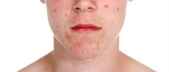
The disease has basic and additional symptoms. The first includes the following:
- the rapid growth of new, recently emerged formations,
- change in the size and structure of the nevus on the leg,
- darkening of the tumor and the appearance of black fragments in it.
Additional symptoms include the development of an inflammatory process along the edge of the pigment spot, as well as a feeling of itching and bleeding of the skin formation. In frequent cases, pathology occurs on the feet of women. The earlier the diagnosis is made, the greater the chances of a favorable outcome. Treatment of superficial melanomas on the feet in the absence of raised areas usually has a positive result.
Nodular formations that are raised above the surface of the epidermis are characterized by an aggressive course. They affect approximately 15 percent of patients. Subungual melanoma on the leg is common. It mainly develops on the big toes.
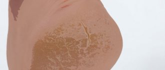
Is it possible to get rid of small age spots on the legs?
Only a doctor can determine the cause of hyperpigmentation and assess the risks to the patient’s health. In most cases, darkening of the epidermis is a safe process. To help slow it down:
- refusal to go to the solarium,
- use of sunscreens,
- taking vitamins, minerals and antioxidants.
After consultation with a doctor, unsightly pigment spots can be lightened or removed. The specialist selects the appropriate method, taking into account the cause and type of tumor.
To exclude dangerous pathologies, if your skin color changes, make an appointment with a therapist at a clinic near the Maryino metro station. The doctor will conduct a comprehensive examination and tell you how to maintain your natural skin color.
All articles
5% discount Print coupon from our website
Ask your question on the website Get professional advice!
Causes of foot melanoma
Many people who are faced with this disease are looking for the reasons for its development. These include the following:
1. Exposure of the skin to ultraviolet rays (vacation in the south up to 1-2 times a year, visiting a solarium, getting sunburn), especially for those with fair skin and hair.
2. Congenital or acquired dysplastic pigmented nevi (moles that increase in size).
3. Chronic injury to the mole - this happens if you constantly wear uncomfortable shoes, expose the nevus to friction with items of clothing, or touch the mole while shaving or during hair removal.
4. Genetic predisposition (multiple dysplastic skin nevi syndrome, a history of melanoma of the foot in a close relative)
5. During pregnancy and menopause, hormonal changes occur, which leads to the degeneration of ordinary moles into malignant ones.
Although in reality, the listed factors only trigger a mechanism that was originally inherent at the genetic level in the human body.
Diagnosis of the disease
Timely diagnosis is the key to the final cure of foot melanoma. It is important to periodically examine the skin yourself, especially if there are moles on the skin. Changes in their shape, shade, edges and growth dynamics should alert you. In generally accepted practice, the international ABCDE rule helps to identify the disease, which is translated from English as: – asymmetry of the neoplasm:
- B – indicates that the edges of the spot are jagged;
- C – change in shade or multicolor (the presence of black, brown, pink and blue colors in one spot);
- D – mole diameter more than 6 mm;
- E - dynamics of changes over time (growth, convexity).
If the above manifestations occur, it is necessary to visit an oncologist, a specialist in the diagnosis and treatment of cancer, as soon as possible.
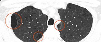
The oncologist examines the patient, his skin, and the melanoma itself on the leg, examines and palpates regional lymph nodes, and collects anamnesis. After which he prescribes an ultrasound of peripheral lymph nodes, the abdominal cavity and retroperitoneal space, the pelvis, and the chest at the time of the absence or development of metastases.
If necessary, computed tomography (CT) of organs and systems with contrast, magnetic resonance imaging (MRI) of the brain with intravenous contrast, or positron emission tomography of the “whole body” is performed. If enlarged peripheral lymph nodes are detected (if metastases are suspected), it is necessary to prescribe the patient a fine-needle aspiration puncture biopsy using cytological examination.
An important diagnostic criterion is called Breslow thickness; it reflects the rate of division of oncogenic cells. The oncologist examines the extent of the lesion and develops a treatment plan. The thinner the Breslow thickness, the greater the chance of recovery. The indicator is detected and calculated after performing a histological examination of tissue samples. If the indicator is less than 1 mm, then the tumor is considered benign and does not require surgical removal of pigmentation. If the indicator is from 1 mm, then immediate removal of the formation is recommended.
Symptoms and course
The process most often begins in the interdigital spaces, mainly between the most closely adjacent 4th and 5th fingers. When you feel a slight itch at the bottom of the interdigital fold, a strip of swollen and slightly flaky epidermis appears. After 2-3 days, a small crack appears here, releasing a small amount of serous fluid. Sometimes the stratum corneum falls off, revealing a pink-red surface. The disease, gradually progressing, can spread to all interdigital folds, the plantar surface of the toes and adjacent parts of the foot itself. The serous fluid seeping onto the surface serves as an excellent nutrient material for further proliferation of fungi.
When fungi enter through the damaged stratum corneum into the deeper parts of the epidermis, the process is complicated by an eczematous reaction. Numerous, very itchy blisters filled with clear liquid appear, which in some places merge and erode, leaving weeping areas.
The process can spread to the back of the foot and fingers, the sole, capturing its arch to the heel. The disease, either weakening or intensifying again, without proper treatment and care, can drag on for many years. Often, a complication with a secondary pyogenic infection occurs: the transparent contents of the vesicles become purulent, the inflammatory redness intensifies and spreads beyond the boundaries of the lesion, the foot becomes swollen, the patient’s movements are difficult or impossible due to severe pain; Subsequent complications may also develop in the form of lymphangitis, lymphadenitis, erysipelas, etc.
In some cases, athlete's foot on the soles is expressed by the appearance on initially unchanged skin of separate groups of itchy, deeply located, dense-to-touch blisters and blisters with transparent or slightly cloudy contents. After their spontaneous opening, the covering of the blisters disappears, remaining in the form of a corolla only at the edges of the lesion; the central parts have a smooth, pink-red color, slightly flaky, less often, weeping surface; Often new bubbles appear on it. Due to their fusion, the lesion expands and can cover large areas of the soles.
The absorption of allergens (fungi and their toxins) is a sensitizing factor for the entire body, increases the sensitivity of the skin, and an allergic rash may appear on it. It is most often observed on the hands (palms). Sharply limited erythematous discs are formed, dotted with a large number of small bubbles with transparent contents, which burst, exposing an erosive, weeping surface, surrounded by a widening rim of swollen and exfoliating epidermis. Fungi are usually not found in these lesions.
Athlete's foot begins mainly in the summer. Increased sweating and insufficient drying of the interdigital spaces after bathing contribute to the introduction of fungus.
Damage to the nails with mycoses of the feet is observed mainly on the 1st and 5th fingers, usually starting from the free edge. The nail is thickened, yellowish in color and has a jagged edge. More or less pronounced subungual hyperkeratosis gradually develops.
Stages of development of melanoma of the foot
The disease proceeds and develops gradually, the following stages are distinguished:
- Stage 1 - damage to an area no larger than 2 mm in size, the occurrence is localized on the surface of the epidermis, there are no depressions or metastases. The risk of secondary occurrence is rare.
- Stage 2 - the thickness of melanoma on the leg is from 2 mm, ingrowth into soft tissues is observed, without the formation of metastases.
- Stage 3 occurs inflammation and an increase in the size of the lymph nodes. Extension beyond the primary lesion is confirmed by a biopsy of the closest lymph node to the affected area.
- Stage 4: the appearance of metastases in tissues, bones and internal organs are affected. The prognosis for cure is unfavorable and varies within 10%.
Treatment methods
Melanoma of the foot develops quickly, metastases form in just a few months. It is difficult to cure the anomaly; it is a multi-step process, including:
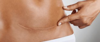
- Stage 1. Removal of the formation by surgery, which may affect healthy tissue.
- Stage 2. Carrying out a biopsy. Removal of lymph nodes if they are affected. Alpha interferon drugs are routinely prescribed to reduce the risk of relapse.
- Stage 3. Removal of the tumor and lymph nodes that are located in close proximity. In this case, immunotherapy, radiation, and chemotherapy are used. Interferon and similar medications are prescribed.
- Stage 4. Difficult to heal. The melanoma on the foot is removed; the operation is often accompanied by amputation of the toes; severe pain is relieved by taking strong analgesics. Chemotherapy and potent medications are prescribed. In order to reduce the tumor and improve the patient’s condition, the doctor prescribes the combined use of immuno- and chemotherapy drugs.
Recurrent melanoma on the leg is treated with carefully selected chemotherapy. Malignant tumors that form from melanocytic cells on the lower extremities account for half of the cases. They arise from benign tumors. There are cases when the disease developed from a single melanocyte.
Forecast and prevention of melanoma development
The further prognosis is determined by the stage of the disease. At the initial stage, melanoma on the leg can be cured. In a complex stage, when the formation has grown into the tissue and the pathology has affected the lymphatic system, the risk of relapse increases markedly, so it is much easier to prevent the disease than to fight it later.
By taking your health seriously, you will avoid most problems, including those whose treatment is not always associated with a favorable prognosis.
To prevent the development of melanomas, it is recommended to sunbathe with caution, especially for those with white sensitive skin. Sunscreen and wearing closed clothing made from breathable natural fabrics on sunny days will help. Regular visits to an oncologist are recommended to promptly identify the development of the pathological process.
It is important to monitor the condition of moles and age spots, monitor the changes that occur and not injure them. Special creams will help to lighten them. A successful preventive remedy that will not allow pigment formation to degenerate into a malignant tumor is Lakshma Maxxi whitening cream. It is one of the most sought after products in the United States and is recommended by the American Dermatological Association. It allows you to eliminate signs of hyperpigmentation on the skin easily, reliably and safely. According to the results of clinical studies, the drug showed high stable results.
Add to cart
Underarm skin brightening treatment
RUB 1,400.00
Add to cart
Whole body skin lightening milk
RUB 3,100.00
Add to cart
Cream Lakshma Maxxi (Lakshma Maxxi)
RUB 1,800.00
Add to cart
Varicose veins
Varicose veins are one of the most common causes of edema. Due to the weakening of the tone of the venous walls, the blood in the vessels stagnates, and the fluid pressure in the vessels increases. The body tries to regulate this situation on its own, and as a result, some of the fluid leaks into the tissue surrounding the vessel.
Varicose swelling usually worsens in the evening. To relieve the condition, you need to lie down and elevate your legs. You can also wear compression stockings (if you have one) or do some exercises to get your blood flowing. It is also useful to make contrast baths.
But if you notice a combination of redness of the skin on your legs and swelling, we recommend that you consult a doctor. This may be a manifestation of thrombophlebitis - a dangerous condition when the lumen of a vein is closed by a thrombus, a blood clot. As a result, blood flow through such a vessel stops.
Thrombophlebitis requires mandatory treatment from a specialist. If blood clots form in the lumens of the vein, it means that a floating thrombus may form in one of the vessels. It is able to move through the bloodstream, and can ultimately clog the pulmonary vessels.
So, if your legs are very red and swollen, or if the redness on your leg is accompanied by pain, rush to the doctor. After all, this may be the result of a minor injury, or it may indicate a somatic disease or the formation of blood clots. Only a specialist will determine the true cause of your condition and help normalize the situation. Treat yourself and your health carefully and feel light and comfortable!