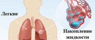
Bullae (false pulmonary cysts) are pathological air cavities in the lungs (from the English blebs - “bubbles”), which can arise due to mechanical damage to the parenchyma, a previous infectious-inflammatory or other disease. Excess air in these bag-like cavities, changes in the structure of the pulmonary matrix, and a reduction in the area of functional areas of the respiratory organ lead to persistent respiratory impairment, and the consequences of such pathological changes can be irreversible. The formation and enlargement of bullae in the lungs leads to a decrease in the gas exchange function of the lungs, and in the case of rupture of a large bulla, it can lead to a life-threatening condition - pneumothorax.
Interesting fact: air travel is dangerous for patients with large bullae in the lungs. The fact is that in flight at an altitude of 2.5 km from the ground, pathological cavities can increase in diameter by 30-40%, which means an episode of pneumothorax and rupture of the bulla may occur.
Bullae are most often diagnosed in people who smoke (usually men over 50 years of age with about 15 years of smoking experience) against the background of centrilobular pulmonary emphysema or COPD. Both of these diseases are in one way or another associated with damage (stretching) of the alveoli, disruption of the structure of the pulmonary parenchyma, and its aging. Sometimes these pathological changes are not associated with smoking, but are a consequence of metabolic disorders, an infectious-inflammatory process, an autoimmune disease, or are genetically determined.
The disease is difficult to treat. If the cause of the formation of bullae is established, their size is small, diagnostic measures are carried out on time and treatment is started, the pathological changes can be treated conservatively. In case of pneumothorax, bullae are removed surgically using endoscopic techniques.
Bullae require dynamic monitoring: CT scan, spirometry, consultation with a pulmonologist.
Where are the lungs located?
The lungs are located on either side of the heart and fill the chest. Each lung of an adult weighs a little more than 400 g
.
The right lung is slightly heavier than the left, since the latter has to share space in the chest with the heart. The lungs are protected by the rib cage
. Between her ribs there are small muscles involved in the breathing process.
Check your lungs
If you smoke, be sure to check your lungs with a simple test. Try blowing out a candle at a distance of a meter.
Beneath the lungs is
the diaphragm
, a dome-shaped muscle formation that separates the chest from the abdominal cavity and is also involved in breathing.
The structure of the human respiratory system
The respiratory system is a set of organs that ensure the supply of oxygen from the surrounding air to the respiratory tract and carry out gas exchange, i.e. bringing oxygen into the bloodstream and removing carbon dioxide from the bloodstream back into the atmosphere. However, the respiratory system is not only about providing the body with oxygen - it is also about human speech, and the capture of various odors, and heat exchange.
Organs of the human respiratory system
conditionally divided into
respiratory tracts,
or
conductors
, through which the air mixture enters the lungs, and
lung tissue
, or
alveoli
.
The respiratory tract is conventionally divided into upper and lower according to the level of attachment of the esophagus. The top ones include:
- nose and paranasal sinuses
- oropharynx
- larynx
The lower respiratory tract includes:
- trachea
- main bronchi
- bronchi of the following orders
- terminal bronchioles.
The nasal cavity is the first boundary when air enters the body. Numerous hairs located on the nasal mucosa stand in the way of dust particles and purify the passing air. The nasal turbinates are represented by a well-supplied mucous membrane and, passing through the convoluted nasal turbinates, the air is not only purified, but also warmed.
The nose is also the organ through which we enjoy the aroma of fresh baked goods, or can accurately determine the location of a public toilet. And all because sensitive olfactory receptors are located on the mucous membrane of the upper nasal concha. Their quantity and sensitivity are genetically programmed, thanks to which perfumers create memorable perfume aromas.
Passing through the oropharynx, air enters the larynx
. How is it that food and air pass through the same parts of the body and do not mix? When swallowing, the epiglottis covers the airway and food enters the esophagus. If the epiglottis is damaged, a person may choke. Inhalation of food requires immediate attention and can even lead to death.
The larynx consists of cartilage and ligaments. The cartilage of the larynx is visible to the naked eye. The largest of the cartilages of the larynx is the thyroid cartilage. Its structure depends on sex hormones and in men it moves forward strongly, forming the Adam's apple
, or
Adam's apple
.
It is the cartilages of the larynx that serve as a guide for doctors when performing tracheotomy or conicotomy - operations that are performed when a foreign body or tumor blocks the lumen of the respiratory tract, and a person cannot breathe in the usual way. Next, the vocal cords get in the way of the air.
It is by passing through the glottis and causing the tense vocal cords to tremble that a person has access to not only the function of speech, but also singing. Some unique singers can make vocal chords tremble at a frequency of 1000 decibels and burst crystal glasses with the power of their voice (in Russia, Svetlana Feodulova, a participant in the show “The Voice-2,” has the widest voice range of five octaves). Air enters the trachea
.
The trachea is anatomically divided into cervical
and
thoracic
parts.
The anatomical landmark is the jugular notch of the sternum
.
The trachea has the structure of cartilaginous semirings
. The anterior cartilaginous part ensures unhindered passage of air due to the fact that the trachea does not collapse. The esophagus is adjacent to the trachea, and the soft part of the trachea does not delay the passage of food through the esophagus.
Then the air travels through the bronchi and bronchioles, lined with ciliated epithelium, to the final section of the lungs - the alveoli
.
Lung tissue, or alveoli, are the final or terminal sections of the tracheobronchial tree
, similar to blindly ending sacs.
Many alveoli form the lungs. The lungs are a paired organ. Nature took care of her careless children, and created some important organs - lungs and kidneys - in duplicate. A person can live with only one lung. The lungs are located under the reliable protection of a frame made of strong ribs, sternum and spine.
Biology. 9th grade. Human. Toolkit. Vertical. Federal State Educational Standard
The methodological manual was prepared for the textbook by M.R. published in accordance with the Federal State Educational Standard. Sapina, N.I. Sonina “Biology. Human. 9th grade." The manual contains detailed lesson plans, including goals, main content of the lesson, planned results (personal, meta-subject, subject), equipment necessary for the lesson, as well as a presentation of the lesson and additional information for the teacher.
Buy Functions of the respiratory system
Interestingly, the lungs are devoid of muscle tissue and cannot breathe on their own. Breathing movements are ensured by the work of the diaphragm and intercostal muscles.
A person makes breathing movements thanks to the complex interaction of various groups of intercostal muscles, abdominal muscles during deep breathing, and the most powerful muscle involved in breathing is
the diaphragm
.
The experience with the Donders model, described on page 177 of the textbook “Biology 9th grade” edited by I.N. Ponomareva, will help you visualize the work of the respiratory muscles.
The lungs and chest are lined with pleura
.
The pleura that lines the lungs is called pulmonary
, or
visceral
.
And the one that covers the ribs is parietal
, or
parietal
.
The structure of the respiratory system
ensures the necessary gas exchange.
When inhaling, the muscles stretch the lung tissue, like a skilled musician playing a button accordion, and the air mixture of atmospheric air, consisting of 21% oxygen, 79% nitrogen and 0.03% carbon dioxide, enters through the respiratory tract to the final section, where the alveoli, entwined with a fine network of capillaries, are ready to receive oxygen and release waste carbon dioxide from the human body. The composition of exhaled air has a significantly higher carbon dioxide content - 4%.
To imagine the scale of gas exchange, just think that the area of all the alveoli in the human body is approximately equal to a volleyball court.
To prevent the alveoli from sticking together, their surface is lined
with surfactant
- a special lubricant containing lipid complexes.
The terminal sections of the lungs are densely woven with capillaries and the wall of the blood vessels is in close contact with the wall of the alveoli, which allows the oxygen contained in the alveoli to differ in concentrations, without the participation of carriers, by passive diffusion into the blood.
If we remember the basics of chemistry, and specifically the topic of solubility of gases in liquids
, the especially meticulous may say: “What nonsense, because the solubility of gases decreases with increasing temperature, but here you are saying that oxygen dissolves perfectly in a warm, almost hot - about 38-39 ° C, salty liquid.” And they are right, but they forget that the red blood cell contains the invader hemoglobin, one molecule of which can attach 8 oxygen atoms and transport them to the tissues!
In the capillaries, oxygen binds to the carrier protein on red blood cells and oxygenated arterial blood returns to the heart through the pulmonary veins. Oxygen participates in oxidation processes, and the cell as a result receives the energy necessary for life.
Breathing and gas exchange are the most important functions of the respiratory system, but they are far from the only ones. The respiratory system maintains thermal balance by evaporating water during breathing. An attentive observer has noticed that in hot weather a person begins to breathe more often. In humans, however, this mechanism does not work as efficiently as in some animals, such as dogs.
Hormonal function through the synthesis of important neurotransmitters
(serotonin, dopamine, adrenaline) are provided by pulmonary neuroendocrine cells (
PNE-pulmonary neuroendocrine cells
). Arachidonic acid and peptides are also synthesized in the lungs.
Set of tables. Biology. 8-9 grades. Man (12 tables)
Educational album of 12 sheets. Types of fabrics. Brain. Spinal cord. Functions of the nervous system. The structure and work of the heart. The connection between blood circulation and lymph circulation. Breath. Digestion. Kidney structure. Structure and functions of the skin. Structure and types of bones. Muscle structure. Perception. Sense organs...
Buy Regulation It would seem that there is nothing complicated here. The oxygen content in the blood has decreased, and here it is - a command to inhale. However, in reality the mechanism is much more complicated. Scientists still have not figured out the mechanism by which a person breathes. Researchers only put forward hypotheses, and only some of them are proven by complex experiments. It is only certain that there is no true pacemaker in the respiratory center, similar to the pacemaker in the heart.
The brainstem contains the respiratory center, which consists of several separate groups of neurons. There are three main groups of neurons:
- the dorsal group
is the main source of impulses that ensure a constant breathing rhythm; - ventral group
- controls the level of ventilation of the lungs and can stimulate inhalation or exhalation depending on the moment of excitation. It is this group of neurons that controls the abdominal and abdominal muscles for deep breathing; - pneumotaxic
center - thanks to its work, there is a smooth change from exhalation to inhalation.
To fully provide the body with oxygen, the nervous system regulates the rate of ventilation of the lungs by changing the rhythm and depth of breathing. Thanks to well-functioning regulation, even active physical activity has virtually no effect on the concentration of oxygen and carbon dioxide in arterial blood.
The following are involved in the regulation of breathing:
- chemoreceptors of the carotid sinus
, sensitive to the content of O2 and CO2 gases in the blood. The receptors are located in the internal carotid artery at the level of the upper edge of the thyroid cartilage; - pulmonary stretch receptorslocated
in the smooth muscles of the bronchi and bronchioles; - inspiratory neuronslocated
in the medulla oblongata and pons (divided into early and late).
What else should I read?
Stages of energy metabolism
Methodological assistance to a biology teacher
Heart stories: 8 interesting facts about the heart
What is photosynthesis? The history of the discovery of the process, the phases of photosynthesis and its significance.
Signals from various groups of receptors located in the respiratory tract are transmitted to the respiratory center of the medulla oblongata, where, depending on the intensity and duration, an impulse to the respiratory movement is formed.
Physiologists have suggested that individual neurons are combined into neural networks to regulate the sequence of changes in inhalation-exhalation phases, registering their flow of information by individual types of neurons, and changing the rhythm and depth of breathing in accordance with this flow.
The respiratory center located in the medulla oblongata monitors the level of blood gas tension and regulates ventilation of the lungs using respiratory movements so that the concentration of oxygen and carbon dioxide is optimal. Regulation is carried out using a feedback mechanism.
How does breathing happen?
Before birth, the baby receives oxygen directly from its mother's blood, so its lungs are filled with fluid and do not work. At the moment of birth, the baby takes his first breath, and from that moment his lungs work without rest. The respiratory center of the brain constantly receives signals about how much oxygen the body requires at any given moment. For example, if a person is sleeping, he needs much less oxygen than when he is running for the bus. The brain sends messages along nerves to the breathing muscles, which help regulate the amount of air entering the lungs. As soon as this signal is received, the diaphragm expands and the muscles stretch the chest outward and upward. This maximizes the volume that the lungs can occupy in the chest. When you exhale, the diaphragm and intercostal muscles relax, reducing the volume of the chest. Due to this, air is pushed out of the lungs.
Symptoms of bullous emphysema
A number of studies published in the open international database PubMed emphasize that factors such as:
- Smoking (15-20 packs);
- Systematic inhalation of toxic substances (unfavorable environment, work in hazardous industries, substance abuse, drug addiction);
- Low level of quality of life (poor and unhealthy diet, hypothermia, illness, lack of qualified medical care).
And only next to these factors in terms of prevalence are past infectious and inflammatory diseases of the respiratory tract, autoimmune diseases, metabolic disorders, and congenital characteristics of the body.
In the vast majority of cases, bullous emphysema is diagnosed in elderly or mature people, but bullae can also be found in young people.
Small bullae do not manifest themselves at all. In general, it is difficult for the patient to even suspect most diseases of the main respiratory organ - the lungs do not hurt, and when shortness of breath, coughing, and lack of air occur, the disease brings the patient to intensive care in a matter of hours.
Pathological symptoms may appear as the bullae enlarge and emphysema develops. Prevention of the disease is possible through regular screening - low-dose CT scan of the lungs. Examination in 3D mode and in high resolution will show even the slightest deviations from the norm.
To determine the (exact stage) of bullous emphysema, the method of spirometry is used - functional diagnosis of the lungs. As a rule, after spirometry, if the indicators are not normal, the pulmonologist sends the patient for a CT scan of the lungs.
Bullae in the lungs can be suspected if the following symptoms are present:
- Shortness of breath and breathing difficulties;
- Resistance to physical activity, fatigue;
- Hypotension, uncontrolled arrhythmias;
- Feeling of constant fatigue;
- Blue lips and fingertips;
- Painful pallor;
- Feeling of incomplete inspiration;
- Deformation of the chest (becomes barrel-shaped if there are large bullae).
Since the enlargement of bullae lasts quite a long time, the patient may not notice a deterioration in health, in particular, it becomes increasingly difficult to hold his breath due to a decrease in the vital capacity of the lungs, and hypoxia generally affects all internal organs - the person literally “fades.”
How does oxygen get into the blood?
Through the thin walls of the alveoli, oxygen enters the blood vessels. Here it is picked up by “transport” - hemoglobin
, which is contained in red blood cells. At the same time, carbon dioxide enters the opposite direction - into the alveoli - from the blood, which is removed from the body when exhaling. Oxygenated blood is sent from the lungs to the left side of the heart, from where it is distributed throughout the body through arteries. As soon as the oxygen in the blood is used up, the blood flows through the veins to the right side of the heart and from there back to the lungs.
Main manifestations of the disease
The course of bullous disease is often asymptomatic. In advanced form, symptoms manifest themselves as complications:
- Pneumothorax (including with bloody, liquid, purulent effusion-exudate);
- pneumomediastinum;
- rigid lung;
- pleural fistula (fistula);
- chronic respiratory failure;
- hemoptysis.
All complications have the same clinical picture:
- Chest pain;
- shortness of breath, lack of air;
- labored breathing;
- cough;
- attacks of suffocation;
- cardiopalmus;
- pale skin.
Additionally: with hemoptysis, scarlet-colored blood discharge from the respiratory tract is observed, often in the form of foam.
In addition, the bulla can grow to gigantic sizes of several centimeters and put pressure on the heart and circulatory system, destabilizing their work.
What else do the lungs do?
Every day, the lungs of an adult person pump about ten thousand liters of air
.
Consequences of smoking
Chronic obstructive pulmonary disease (COPD) is rightfully considered the most obvious consequence of smoking. Moreover, it is incurable. Find out how to prevent the disease.
With each breath, not only oxygen enters them, but also dust, germs and other foreign objects.
Therefore, the lungs also perform the function of physical and chemical defense against unwanted objects from the air. On the walls of the bronchi there are tiny villi that trap dust and germs
. In the walls of the airways, special cells produce mucus that helps clean and lubricate these villi. Contaminated mucus is removed through the bronchi to the outside and coughed up.
Surgery
Surgical treatment is called bullectomy. If a person is diagnosed with a giant bulla, then this is an indication for surgical treatment. Small bullae are difficult to treat surgically, and the effect of the operation is insignificant. Shortness of breath rarely improves.
Bullae that are not accompanied by clinical symptoms (shortness of breath) or complications (pneumothorax, suppuration) are not recommended to be removed.
Contraindications to bullectomy include continued smoking, severe cardiovascular disease, and diffuse emphysema with low compression of the surrounding lung tissue.
The indication for surgical treatment may be recurrent pneumothorax against the background of bullous changes. In this case, bullectomy and pleurodesis are performed simultaneously.
1.General information
The human respiratory system, it would seem, is ideally adapted and “designed” by evolution for gas exchange processes, and the only thing that can reduce its efficiency is insufficient oxygen content in the air. However, this, unfortunately, is far from the case. The respiratory organs are complex in structure and are susceptible to numerous diseases, some of which are associated not with a deficiency, but with an excess of air, airing of the internal cavities of the pulmonary-bronchial gas exchange system.
Many people know the word “emphysema,” which means excess content or accumulation of air where there should not be any at all - as, for example, with pulmonary emphysema (pathological expansion of the alveoli) or subcutaneous emphysema (occurring with certain types of pulmonary injury). Sometimes they also talk about bullous emphysema, or simply bullous disease (pulmonary bullae); you can find the terms “blebs” (from the English blebs - bubbles), “bullous lung”, “false pulmonary cyst”, etc. In total, over 20 nosological definitions that are very close in meaning are used, which creates some confusion, since each of the terms implies certain nuances of similar, but not identical, pathological changes.
According to one of the most precise definitions, which was given at the international CIBA symposium in 1958, a bulla should be considered an air cavity larger than 1 cm in size (as opposed to smaller blebs, which are accumulations of air under the pleura and in the lung tissue) . Thus, bullae are a pathological expansion of the pulmonary alveoli, which have largely lost their elasticity and ability to contract to normal sizes; bullous disease (bullous emphysema) - expansion, enlargement of the lungs due to the formation of bullae, giant versions of which can reach 10 cm in diameter.
Statistical data reveals a number of age and gender trends in the structure of morbidity. In particular, an almost twofold predominance of men among patients with bullous disease was established, a direct correlation of the frequency of detection with the age of the examined, etc.
A must read! Help with hospitalization and treatment!