Autoimmune hepatitis (AIH) is a chronic inflammatory liver disease characterized by the loss of the body's immunological tolerance to tissue antigens [1, 2].
For the first time, information about severe liver damage with severe jaundice and hyperproteinemia appeared in the 30–40s. XX century. In 1950, the Swedish doctor Jan Waldenström observed chronic hepatitis with jaundice, telangiectasia, increased ESR, and hypergammaglobulinemia in 6 young women. Hepatitis responded well to treatment with corticotropin [3]. Due to the similarity of laboratory changes with the picture of systemic lupus erythematosus (the presence of antinuclear antibodies in the serum, positive results of the LE test), one of the names of the pathology became “lupoid hepatitis”.
Currently, autoimmune hepatitis is defined as chronic, predominantly periportal hepatitis with lymphocytic-plasmacytic infiltration and stepwise necrosis (Fig. 1). Characteristic manifestations: hypergammaglobulinemia, the appearance of autoantibodies in the blood.
Classification
Depending on the type of autoantibodies, three types of disease are distinguished:
- Type 1 AIH is the most common and is characterized by the appearance in the blood of antinuclear antibodies (ANA) and/or smooth muscle antibodies (SMA).
- In type 2 AIH, autoantibodies are formed to microsomal antigens of the liver and kidneys (anti-liver kidney microsomal type-1 antibodies, anti-LKM-1).
- Type 3 AIH is associated with the formation of autoantibodies to soluble liver antigen, liver and pancreas tissue (anti-soluble liver antigen/liver-pancreas antibodies, anti-SLA/LP).
- Some authors combine AIH 1 and AIH 3 due to the similarity of clinical and epidemiological features [4]. There are also crossover forms (overlap syndrome) of various autoimmune liver pathologies, including AIH: AIH + PBC (primary biliary cirrhosis), AIH + PSC ( primary sclerosing cholangitis). It is not yet entirely clear whether these diseases should be considered parallel ongoing independent nosologies or parts of a continuous pathological process.
AIH that developed de novo after liver transplantation performed for liver failure associated with other diseases is considered as a separate nosology [1, 5].
Autoimmune hepatitis is found everywhere. The prevalence of AIH in European countries is about 170 cases per 1 million population. Moreover, up to 80% of all cases are type 1 AIH. Type 2 AIH is unevenly distributed - up to 4% in the USA and up to 20% in Europe.
Mostly women are affected (the sex ratio among patients in European countries is 3–4:1). The age of the patients ranges from 1 year to 80 years, the average age is about 40 years [6].
Causes
The cause of autoimmune hepatitis (in other words, the reason why the immune system attacks and destroys healthy liver cells) is still not clear. There is an assumption that inflammatory-necrotic liver damage may be associated with an imbalance of effector and regulatory T cells (cells of the immune system), and may arise as a result of adverse environmental influences or due to genetic predisposition.
Without appropriate treatment, the disease progresses and the following complications may develop: ascites (accumulation of fluid in the abdominal cavity), gastrointestinal bleeding due to impaired blood flow in the esophagus and stomach, cirrhosis of the liver (and liver cancer due to the increased risk associated with cirrhosis) , kidney failure.
Etiopathogenesis
The etiology of AIH is unknown, but the development of the disease is thought to be influenced by both genetic and environmental factors.
An important link in the pathogenesis may be certain alleles of the HLA II genes (human leukocyte antigen type II, Human leucocyte antigen II) and genes associated with the regulation of the immune system [7, 8].
It is worth mentioning separately about AIH, which is part of the clinical picture of autoimmune polyendocrine syndrome (autoimmune polyendocrine syndrome, autoimmune polyendocrinopathy-candidiasis-ectodermal dystrophy, APECED). This is a monogenic disease with autosomal recessive inheritance associated with a mutation in the AIRE1 gene. Thus, in this case, genetic determination is a proven fact [4, 9].
The autoimmune process in AIH is a T-cell immune response, accompanied by the formation of antibodies to autoantigens and inflammatory tissue damage.
Pathogenesis factors of AIH:
- pro-inflammatory factors (cytokines) produced by cells during the immune response. Indirect confirmation may be that autoimmune diseases are often associated with bacterial or viral infections;
- inhibition of the activity of regulatory T cells, which play a critical role in maintaining tolerance to autoantigens;
- dysregulation of apoptosis, normally a mechanism that controls the immune response and its “correctness”;
- molecular mimicry is a phenomenon where the immune response against external pathogens can also affect its own components that are structurally similar to them. Viral agents may play an important role in this. Thus, several studies have shown the presence of a pool of circulating autoantibodies (ANA, SMA, anti-LKM-1) in patients suffering from viral hepatitis B and C [2,4];
- factor of toxic drug effects on the liver. Some researchers associate the manifestation of AIH with the use of antifungal drugs and nonsteroidal anti-inflammatory drugs.
Clinic
In approximately a quarter of patients, AIH begins acutely, and even rare cases of acute liver failure have been described. Acute hepatitis with jaundice is more common in children and young people; in this same group, a fulminant course of the disease is more often observed [6].
It should be noted that some patients with symptoms of acute AIH in the absence of treatment may experience spontaneous improvement and normalization of laboratory parameters. However, a repeat episode of AIH usually occurs within a few months. Histologically, a persistent inflammatory process in the liver is also determined [6].
More often, the clinical picture of AIH corresponds to the clinical picture of chronic hepatitis and includes symptoms such as asthenia, nausea, vomiting, pain or discomfort in the right upper quadrant of the abdomen, jaundice, sometimes accompanied by skin itching, arthralgia, and less commonly, palmar erythema, telangiectasia, hepatomegaly [2, 6]. With developed liver cirrhosis, symptoms of portal hypertension and encephalopathy may prevail.
AIH can be associated with autoimmune diseases of various profiles:
- hematological (thrombocytopenic purpura, autoimmune hemolytic anemia);
- gastroenterological (inflammatory bowel diseases);
- rheumatological (rheumatoid arthritis, Sjogren's syndrome, systemic scleroderma);
- endocrine (autoimmune thyroiditis, diabetes mellitus);
and other profiles (erythema nodosum, proliferative glomerulonephritis) [1, 2].
Kishkun A.A. LABORATORY MEDICINE No. 9/2008
Autoimmune mechanisms play an important role in the pathogenesis of a number of liver diseases. An important sign of impaired immunity in chronic active liver diseases is the appearance in the blood of autoantibodies that react with various antigenic components of cells and tissues. The literature review is devoted to the laboratory diagnosis of autoimmune liver diseases, incl. chronic autoimmune hepatitis, primary biliary cirrhosis, primary sclerosing cholangitis, autoimmune cholangitis. Information is provided on diagnostic tests developed to confirm the autoimmune nature of liver damage (antimitochondrial antibodies, antibodies to smooth muscle, antibodies to liver-specific lipoprotein and liver membrane antigens, antibodies to microsomal antigen of the liver and kidneys, antibodies to neutrophils, etc.), the main methods for detecting antibodies in autoimmune liver diseases, an algorithm for diagnosing symptomatic and asymptomatic liver diseases is proposed. It is indicated that understanding the above features of detecting autoantibodies will allow the doctor to best use the results obtained to diagnose the disease and assess the effectiveness of treatment in each individual patient.
Autoimmune diseases are considered to be those diseases that are based on an immune reaction to one’s own (autologous) antigens of tissues and organs. Normally found in small quantities, natural autoantibodies, usually of the IgM class, do not cause pathological processes, but stimulate tissue regeneration. Immune autoaggression requires not only an increase in their number, but also qualitative changes - increased antigen specificity, increased avidity, etc. The criteria for autoimmune diseases were formulated in 1961 by L.Witebsky and include the presence of autoantibodies or sensitized lymphocytes in all cases of the disease, or at least at some of its stages. Autoimmune mechanisms play an important role in the pathogenesis of a number of liver diseases: chronic active hepatitis, chronic autoimmune hepatitis, primary biliary cirrhosis, primary sclerosing cholangitis, autoimmune cholangitis.
An important sign of impaired immunity in chronic active liver diseases is the appearance in the blood of autoantibodies that react with various antigenic components of cells and tissues. Autoimmune chronic hepatitis (a variant of chronic active hepatitis) is a heterogeneous group of progressive inflammatory liver diseases. Autoimmune chronic hepatitis syndrome is characterized by clinical symptoms of liver inflammation lasting more than 6 months and histological changes, including mole-shaped necrosis and infiltrates of the portal fields.
Autoimmune chronic hepatitis is characterized by the presence of the following factors:
- the disease occurs predominantly in young women (85% of all cases);
- changes in the results of traditional laboratory parameters manifest themselves in the form of accelerated ESR, moderately expressed leukopenia and thrombocytopenia, anemia of mixed origin - hemolytic (positive direct Coombs test) and redistribution;
- changes in the results of liver tests characteristic of hepatitis (bilirubin is increased by 2-10 times, transaminase activity is 5-10 times or more, the de Ritis coefficient is less than 1, the level of alkaline phosphatase is increased slightly or moderately, there is an increase in the level of alpha-fetoprotein, correlating with biochemical activity of the disease);
- hypergammaglobulinemia exceeding the norm by 2 times or more (usually polyclonal with a predominant increase in IgG);
- negative serological markers of hepatitis A and B;
- absence of hepatitis C virus RNA;
- negative or low titer of antibodies to mitochondria.
In patients with autoimmune chronic hepatitis, initial liver damage, of unknown etiology, includes the autoimmune mechanism as the main one due to a genetically determined defect in T-suppressors for their own liver-specific lipoproteins and hepatocyte membrane antigens. As a result of T-cell stimulation, B-lymphocytes are activated, causing the production of antibodies to the membrane antigen. These antibodies, by binding to the surface of hepatocytes, create conditions for the development of antibody-dependent cytolysis mediated by killer T cells.
Autoimmune liver diseases also include primary biliary cirrhosis (PBC), which manifests itself in the form of low-symptomatic chronic destructive non-purulent cholangitis, which then goes through the stage of cholestasis, which ends with the formation of cirrhosis. If PBC was previously considered a rare disease, its prevalence has now become very significant. The increase in the diagnosis of PBC is explained by the introduction of modern laboratory research methods into clinical practice. The most characteristic of PBC is an increase in the level of alkaline phosphatase, usually more than 3 times (in some patients it may be within normal limits or slightly increased), GGTP, PAP, 5-nucleotidase (reflecting cholestasis syndrome). Alkaline phosphatase level has no prognostic value, but a decrease in its level reflects a positive response to treatment. The activity of AST and ALT is moderately increased (transaminase activity 5-6 times higher than normal is not typical for PBC).
Primary sclerosing cholangitis (PSC) is a chronic cholestatic liver disease of unknown etiology, characterized by non-purulent destructive inflammation, obliterating sclerosis and segmental dilatation of the intra- and extrahepatic bile ducts, leading to the development of biliary cirrhosis, portal hypertension and liver failure. The concept of PSC as an autoimmune disease with a genetic predisposition is based on the identification of familial cases of PSC, the combination of PSC with other autoimmune diseases (most often with ulcerative colitis), disorders in cellular and humoral immunity, and the identification of autoantibodies (antinuclear, anti-smooth muscle, neutrophil cytoplasm). PCS is characterized by a stable cholestasis syndrome (usually no less than a twofold increase in the level of alkaline phosphatase), the level of transaminases in the blood is increased in 90% of patients (no more than 5 times). Autoimmune cholangitis is a chronic cholestatic liver disease caused by immunosuppression. The histological picture of the liver tissue in this disease is almost similar to primary biliary cirrhosis, and the spectrum of antibodies includes increased titers of antinuclear and antimitochondrial antibodies.
Autoimmune cholangitis does not appear to be a variant of primary sclerosing cholangitis
Laboratory tests for the diagnosis of autoimmune liver diseases For the first time, the concept of autoimmune liver damage was confirmed by the detection of antinuclear antibodies (antinuclear factor) in the blood serum of patients. These antibodies are detected in 50–70% of cases of active chronic (autoimmune) hepatitis and in 40–45% of cases of PBC. The presence of antinuclear antibodies (ANA) in patients with chronic autoimmune hepatitis is one of the main indicators that distinguishes this disease from protracted viral hepatitis. At the same time, low titers of antinuclear antibodies can be found in practically healthy people and their titer increases with age. They may appear after taking certain medications, such as procainamide, methyldopa, certain anti-tuberculosis and psychotropic drugs. Very often, the titer of antinuclear antibodies increases in healthy women during pregnancy. Subsequently, to confirm the autoimmune nature of liver damage and carry out differential diagnosis of various forms of autoimmune hepatitis and PBC, diagnostic tests were developed to determine antimitochondrial antibodies, antibodies to smooth muscle, antibodies to liver-specific lipoprotein and liver membrane antigens, antibodies to liver microsomal antigen and kidneys, antibodies to neutrophils, etc. The main method for detecting antibodies in autoimmune liver diseases is the indirect immunofluorescence (IIF) method. The method of conducting the study is that the patient’s blood serum, which may contain antibodies to this antigen, is applied to tissue sections containing the corresponding antigens. After washing off unbound proteins, fluorescein isocyanate-labeled anti-human γ-globulin antiserum is applied. If there are antibodies to the corresponding tissue antigen in the patient's serum, a specific glow is detected.
The disadvantage of the indirect immunofluorescence method is the subjectivity of the method for assessing the analysis result. In this regard, in recent years, test systems for enzyme-linked immunosorbent assay (ELISA) have been developed, in which the corresponding antigens are applied to a special sorbent. The ELISA method allows quantitative expression of research results. Antimitochondrial antibodies (AMA) are produced against an antigen in the inner membrane of mitochondria. The structure of the antigen is a lipoprotein involved in the transport functions of the membrane. The diagnostic test systems available in laboratories make it possible to determine both total AMA and their individual subtypes. Antimitochondrial antibodies are not normally detected in serum by NIF; when using the ELISA method, normal values are less than 20 IU/ml, 20–25 IU/ml are borderline values. An increased titer of total AMA (1:160 and above) is characteristic of PBC (more than 90% of patients).
A very small proportion of patients with PBC are AMA negative. In secondary biliary cirrhosis, AMAs are detected in low titers or, more often, not detected at all. Low AMA titers can also be observed in chronic active hepatitis, chronic autoimmune hepatitis (up to 20% of cases), alcoholic or viral hepatitis. Currently, there are 4 subtypes of AMA. Antibodies to the mitochondrial antigen M-2 (which is a complex of enzymes on the inner membrane of mitochondria) are considered specific for PBC. The presence of antibodies against the M-2 antigen can be detected by ELISA test systems. The diagnostic sensitivity of test systems for detecting PBC is 98%, specificity is 96%. This means that only 5% of patients with PBC do not have AMA M-2 in their blood. An AMA M-2 level of more than 25 IU/ml is considered elevated. Along with anti-M2 antibodies, in PBC there are, in most cases simultaneously, anti-M9, anti-M4 and anti-M8 antibodies, which react with various epiotypes of the mitochondrial membrane. There is a relationship between the AMA profile and the prognosis of PBC. Isolated detection of anti-M9 and/or anti-M2 in serum correlates with a good prognosis of PBC. A progressive course of the disease is observed in patients with anti-M2, anti-M4 and/or anti-M8, in combination with elevated bilirubin levels. This means that using the AMA profile it is possible to identify inactive (early) and active (progressive) forms of PBC and, accordingly, identify groups of patients with low and high risk.
Antismooth muscle antibodies (ASMA) are antibodies to the protein actin or non-actin components (tubulin, vimentin, desmelin and skeletin) and appear in response to damage to hepatocytes. They refer to IgG or IgM in the presence of cholestasis. AGM is detected by indirect immunofluorescence. The substrate for indirect immunofluorescence is usually tissue containing smooth muscle of human or animal origin (smooth muscle layer of the rat gastric mucosa, smooth muscle layer of the outer lining of the rat stomach, smooth muscle elements of the rat kidney glomeruli, smooth muscle layer of the rat kidney vessels). AHM is detected in 60–80% of cases of autoimmune (lipoid) hepatitis (titer 1:80 and above), in 50% of cases of PBC and is not detected in SLE and extrahepatic lesions of the biliary tract. AHMs are present in 70% of patients with chronic active hepatitis and belong to the IgG class. AGMs are detected in acute viral hepatitis and disappear upon recovery. In some cases, AGM can be detected in low titers in infectious mononucleosis, cytomegalovirus infection, mycoplasma pneumonia, lymphoproliferative diseases, drug addiction, malignant neoplasms, in women suffering from infertility and sometimes in healthy people. In these groups of patients, AGM belongs to the IgM class. Antibodies to liver-specific lipoprotein (anti-LSP) are determined by indirect immunofluorescence. LSP antigen is a heterogeneous material from hepatocyte membranes containing 7–8 antigenic determinants, some of them liver-specific, others nonspecific. It is antibodies to LSP that cause an autoimmune reaction with the development of antibody-dependent cytolysis of hepatocytes and provoke a relapse when glucocorticosteroids are withdrawn in patients with chronic autoimmune hepatitis. The presence of anti-LSP in the blood serum is a hallmark of autoimmune hepatitis and is detected only in 5% of cases among patients with HBs4-positive chronic active hepatitis. However, a large number of clinical observations have shown that anti-LSP are also detected in chronic liver diseases of viral etiology (in 48–97% of cases). Antibodies against liver and kidney microsomal antigen (LKM) are a heterogeneous group of autoantibodies that are divided into three subtypes based on their target antigens. A component of cytochrome P4450IID6 with a molecular weight of 50,000 has been identified as the main antigen for LKM type 1 (LKM-1), LKM-2 is directed to cytochrome P4450IIC9 and was found in patients taking ticrynafen (a thienylic acid diuretic, not currently used), LKM- 3 were identified in the blood serum of patients with chronic viral hepatitis D (found in 5–13% of patients), but the antigen has not yet been identified. They can occur in patients with autoimmune hepatitis type II (in 10% of patients). The level of antibodies to the microsomal antigen of the liver and kidneys in the serum is normal - less than 20 IU/ml, 20-25 IU/ml are borderline values. The basis for determining the level of antibodies to liver and kidney microsomes (LKM-1) is the ELISA method. This study is an addition to existing methods for diagnosing autoimmune hepatitis. Antineutrophil cytoplasmic antibodies (ANCA) are a complex of antibodies specific to various granulocyte, monocyte and, possibly, endothelial cytoplasmic antigens. When ANCA is determined by indirect immunofluorescence using neutrophils from healthy donors, two different types of fluorescence can be observed - classical diffuse (c-ANCA) and perinuclear (p-ANCA). These types of fluorescence are due to the different antigenic targeting of ANCA. Antibodies with classical diffuse fluorescence are in most cases directed against protein kinase-3 and the neutrophil protein that enhances the bactericidal effect. Detection of c-ANCA in blood serum is a highly specific sign for Wegener's granulomatosis. P-ANCA are directed against a wide range of cytoplasmic antigens: myeloperoxidase, elastase, lactoferrin, cathepsin G and other polypeptides. Most often, p-ANCA is detected in primary sclerosing cholangitis (in 60–85% of patients), ulcerative colitis (in 60–75%), chronic autoimmune active hepatitis (in 60–70%), primary biliary cirrhosis (in 30–40 %), Crohn's disease (in 10–20% of patients). In patients with primary sclerosing cholangitis, the presence of p4ANCA does not correlate with the clinical activity of liver damage.
Classification of autoimmune hepatitis and diagnostic criteria Currently, a subclassification of autoimmune hepatitis is used in clinical practice, which, in accordance with the serological profile, provides for the identification of 4 types of autoimmune hepatitis (Table 1).
Table 1 Antibody profile in autoimmune hepatitis
| Type of hepatitis | ANA | LKM-1 | AGM | HCVAb |
| I | + | – | + | – |
| IIa | – | + | – | – |
| IIb | – | + | – | + |
| III | – | – | ± | – |
| IV | – | – | + | – |
- The first type of autoimmune hepatitis is characterized by the presence of antinuclear antibodies (in 70–80% of patients) and smooth muscle antibodies (in 50–70% of patients), often in combination with antineutrophil cytoplasmic antibodies. Although AMAs are a sensitive and specific marker of PBC, they can be detected in up to 15% of patients with clinical and histological signs of autoimmune hepatitis, including cases of clinical improvement in response to immunosuppressive therapy. Such patients are regarded as having an “overlap syndrome” of signs of autoimmune hepatitis and PBC. The AMA titer in this subgroup of autoimmune hepatitis is rarely more than 1:40. The antigenic specificity of these AMAs is similar to that observed in classical PBC (AMA M-2). In such cases, a repeat biopsy and further observation of the patient is performed. The titer of antibodies to the asialoglycoprotein receptor (ASGP-R), a glycoprotein of the plasma membrane of hepatocytes, correlates with the activity of autoimmune hepatitis type I. This is because tissue expression of ASGP-R is most pronounced in the periportal zone, where stepwise necrosis is usually observed, reflecting disease activity.
- The second type of autoimmune hepatitis, which is characterized by the presence of antibodies to liver and kidney microsomes (in 100% of patients), is divided into two subgroups (subtypes 2a and 2b). Typically, patients with the second type are negative for ANA and AGM, but they often have antibodies to the microsomal fraction of the thyroid gland, thyroglobulin and parietal cells. Subtype 2a is detected in young women who have high titers of anti-LKM-1 (the main diagnostic marker of this subtype) and antibodies to liver cytosolic antigen (anti-LC-1), a negative serological reaction to hepatitis C, and active disease responsive to corticosteroid therapy. Subtype 2b occurs in older patients and the “preference” for women is less pronounced. These patients have low titers of LKM-1, the clinical manifestations of the disease are less pronounced, and antibodies to the hepatitis C virus (anti-HCV) are often detected; When treated, subtype 2b responds worse to immunosuppressive therapy compared to subtype 2a. The presence of anti-HCV in patients with chronic autoimmune hepatitis type 2 is a false positive, and there is a relationship between the frequency of false-positive anti-HCV results and the concentration of gammaglobulins in such patients. The higher the serum gammaglobulin concentration, the more often false-positive anti-HCV test results are detected. In such cases, it is necessary to determine HCV RNA in serum or lymphocytes (by PCR). A number of authors provide data that in approximately 3% of patients with viral hepatitis C, antibodies to liver and kidney microsomes can be detected, but their titer is significantly lower than in patients with chronic autoimmune hepatitis type 2. In patients with type 2 hepatitis, the level of IgA in the blood serum is lower than in patients with type I of the disease. A number of studies provide evidence that autoimmune hepatitis type 2 is accompanied by the development of liver cirrhosis more often than hepatitis type 1.
- A third subtype of autoimmune hepatitis is distinguished based on the detection in serum of antibodies to soluble liver antigens (anti-SLA), which have more recently been shown to be identical to liver-pancreatic antigen (LP). Anti-SLA/LP bind to hepatic cytokeratins 8 and 18. These antibodies are formed only in autoimmune hepatitis and serve as an important serological marker (they have 100% specificity and in 15% of patients with autoimmune hepatitis they are the only detectable antibodies). Test systems for the detection of anti-SLA/LP have now been developed and are available as routine testing in the laboratory.
- High titers of AGM directed against F-actin characterize the fourth subtype of autoimmune hepatitis, which is often observed in children.
It should be noted that the identification of 4 types of chronic autoimmune hepatitis does not have much clinical significance from the point of view of treatment tactics, since the majority of patients, regardless of the type to which they belong, respond well to immunosuppressive therapy.
The diagnosis of chronic autoimmune hepatitis is considered definitely established if:
- in the blood serum, titers of antinuclear antibodies, antibodies to smooth muscles, antibodies to microsomal antigen of the liver and kidneys are increased in a titer of more than 1:80;
- serum IgG concentration exceeds the upper limit of normal by 1.5 times;
- there is no history of taking hepatoxic medications or alcohol abuse;
- There is no HCV RNA in the blood serum of patients.
For a more accurate diagnosis of autoimmune hepatitis, you can use an assessment of all indicators in points (Table 2).
Table 2 Diagnostic criteria for autoimmune hepatitis
| Criteria | Number of points |
| Floor: | |
| male | 0 |
| female | +2 |
| Alkaline phosphatase/ALT activity ratio: | |
| <1,5 | +2 |
| 1,5-3 | 0 |
| >3,0 | –2 |
| Presence of autoantibodies: antinuclear, to smooth muscle, liver and kidney microsomal antigen ( LKM -1), titer: | |
| >1:80 | +3 |
| 1:80 | +2 |
| 1:40 | +1 |
| <1:40 | 0 |
| Presence of antimitochondrial autoantibodies | — 4 |
| Medicinal history: | |
| positive | –4 |
| negative | +1 |
| Alcohol consumption: | |
| <25 g per day | +2 |
| >60 g per day | –2 |
| Markers of viral infections (including IgM to the hepatitis A virus, HBsAg IgM antibodies to HBc - hepatitis B nuclear antigen, detection of hepatitis C virus RNA by PCR): | |
| positive | –3 |
| negative | +3 |
| Presence of other autoimmune diseases | +2 |
| Liver histology: | |
| stepwise necrosis and lobular hepatitis | +3 |
| pronounced lymphocytic infiltration | +1 |
| rosette liver cells | +1 |
| none of the above changes | –5 |
| changes in the bile ducts | –3 |
| other changes | –3 |
| Additional research: | |
| positive research results | |
| other autoantibodies | +2 |
| presence of immunogenetic markers: | |
| HLA-DR3 or DR4 | +1 |
| Response to immunosuppressive therapy: completely weakened | +2 +3 |
The final assessment is carried out as follows
- with a total score of more than 15 before treatment and more than 17 after treatment, the presence of autoimmune hepatitis can be considered established;
- with a score of 10 to 15 before and 12 to 17 after treatment, the presence of autoimmune hepatitis is possible (cannot be excluded);
- with a lower score, autoimmune hepatitis is excluded.
Methodological features of assessing the results of autoimmune tests When assessing the results of autoimmune tests, it is necessary to understand that autoantibodies are immunoglobulins of the IgM, IgA and IgM classes, specifically binding to antigenic epitopes of molecules of cells and tissues of the human body. These autoantibodies, which react with cellular antigens, appear in the blood as a result of the normal functioning of the immune system and can be found in the serum of almost all healthy people of any age. At the same time, autoantibodies are detected in many diseases and pathological conditions, chronic intoxication (alcoholism); they can be transiently induced by an infectious process, a tumor, or taking medications. The incidence of many autoantibodies increases with age.
However, due to the high occurrence of some autoantibodies in certain pathological processes in the liver, they can serve as specific laboratory markers of autoimmune diseases. A certain spectrum of autoantibodies in certain autoimmune diseases indicates the peculiarities of the functioning of the immune system and reflects its special predisposition to the formation of autoimmune responses. The production of autoantibodies is not constant. The incidence of autoantibodies may increase with the duration of autoimmune liver disease. Autoantibodies are synthesized as part of the normal pool of serum immunoglobulins by plasma cells. Each type of autoantibody, even directed to the same antigen, is a polyclonal population of autoantibodies, represented by immunoglobulin molecules with different physicochemical properties (polyclonal antibodies react with different epitopes on the surface of the antigen). Ideally, laboratory methods should detect not just one type of molecule (autoantibody), but a whole spectrum of autoantibodies directed to different epitopes of the antigen. Currently, there is no absolutely correct approach (reference method) for identifying autoantibodies in autoimmune diseases. The main reasons for this are as follows:
- most methods are based on the binding of autoantibodies and the target antigen, so the result of the method depends on the concentration of autoantibodies in the blood serum, as well as the affinity of autoantibodies and antigen;
- In some autoimmune diseases, levels of autoantibodies may be low or they may be selectively lost (for example, in kidney damage) in the urine, leading to false-negative test results;
- or vice versa, a high concentration of low-affinity autoantibodies with low high-affinity autoantibodies in the blood can lead to a false-positive response;
- an increase in the level of autoantibodies can be triggered by an inflammatory, infectious, oncological process or taking a number of medications;
- to distinguish transient induction from the chronic presence of autoantibodies in autoimmune diseases, it is necessary to re-determine autoantibodies after 6 months (3 months half-life of autoantibodies);
- cross-reaction of autoantibodies - often occurs in various autoimmune diseases, therefore, when assessing test results, it is necessary to use the so-called “borderline” or “gray” zone, when the answer is neither positive nor negative;
- Most test systems use animal tissue antigens as antigens - this is another possible reason for an incorrect result;
- for early diagnosis of autoimmune diseases, it is necessary to use test systems with high sensitivity, a negative result in which makes it possible to exclude the disease with a high probability;
- highly sensitive tests usually have low specificity, so confirmatory tests must be used.
Thus, diagnosing autoimmune liver diseases is a rather difficult task. Understanding these features of autoantibody detection will allow the doctor to make the best use of the results obtained to diagnose the disease and assess the effectiveness of treatment in each individual patient.
Diagnosis of AIH
Diagnosis of autoimmune hepatitis is based on:
- research results: clinical, serological and immunological;
- excluding other liver diseases that occur with or without an autoimmune component (chronic viral hepatitis, toxic hepatitis, non-alcoholic steatosis, Wilson's disease, hemochromatosis, and cryptogenic hepatitis).
AIH types 1 and 3 are similar in terms of patient demographics, HLA profile, inflammatory activity, and response to therapy. AIH type 2 has significant differences, affecting more often children and adolescents and usually having an acute onset and a more severe course.
It is important to be aware of the possibility of AIH in patients with elevated liver enzyme levels, as well as in patients with liver cirrhosis. If there are signs of cholestasis, primary biliary cirrhosis and primary sclerosing cholangitis should be included in the range of pathologies for differential diagnosis.
The clinical search includes the determination of such laboratory parameters as the activity of alanine aminotransferase and aspartate aminotransferase (ALT and AST), alkaline phosphatase (ALP), level of albumin, gamma globulin, IgG, bilirubin (bound and unbound). It is also necessary to determine the level of autoantibodies in the blood serum and obtain histological examination data [9].
For patients who failed to achieve remission and stop the development of cirrhosis within 4 years, the method of choice is liver transplantation. The 10-year survival rate in patients who have undergone this operation is quite high (75–85%), however, the rate of relapses reaches 11–41%. The persistence of autoantibodies in the blood is not a sign of relapse of AIH and does not predict its development.
Visual diagnostic methods (ultrasound, CT, MRI) do not make a decisive contribution to the diagnosis of AIH, however, they make it possible to establish the fact of progression of AIH and the outcome in cirrhosis of the liver, as well as exclude the presence of focal pathology. In general, diagnosis is based on 4 points [10]:
- Hypergammaglobulinemia is one of the most accessible tests. An increase in the level of IgG with normal levels of IgA and IgM is indicative. However, there are difficulties when working with patients with initially low IgG levels, as well as with patients (5–10%) with normal IgG levels in AIH. In general, this test is considered useful in monitoring disease activity during treatment [6].
- Presence of autoantibodies. At the same time, antibodies of the ANA and SMA types are not a specific sign of autoimmune hepatitis, nor are anti-LKM-1 antibodies, which are found in 1/3 of children and a small proportion of adults suffering from AIH. Only anti-SLA/LP antibodies are specific for AIH. Antibodies to double-stranded DNA can also be detected in patients.
- Histological changes are assessed in conjunction with previous indicators. There are no strictly pathognomonic signs of AIH, but many changes are quite typical. The portal fields are infiltrated to varying degrees by T lymphocytes and plasma cells. Inflammatory infiltrates are capable of “cutting off” and destroying individual hepatocytes, penetrating into the liver parenchyma - this phenomenon is described as stepwise (fine-focal necrosis), borderline hepatitis. Balloon degeneration of hepatocytes occurs inside the lobules with their swelling, formation of rosettes and necrosis of individual hepatocytes - Fig. 2. The fulminant course is often characterized by centrilobular necrosis. Bridge-like necrosis connecting adjacent periportal fields may also be observed [2, 6].
- Absence of markers of viral hepatitis.
- The International AIH Research Group has developed a scoring system to assess the reliability of the diagnosis - Table. 1.
Other autoimmune variants of liver damage are PBCP and PSC
Primary autoimmune cholestatic variants of liver damage - PBCP and PSC - are slowly progressive diseases characterized by obligatory damage to the bile ducts with the development of chronic non-purulent destructive cholangitis. For PBCP, granulomatous non-purulent destructive cholangitis with damage to the interlobular and septal bile ducts is typical, and for PSC, fibrous obliterating non-purulent destructive cholangitis is typical, affecting both intra- and extrahepatic bile ducts. In PBCP, along with cholestasis syndrome, AMAs are detected, the most specific are M2-AMAs, directed against the E2 subunit of pyruvate dehydrogenase, increased serum IgM and immune-mediated extrahepatic syndromes - Hashimoto's thyroiditis, Sjögren's syndrome, fibrosing alveolitis, tubulointerstitial nephritis, celiac disease, as well as a combination with diseases rheumatic circle - systemic scleroderma, rheumatoid arthritis, systemic lupus erythematosus. ANA is detected in 52% of patients with PBCP, reflecting the universal nature of these antibodies in various immune-mediated diseases. PBCP occurs predominantly in middle-aged and elderly women. There is a variant of PBCP without AMA - autoimmune cholangitis without AMA. PSC develops more often in men (2:1), and is often (in 70–90% of cases) combined with inflammatory bowel diseases - ulcerative colitis or Crohn's disease. Along with cholestasis syndrome, typical for PSC is a change in the biliary tree during endoscopic retrograde cholangiopancreatography (ERCP) or magnetic resonance imaging (MRI) cholangiography of the “rosary” or “bead” type; in 70% of patients, perinuclear antineutrophil cytoplasmic antibodies are detected in the blood serum.
Treatment of autoimmune hepatitis
AIH refers to diseases for which treatment can significantly increase patient survival.
Indications for starting treatment are:
- an increase in AST activity in the blood serum by 10 times compared to the norm or 5 times, but in combination with a twofold increase in the level of gamma globulin;
- the presence of bridge-like or multilobular necrosis during histological examination;
- pronounced clinic - general symptoms and symptoms of liver damage.
Less pronounced deviations in laboratory parameters in combination with a less pronounced clinical picture are a relative indication for treatment.
In case of inactive cirrhosis of the liver, the presence of signs of portal hypertension in the absence of signs of active hepatitis, and “mild” hepatitis with stepwise necrosis and without clinical manifestations, treatment is not indicated [1, 9]. The general concept of therapy for AIH involves achieving and maintaining remission. The basic immunosuppressive therapy is glucocorticosteroids (prednisolone) as monotherapy or in combination with azathioprine [2, 6, 9, 11]. Therapy continues until remission is achieved, and it is important to achieve histologically confirmed remission, which may lag behind the normalization of laboratory parameters by 6–12 months. Laboratory remission is described as normalization of the levels of AST, ALT, gamma globulin, IgG [2].
Maintenance therapy with lower doses of immunosuppressive drugs to reduce the likelihood of relapse after achieving remission is carried out for at least 2 years.
In addition, the possibility of using ursodeoxycholic acid (UDCA) drugs for autoimmune hepatitis as concomitant therapy or even monotherapy is being discussed [11]. AIH in patients receiving UDCA drugs in mono- and combination therapy was characterized by a milder course and accelerated normalization of laboratory parameters.
Symptoms
Symptoms of autoimmune hepatitis vary from person to person and may be mild or sudden in the early stages.
Symptoms of AIH include:
- increased fatigue, tiredness;
- discomfort and pain in the abdomen;
- skin rash, itching, formation of arachnoid angiomas;
- yellowness of the skin and visible mucous membranes (jaundice);
- joint pain;
- dark colored urine;
- amenorrhea (cessation of menstruation);
- hepatomegaly;
- splenomegaly.
Response to treatment
The results of treatment with prednisone and azathioprine for AIH may be as follows:
- A complete response is normalization of laboratory parameters, which persists for a year during maintenance therapy. At the same time, the histological picture is normalized (with the exception of small residual changes). The full effectiveness of treatment is also indicated in cases where the severity of clinical markers of autoimmune hepatitis significantly decreases, and during the first months of therapy laboratory parameters improve by at least 50% (and in the next 6 months they do not exceed the normal level by more than 2 times) .
- Partial response—there is an improvement in clinical symptoms and a 50% improvement in laboratory values within the first 2 months. Subsequently, the positive dynamics are maintained, but complete or almost complete normalization of laboratory parameters does not occur during the year.
- Lack of therapeutic effect (ineffectiveness of treatment) - an improvement in laboratory parameters by less than 50% in the first 4 weeks of treatment, and no further decrease (regardless of clinical or histological improvement) occurs.
- An unfavorable outcome of therapy is characterized by a further deterioration in the course of the disease (although in some cases there is an improvement in laboratory parameters).
Disease relapse is said to occur when, after achieving a complete response, clinical symptoms reappear and laboratory parameters deteriorate.
Typically, therapy has a good effect, but in 10–15% of patients it does not lead to improvement, although it is well tolerated. The reasons for the ineffectiveness of therapy can be [6]:
- lack of response to the drug;
- lack of compliance and adherence to therapy;
- drug intolerance;
- presence of cross syndromes;
- hepatocellular carcinoma.
Other immunosuppressants are also used as alternative drugs for the treatment of autoimmune hepatitis: budesonide, cyclosporine, cyclophosphamide, mycophenolate mofetil, tacrolimus, methotrexate [1, 2, 6, 11].
Clinical case
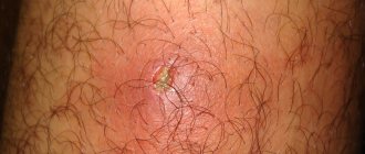
An eight-year-old girl was observed for skin rashes (erythematous and nodular elements without any discharge on the lower extremities) that had been bothering her for 5–6 months.
I used topical remedies for eczema for two months, there was no improvement. Later, discomfort in the epigastric region, weakness, and occasional nausea and vomiting arose. The rashes were localized on the legs. Histological examination of a skin biopsy revealed infiltration of subcutaneous fatty tissue with lymphocytes without signs of vasculitis. These phenomena were regarded as erythema nodosum. Upon examination, mild hepatosplenomegaly was revealed, otherwise the somatic status was without pronounced features, the condition was stable.
According to the results of laboratory tests:
- CBC: leukocytes - 4.5×10⁹/l; neutrophils 39%, lymphocytes 55%; signs of hypochromic microcytic anemia (hemoglobin 103 g/l); platelets - 174,000/μl, ESR - 24 mm/h;
- biochemical blood test: creatinine - 0.9 mg/dl (normal 0.3–0.7 mg/dl); total bilirubin - 1.6 mg/dl (normal 0.2-1.2 mg/dl), direct bilirubin - 0.4 mg/dl (normal 0.05-0.2 mg/dl); AST - 348 U/l (normal - up to 40 U/l), ALT - 555 U/l (normal - up to 40 U/l); alkaline phosphatase - 395 U/l (normal - up to 664 U/l), lactate dehydrogenase - 612 U/l (normal - up to 576 U/l).
Indicators of the hemostasis system (prothrombin time, international normalized ratio), the level of total protein, serum albumin, gamma globulin, the level of ferritin, ceruloplasmin, alpha-antitrypsin, gamma-glutamyl transpeptidase are within the reference values.
No HBs antigen, antibodies to HBs, HBc, or antibodies to hepatitis A and C virus were detected in the blood serum. Tests for cytomegalovirus, Epstein-Barr virus, toxoplasma, and brucella were also negative. The ASL-O titer is within the normal range. There is no evidence for diabetes mellitus, thyroiditis, Graves' disease, proliferative glomerulonephritis.
Ultrasound of the abdominal organs visualized an enlarged liver with an altered echostructure, without signs of portal hypertension and ascites. Ophthalmological examination did not reveal the presence of a Kayser-Fleischer ring.
Anti-SMA antibody titer - 1:160, ANA, AMA, anti-LKM-1 antibodies were not detected. The patient is positive for haplotypes HLA DR3, HLA DR4.
Histological examination of the liver biopsy revealed fibrosis, lymphocytic infiltration, formation of hepatocyte rosettes and other signs of chronic autoimmune hepatitis.
The patient was diagnosed with autoimmune hepatitis type 1 associated with erythema nodosum, and therapy with prednisolone and azathioprine was started. The IAIHG score was 19 points before treatment, which is characterized as “definite AIH.”
After 4 weeks of therapy, relief of general clinical symptoms and normalization of laboratory parameters were noted. After 3 months of therapy, the skin rashes completely regressed. 6 months after the end of therapy, the patient’s condition was satisfactory, laboratory parameters were within the reference values.
Adapted from Kavehmanesh Z. et al. Pediatric Autoimmune Hepatitis in a Patient Who Presented With Erythema Nodosum: A Case Report. Hepatitis monthly, 2012. V. 12. - N. 1. - P. 42–45.
Thus, AIH is a fairly rare and relatively treatable disease. With the introduction of modern immunosuppressive therapy protocols, the 10-year survival rate of patients reaches 90%. The prognosis is less favorable in patients with autoimmune hepatitis type 2, especially in children and adolescents, in whom AIH progresses much faster, and the effectiveness of therapy is generally lower. You should also be aware of the risk of developing hepatocellular carcinoma (4–7% within 5 years after diagnosis of liver cirrhosis).
List of sources
- Viral hepatitis and cholestatic diseases / Eugene R. Schiff, Michael F. Sorrell, Willis S. Maddray; lane from English V. Yu. Khalatova; edited by V. T. Ivashkina, E. A. Klimova, I. G. Nikitina, E. N. Shirokova. - M.: GEOTAR-Media, 2010.
- Kriese S., Heneghan MA Autoimmune hepatitis. Medicine, 2011. V. 39. - N. 10. - P. 580–584.
- Autoimmune hepatitis and its variant forms: classification, diagnosis, clinical manifestations and new treatment options: a manual for doctors. T. N. Lopatkina. - M. 4TE Art, 2011.
- Longhia MS et al. Aethiopathogenesis of autoimmune hepatitis. Journal of Autoimmunity, 2010. N. 34. - P. 7–14.
- Muratori L. et al. Current topics in autoimmune hepatitis. Digestive and Liver Disease, 2010. N. 42. - P. 757–764.
- Lohse AW, Mieli-Vergani G. Autoimmune hepatitis. Journal of Hepatology, 2011. V. 55. - N. 1. - P. 171–182.
- Oliveira LC et al. Autoimmune hepatitis, HLA and extended haplotypes. Autoimmunity Reviews, 2011. V. 10. - N. 4. - P. 189–193.
- Agarwal, K. et al. Cytotoxic T lymphocyte antigen-4 (CTLA-4) gene polymorphisms and susceptibility to type 1 autoimmunehepatitis. Hepatology, 2000. V. 31. - N. 1. - P. 49–53.
- Manns MP et al. Diagnosis and Management of Autoimmune Hepatitis. Hepatology, 2010. V. 51. - N. 6. - P. 2193–2213.
- Lohse AW, Wiegard C. Diagnostic criteria for autoimmune hepatitis. Best Practice & Research Clinical Gastroenterology, 2011. V. 25. - N. 6. - P. 665–671.
- Strassburg CP, Manns MP Therapy of autoimmune hepatitis. Best Practice & Research Clinical Gastroenterology, 2011. V. 25. - N. 6. - P. 673–687.
- Ohira H., Takahashi A. Current trends in the diagnosis and treatment of autoimmune hepatitis in Japan. Hepatology Research, 2012. V. 42. - N. 2. - P. 131–138.
Orthotopic liver transplantation for AIH
In most cases, successful immunosuppression allows long-term survival of patients with AIH and rarely requires liver transplantation. In the final stage of AIH, liver transplantation maintains high survival rates for patients and does not threaten their lives. Although AIH can recur in the graft, long-term survival is significantly better than other liver diseases. Immunosuppression without corticosteroids is feasible in the future of transplantation, but effective immunosuppression with steroids remains necessary to prevent serious relapse of autoimmune liver disease. As an effective treatment option, orthotopic liver transplantation for AIH remains very attractive for research. The interaction of the donor organ with the host’s immune system and bone marrow leads to very intense chimerism. After years, the “foreign” donor liver becomes “our own”. A significant number of hepatocytes from the donor liver are replaced by host liver cells, in most cases likely derived from host bone marrow stem cells. The microenvironment in the liver after transplantation may be important in case of disease relapse or loss of tolerance to the transplanted organ. In this light, the fact of de novo development of AIH in the donor liver seems very important. Studying this phenomenon may provide additional clues to the events underlying loss of tolerance and the autoimmune process directed against one's own tissues.
Literature
1. Aprosina Z.G. History of the study and modern aspects of autoimmune hepatitis // Clinical hepatology. 2005. No. 1. P. 5–16. 2. Filatova A.L. Clinical characteristics of autoimmune hepatitis // Clinical hepatology. 2005. No. 1. P. 28–30. 3. Czaja AJ, Freese DK. Diagnosis and Treatment of Autoimmune Hepatitis. Hepatology2002;36:479–97. 4. Yeoman AD, Westbrook RH, Al-Chalabi T, et al. Diagnostic Value and Utility of the Simplified International Autoimmune Hepatitis Group(IAIHG) Criteria in Acute and Chronic liver Disease. Hepatology 2009;50:538–45.5. Hennes EM, Zeniya M, Czaja AJ, et al. SimplifiedCriteria for the Diagnosis of AutoimmuneHepatitis. Hepatology 2008;48:169–76. 6. Krawitt EL. Autoimmune Hepatitis. N Engl J Med2006;354:54–66. 7. Filatova A.L. Clinical significance of serum autoantibodies in liver diseases // Clinical Hepatology. 2005.No. 1. P. 45–47. 8. Burnevich E.Z., Arion E.A. Variant forms of autoimmune liver diseases//Hepatology forum. 2009. No. 2. P. 6–13. 9. Leuschner U. Autoimmune liver diseases and cross syndrome. M., 2005. 10. Ivashkin V.T., Bueverov A.O. Cross syndromes as atypical manifestations of autoimmune hepatitis // Clinical perspectives of gastroenterology and hepatology 2003. No. 1. pp. 20–25. 11. Mukhin N.A., Lopatkina T.N., Burnevich E.Z. et al. Variant forms of autoimmune hepatitis // Doctor. 2010. No. 1. P. 1–6. 12. Csepregi A, Rocken C, Treiber G, et al. Budesonide induces complete remission of autoimmune hepatitis. World J Gastroenterol2006;12(9):1362–66. 13. Czaja AJ. Difficult treatment dicisions in autoimmunehepatitis. World J Gastroenterol2010;16(8):934–47.