What is nephrolithiasis
The content of the article
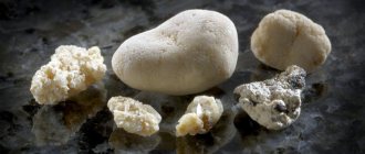
Nephrolithiasis, also known as urolithiasis, develops when chemicals in the kidneys and/or other parts of the urinary tract are activated to form deposits (stones). Usually the first sign that a person has developed kidney stones is an attack of renal colic.
Kidney stones
Urolithiasis occurs four times more often in men than in women. Men aged 30-50 years are most often affected. Women are more at risk of developing urolithiasis during pregnancy, as stagnation of urine and bacterial urinary tract infections may occur during this period.
Bacteria alkalinize the urine, and calcium precipitates more quickly in an alkaline environment, which contributes to the formation of stones.
Modern approach to the treatment of urate nephrolithiasis
The problem of treating urolithiasis remains one of the most pressing in modern urology. Urate urolithiasis is one of the types of urolithiasis, the frequency of which has increased significantly in recent years - from 5–10% in the 50s of the last century to 20–30% currently, which is associated with the increasing impact of a number of environmental factors leading to the accumulation of excess lead in the body, as well as increased alcohol consumption.
The etiopathogenesis of urate lithiasis is the most studied and is associated with complex physicochemical processes occurring both in the body as a whole and at the level of the urinary system of a congenital or acquired nature. The biochemical basis for disorders of purine metabolism are hyperuricemia and hyperuricuria, leading to the formation of stones consisting of uric acid, as well as sodium, ammonium and calcium (very rarely) salts of this acid. The process of stone formation goes through a number of stages - from saturation and supersaturation of urine with salts and further to the phases of enucleation, crystallization and crystal growth to clinically significant sizes, when mechanisms for inhibiting crystal growth are ineffective or absent.
Hyperuricuria creates the preconditions for crystallization of uric acid, mainly in the area of the terminal nephron and at the apex of the renal papilla, similar to Randall's plaques. Uric acid crystals can also lead to the development of aseptic necrosis of the tubular epithelium, which, when rejected, under conditions of hyperuricemia, can become the core of a future calculus. Hyperuricemia leads to the accumulation of uric acid crystals in the interstitial tissue of the kidney and causes interstitial nephritis or early changes in small renal vessels with the development of arterial hypertension.
The genesis of urate nephrolithiasis has both common causes for stone formation and a characteristic feature unique to it, which is that the formation of urate stone requires high acidity of urine, since uric acid dissolves only in slightly acidic and alkaline environments. When the pH of the urine is above 6.5, crystallization of uric acid does not occur, and it is released in a dissolved state; a decrease in the pH of the urine below 5.5 leads to a supersaturation of the urine with uric acid crystals, which precipitate and serve as a framework for the formation of stones.
An increase in the concentration of uric acid in the blood is associated with excess nutrition (especially with an increase in the proportion of food rich in protein in the diet), prolonged fasting, physical inactivity, frequent consumption of alcohol and caffeine, and the use of certain medications: laxatives and diuretics, antibiotics, corticosteroid hormones. In some cancers and malignant blood diseases, hyperuricemia can also develop.
Attempts to explain the development of urolithiasis by any one reason have led to nothing, therefore, in each specific case, it is necessary, before prescribing treatment, to conduct an examination in order to find out all the possible causes of the development of the disease in a given patient.
The examination of patients involves the collection of anamnestic data, laboratory (with mandatory testing of the level of calcium and uric acid in the blood, excretion of calcium, oxalates, phosphates, uric acid), ultrasound, X-ray (survey and excretory urography) examination, as well as bacteriological analysis of urine. Additional information can be obtained from helical computed tomography with a bolus of radiocontrast or magnetic resonance imaging. When preparing for surgery, it is also necessary to consult a therapist, an anesthesiologist, and, if indicated, other specialists. After spontaneous passage or removal of the stone in one way or another, a study of the chemical composition of the stones is carried out.
The examination results reveal various variants of purine metabolism disorders: an increase in both the level of uric acid in the blood and its daily excretion in an average of 25% of patients; increased levels of uric acid in the blood with normal daily excretion in 20% of patients; normal levels of uric acid in the blood with increased daily excretion in 15% of patients.
In other patients, the level of uric acid in the blood and its daily excretion may be normal, despite the urate composition of the stones.
More than 50% of patients with urate nephrolithiasis belong to the age category of 50 years and older and have various concomitant diseases (obesity, coronary artery disease, arterial hypertension, diabetes mellitus, gout, etc.), which influence the choice of treatment method and necessitate the need for them additional preoperative preparation.
Modern medicine has a whole arsenal of conservative, surgical and combined methods for treating urate urolithiasis. The choice of treatment method is determined by the number of stones, the location of the stones, their size and shape, the duration of the disease, the presence of concomitant urinary tract infection, the functional capacity of the kidney, the presence of concomitant diseases, the general condition of the patient, the anatomy of the upper urinary tract and other features. It is important to note that treatment is carried out for both emergency and planned indications.
Open surgical interventions (pyelolithotomy, nephrolithotomy), although they have not lost their importance to date for nephrolithiasis, are not of decisive importance and are used in 3–5% of patients. They should be considered as a forced measure and used in specific circumstances and when other treatment methods are impossible. Basically, these operations are performed in emergency situations in acute obstructive pyelonephritis, caused by blockage of the urinary tract with large kidney stones, or when destructive forms of this disease occur. In a planned manner, open operations are indicated in the presence of secondary kidney stones in combination with various anomalies of the upper urinary tract, which cannot be corrected otherwise than by resorting to surgical correction (stricture, periureteritis, etc.).
In recent decades, laparoscopic stone removal and retroperitoneal pyelolithotomy (endoscopic stone removal using percutaneous retroperitoneal access) have competed with open operations for nephrolithiasis.
The main interventions for the purpose of removing urate kidney stones are extracorporeal lithotripsy (ESLT), percutaneous nephrolithotripsy (PCNL), which can be combined and combined with litholytic therapy.
The introduction of percutaneous nephrostomy into practice has revived interest in local litholysis, which refers to the direct application of a dissolving drug to the stone. An optimal litholytic effect can be achieved with uric acid and struvite stones. The method can be combined with percutaneous stone removal or EBRT.
Percutaneous X-ray endoscopic surgery in the “era of DLT” is used to solve complex clinical cases of urate nephrolithiasis: unsuccessful DLT, in the presence of contraindications for DLT, and also as an independent or combined with DLT (“sandwich therapy”) method in the treatment of large, multiple stones , stones in abnormal, repeatedly operated kidneys, in a single kidney, bilateral stones, as well as when removing coral stones.
EBRT is successfully used for kidney and ureteral stones up to 2.5 cm in size. However, if for relatively small stones (up to 1.5 cm maximum linear size) it is indicated as monotherapy, then for larger stones it must be combined with kidney catheterization, installation internal stent or, less commonly, percutaneous puncture nephrostomy (PPNS).
With DLT, only the destruction of the stone occurs. The most responsible period is the period of spontaneous passage of fragments after fragmentation, when periods of disturbance in the passage of urine from the kidney exposed to shock waves are observed. The main methods of kidney drainage used in the postoperative period in case of DLT for ureteral obstruction or attack of pyelonephritis are also ultrasound-guided PPNS, installation of an internal stent, and renal catheterization.
Currently, DLT is the method of choice in the treatment of patients with various clinical forms of urate nephrolithiasis that has not responded to litholysis. Alternative treatment methods (endoscopic or open surgery) should be used only in case of contraindications or prognostic ineffectiveness. The importance of EBRT is especially increasing in older patients with urate nephrolithiasis, since a comprehensive assessment of the invasiveness, clinical effectiveness and impact on the quality of life of all modern treatment methods in the elderly allows us to consider EBRT as the method of choice for stones up to 2.5 cm in size. Introduction of EBRT , undoubtedly, has significantly changed the approach to the removal of urinary stones. However, it is specifically for urate stones that the implementation of this method has so far been associated with certain difficulties, since visualization, and therefore the conduct of an EBRT session as monotherapy for urate stones, is possible only under ultrasound guidance. Since ultrasound guidance and crushing of urate stones are limited to their localization in the kidney, pelvic and prevesical sections of the ureter, after crushing, endoscopic contact lithotripsy of fragments and “stone paths” that caused occlusion in the middle sections of the ureter is most often used, due to the fact that their visualization Ultrasound and DLT are not possible. That is why, more and more often, works are appearing whose authors recommend carrying out DLT and litholytic therapy against the background of installing an internal stent in the preoperative period.
External shock wave lithotripsy for urate lithiasis should be performed under ultrasound guidance. As an alternative method, especially in case of ureteral obstruction in the postoperative period of DLT, X-ray guidance can be used using the following techniques: intravenous administration of a radiocontrast agent before DLT, provided that the renal pelvis is “filled” with a contrast agent, indicating the location of the “stone path” in the ureter ; retrograde insertion of a catheter to a stone in the ureter or pelvis with the introduction of a radiopaque substance into the renal cavity system; antegrade administration of contrast through nephrostomy drainage in the absence of an internal or external stent or ureteral catheter.
Our experience in the use of DLT for the treatment of patients with urate nephrolithiasis indicates that for stones with a maximum linear size of more than 1.5 cm (especially more than 2 cm), DLT gives better results in conditions of kidney drainage, which is preferably carried out by installing an internal stent, since this significantly reduces the incidence of postoperative complications. DLT of uric acid stones in the form of monotherapy can be carried out for relatively small stone sizes - up to 1.5 cm. Litholytic therapy with citrate mixtures, carried out for 1 month and which did not allow dissolution of the stone during subsequent DLT, significantly improves treatment results (the number of complications and time frame for the kidney to be freed from stone fragments). The administration of citrate mixtures can be recommended as preoperative preparation for large urate kidney stones - more than 1.5 cm.
However, the “gold standard” for the treatment of urate stones should be recognized as oral administration of citrate mixtures, which provide dose-dependent alkalinization of urine without changing the acid-base balance of the blood. With concomitant hyperuricemia, treatment must be supplemented with uricostatics to reduce the level of uric acid in the blood. The daily dose of citrate mixtures is selected individually within the range of 6–18 g, evenly distributed throughout the day in 2–3 doses to maintain urine pH at 6.2–6.8, and it does not affect the level of potassium, sodium, oxygen, carbon dioxide and bicarbonate in the blood. The mechanism of action of citrate mixtures is based on reducing the processes of crystallization in the urine and the binding of calcium ions from the gastrointestinal tract to the urinary tract, where this effect is maximally manifested due to the highest concentration of citrate. Thus, the complex effect of citrate on the physicochemical state of urine leads to an increase in the solubility of urates, calcified oxalates, complex magnesium-ammonium phosphates and some other salts, helping to inhibit stone formation and dissolve already formed stones.
There is sufficient data in domestic and foreign literature on the effectiveness of litholytic therapy for urate stones. It should be noted that in the treatment of large urate stones, the effectiveness of litholytic therapy of urate stones increases significantly after their fragmentation by DLT, since after fragmentation the area of contact of urine with the surface of the stone increases hundreds of times.
In addition, for lithokinetic purposes, conservative therapy for urate lithiasis may include the prescription of antispasmodics, anti-inflammatory and antibacterial drugs.
Litholytic therapy for urate nephrolithiasis should be comprehensive and aimed at reducing the content of uric acid in the extracellular space. For this purpose, drugs that have a uricostatic effect and diuretics, including official ones, are used. Patients are prescribed a diet with limited protein-rich foods. Litholysis of urate stones is carried out under ultrasound control. It is important to note that only uric acid stones are reliably soluble during litholytic therapy. Stones consisting of uric acid salts: sodium urate, potassium urate - dissolve less well. Stones consisting of ammonium urate are practically insoluble, so it is advisable to additionally prescribe potassium preparations, which promote the conversion of uric acid salts into potassium urate salts (the solubility of these urate salts is higher).
Citrate therapy in patients with urate nephrolithiasis is carried out provided that the patient follows a diet with limited protein-rich foods, such as meat products, their derivatives and by-products, fatty fish, mushrooms, legumes, lentils; strong alcoholic drinks, red wine, dark beer, chocolate, strong tea and coffee, pickles, smoked meats, raspberries, figs, alkaline mineral waters (Borjomi), sorrel, spinach, celery, peppers, radishes, cauliflower. Patients with impaired purine metabolism are additionally prescribed uricostatics (allopurinol), in the presence of metabolic disorders in the form of oxaluria + hyperuricuria, magnesium oxide is used, in the presence of metabolic disorders in the form of hypercalciuria + hyperuricuria, hypothiazide is added in normal dosages.
In general, when litholytic therapy is carried out correctly by patients, it is possible to dissolve up to 60% of all stones, and in 40% of cases it is necessary to resort to alternative methods of removing stones from the kidneys, and primarily to DLT, the absolute indications for which are: urate stone, in which it is possible to achieve the optimal pH value or there is no effect from long-term therapy; a calculus that caused occlusion of the ureteropelvic segment and an attack of acute pyelonephritis; kidney stone, which disrupts the passage of urine and causes pronounced dilatation of the collecting system; frequent gross hematuria; pain that renders the patient unable to work.
A relative contraindication to DLT of urate stones is the combination of urolithiasis with the active stage of chronic pyelonephritis and constant aching pain in the lumbar region. Relative contraindications include clinical situations such as intolerance to an internal catheter such as a stent (dysuria, hematuria, vesicorenal reflux).
Thus, the currently existing methods of treating urate nephrolithiasis can basically solve the problem of removing urate kidney stones without changing the fundamental principles and principles of treatment, achieving results with a significantly lower risk for the affected organ and the patient’s body as a whole.
It should be noted that patients with urate nephrolithiasis with impaired purine metabolism after stone removal require clinical observation and therapy aimed at preventing relapse of the disease.
Thus, urate urolithiasis is one of the most complex types of urolithiasis; With this disease, successful use of conservative therapy (litholysis) is possible. Indications for surgical treatment have been developed. EBRT in the form of monotherapy is the most favorable method of surgical treatment of urate stones up to 1.5 cm; EBRT in patients with urate nephrolithiasis with stones larger than 1.5 cm gives optimal results in conditions of kidney drainage, which is preferably carried out by installing an internal stent, as this significantly reduces the incidence of postoperative complications. For urate stones larger than 1.5 cm, litholytic therapy with citrate mixtures as preoperative preparation for 1 month before DLT also increases its effectiveness; after DLT of urate stones of any size, litholytic therapy significantly increases the effectiveness of treatment of urate lithiasis, increasing the area of contact of urine with the surface of the stone; the combined use of percutaneous surgery and radiotherapy also maximizes the release of the kidney from stone fragments in the most severe forms of this disease (large stones and coral nephrolithiasis), thereby increasing the effectiveness of treatment. Open surgical interventions are currently rarely performed and are performed mainly for emergency reasons or when other treatment methods are ineffective.
Literature
- Alyaev Yu. G., Kuzmicheva G. M., Rapoport L. M., Rudenko V. I. Modern aspects of citrate therapy in patients with urolithiasis // Medical class. 2004. No. 4. P. 20-24.
- Alyaev Yu. G., Rapoport L. M., Rudenko V. I. Citrate therapy to prepare for extracorporeal lithotripsy // Materials of the plenum of the board of the Russian Society of Urologists. M., 2003. P. 59-60.
- Alyaev Yu. G., Rapoport L. M., Rudenko V. I. et al. 1st International Consultation on Stone Disease. aris, 3-4 July 2001. Book of abstracts; 63:63.
- Baibarin K. A. Surgical methods of treating nephrolithiasis in gerontological patients: Dis. ...cand. honey. Sci. M., 2004.
- Beshliev D. A. Dangers, errors, complications of extracorporeal lithotripsy, their treatment and prevention: Dis. ...Dr. med. Sci. M., 2003. 356 p.
- Bugaeva N.V., Balkarov I.M. Arterial hypertension and disorders of purine metabolism // Ter. arch. 1996. No. 1. P. 36-39.
- Dzeranov N.K., Grishkova N.V., Boyko T.F., Golovanov S.A. Conditions for performing external lithotripsy with different physicochemical composition of urinary stones // Urology and Nephrology. 1994. No. 6. P. 10-13.
- Dutov V.V. Modern aspects of the treatment of some forms of urolithiasis: Dis. ...Dr. med. Sci. M., 2000.
- Kaprin A.D., Ivanenko K.V., Ivanov S.A. Treatment of patients with urate nephrolithiasis with disorders of purine metabolism. M., 2003.
- Martov A. G. X-ray endoscopy and external shock wave lithotripsy in the combined treatment of nephroureterolithiasis: method. recommendations. M., 1994. pp. 11-14.
- Nephrology: A Guide for Doctors /ed. I. E. Tareeva. M., 1995. T. 2.
- Pytel Yu. A., Zolotarev I. I. Urate nephrolithiasis. M., 1995. 176 p.
- Pytel Yu. A., Chakaleva I. I., Shemyakin F. M. About some biochemical changes in the body during citrate therapy of patients with urate nephrolithiasis // Urology and Nephrology. 1972. No. 6. P. 28-33.
- Pytel Yu. A., Zolotarev I. I. On the issue of the use of conservative therapy in patients with urate nephrolithiasis // 4th Lithuanian Conf. urologists. Kaunas, 1987. pp. 66-68.
- Rudenko V.I. Urolithiasis. Current issues of diagnosis and choice of treatment method: Dis. ...Dr. med. Sci. 2004.
- Sergienko N. F., Shaplygin L. V., Kuchits S. F. Citrate therapy in the treatment of urate nephrolithiasis // Urology and nephrology. 1999. No. 2. P. 34-36.
- Buck AC The epidemiology, formation, composition and medical management of idiophatic stone disease//Curr. Opin. Urol. 1993; 3: 316-322.
- Grases F., Ramis M., Villacampa Al., Costa-Bauza A. Uric acid urolithiasis and crystallization inhibitors // Urol. Int. 1999; 62(4): 201-204.
- Hoffman N., McGee SM, Hulbert JC Resolution of ephedrine stones with dissolution therapy//Urology/2003/may: 61(5): 1035.
N. K. Dzeranov , Doctor of Medical Sciences, Professor, Academician of the Moscow Aviation Institute D. A. Beshliev , Doctor of Medical Sciences R. I. Bagirov K. A. Baibarin , Candidate of Medical Sciences RMAPO, Moscow
Attack of renal colic
Severe pain accompanied by nausea, vomiting and hematuria are symptoms of an attack of renal colic, which portends acute urolithiasis. The stone gets stuck in the urinary tract, preventing urine from entering the bladder. An infection then develops, leading to acute renal failure.
The attack begins suddenly, for example, after great physical effort, a long car journey, or after alcohol abuse. Pain appears in the lumbar region, radiating along the ureter to the lower abdomen and external genitalia.
Attack of renal colic
The attack is accompanied by flatulence and painful urination. The pain can be so severe that patients lose consciousness. Relieve pain with strong analgesics.
Causes of kidney stones
Urinary stones can form both from physiological compounds in the urine and from pathological ones. The kidneys filter the blood, that is, they separate harmful or toxic substances from it, and then remove them from the body along with urine. However, it happens that some deposits settle at the bottom of the kidney cup. A layer of sediment forms.
If the stone is small, it can be removed naturally, that is, through the urinary tract to the bladder and then out through the urethra.
If the stone is large or the stones are immobile, they remain and are overgrown with new layers. This occurs when the concentration of compounds that can form kidney stones exceeds the solubility threshold in the body.
This is facilitated by:
- heredity;
- defects in the structure of the urinary system;
- urinary tract infections;
- taking certain medications, such as glucocorticosteroids;
- hyperparathyroidism;
- gastrointestinal diseases - inflammatory bowel diseases, for example Crohn's disease, malabsorption syndrome;
- long-term treatment of peptic ulcer with alkalizing drugs to reduce the secretion of gastric acid;
- excessive thickening of urine, caused, for example, by limiting drinking, dehydration;
- overdose of vitamin D3, vitamin C, calcium;
- wrong diet.
Hyperparathyroidism
2. Reasons
The causes of stone formation in general, and the formation of coral stones in particular, remain the subject of research. The role of a number of factors has been established: heredity, endocrine disorders (in relation to coral stones, for example, the hypothesis of a connection with dysfunction of the parathyroid glands is actively considered and discussed), poor nutrition and resulting metabolic disorders, insufficient fluid intake with an inevitable shift in the water-salt balance ( it is known that the composition of stones is dominated by phosphates, urates, oxalates and other salts), disturbances of natural urodynamics, gastrointestinal diseases, chronic inflammatory processes, etc. However, none of these reasons is an obligate trigger (i.e., a factor in the presence of which stone formation is triggered in 100% of cases). Perhaps nephrolithiasis should really be considered a polyetiological disease, developing only under a certain confluence of unfavorable objective and subjective conditions, and their “trigger” combination in each case, again, is individual.
Visit our Nephrology page
Symptoms of kidney stones
Nephrolithiasis does not cause any symptoms for many years, and can be discovered incidentally, for example, during an x-ray or ultrasound of the abdominal cavity. The patient usually finds out that he has a problem with kidney stones only when attacks of renal colic begin.
This occurs when a stone blocks the urinary tract, causing swelling of the kidneys and triggering an attack of renal colic. This condition requires immediate intervention by a urologist.
- First, you need to quickly relieve the unbearable pain - its intensity is so great that it is often accompanied by nausea and vomiting.
- Secondly, prolonged anuria leads to kidney damage.
Very severe colic pain usually affects the abdomen, side, groin, and, depending on gender, the testicles or labia. In addition, there is hematuria (blood in the urine), pain when urinating, frequent urination, and a constant urge to urinate. An increase in temperature, pale skin, and a feeling of anxiety are also usually observed.
1.General information
The problem of nephrolithiasis, or kidney stones, remains quite acute throughout the world. At the same time, intensive research into the causes and risk factors of stone formation, the chemical composition of stones, various variants of their dynamics and clinical picture, as well as the ongoing development of medical technology and methodology - together, all this has made it possible to radically change the situation compared to what it was another half century back.
Coral stones are one of the most common types of nephrolithiasis, which has certain etiopathogenetic and clinical specificity. Obviously, coral-shaped stones got their name due to their purely external resemblance to the waste product of coral polyps; There is, of course, no other connection here. A distinctive feature of a coral stone is that it is formed as an internal “cast” or a filling “cast” of the intimate renal cavities, which in the later stages of stone formation leads to pronounced changes in the entire renal parenchyma and, accordingly, to progressive renal failure.
According to various statistical estimates, the share of coral stones in the total volume of registered nephrolithiasis is 10%-30%. It is known that women suffer up to twice as often as men (however, it is not yet entirely clear why this is so). The age range in which this type of nephrolithiasis is usually diagnosed - when the symptoms reach the level of clinical significance and forces one to seek specialized help - is 25-50 years.
A must read! Help with treatment and hospitalization!
Types of Kidney Stones
Table 1. Characteristics of various kidney stones
| Types of stones | Color | Structure | Density |
| Phosphates | White, light gray | Smooth | Soft |
| Oxalates | Grey, black | With sharp edges | Dense, hard |
| Urats | Brick color | Smooth | Dense |
| Cystines | Yellowish, light brown | Smooth | Soft |
| Protein stones | White, light | Smooth | Soft |
Cystine stones
. Diagnosed in 1% of patients.
- Causes: congenital defect resulting in impaired absorption of cystine (amino acid).
- Treatment: The basis is a diet that limits cystine and methionine. Penicillamine, which forms soluble compounds with cystine, can be administered.
- Diet: the basis should be milk and its products, as well as plant products. You need to drink plenty of fluids. Consumption of meat and meat products should be limited.
Phosphate stones (struvite)
. Diagnosed in 5-10% of patients.
- Causes: Frequent urinary tract infections with urease-producing bacteria, that is, Proteus, Klebsiella and Pseudomonas. Urease is the breakdown of urea into ammonia and the alkaline reaction of urine.
- Treatment: The most important is acidification of the urine. However, this cannot be achieved through diet alone. It is necessary to take acidifying drugs.
- Diet: consists of avoiding foods rich in calcium and phosphates, that is, legumes, potatoes, large amounts of vegetables, fruits, milk and eggs. Recommended are fish, cereals, pasta, bread, butter, meat.
Oxalate urolithiasis (calcium oxalate).
Diagnosed in 70-80% of patients with urolithiasis.
- Reasons: it may be hyperparathyroidism, an overdose of vitamin D3, excessive excretion of calcium, citrates and oxalates from the body.
- Treatment: increase fluid intake to 4-5 liters per day. Drugs that retain calcium in the digestive tract and reduce urinary excretion.
- Diet: Mainly consists of limiting oxalate intake. You cannot eat chocolate, cocoa, spinach, rhubarb, sorrel, lemons, beets, hot spices, alcohol, oranges. Coca-Cola, coffee and tea are prohibited. In very small quantities you can eat potatoes, tomatoes, peas, plums, and strawberries. Recommended foods include yeast, eggs, wholemeal bread, cereals, lean meats, fish, poultry, cucumbers, lettuce, butter, onions and fruits.
Cholelithiasis gout. Diagnosed in 5-10% of patients.
- Causes: Occurs when the urine pH is acidic or the body produces excess uric acid. This also affects patients with lymphoma, leukemia and bone marrow diseases.
- Treatment: These stones can be dissolved by taking appropriate tablets. Agents that change the acidity of urine to alkaline are also used.
- Diet: it is necessary to reduce the acidity of urine. This can be achieved by consuming dairy products and plenty of vegetables and fruits. The second rule is to limit foods that are sources of purine (purine bases), which are found in offal (liver, lungs, tongues, kidneys, brain and heart), decoctions, jellies, meat sauces, sardines and herring. Legumes (peas, beans, lentils and beans) and mushrooms are rich in purines. They also contain cocoa, coffee and tea. Pork, beef and lamb should be avoided. It is best to cook meat by steaming and in plenty of water. Lean poultry and veal are recommended. However, their amount in the daily diet should not exceed 100-150 g. Vegetables, fruits, sugar, butter in small quantities, milk, low-fat cheese, and potatoes are recommended. The total daily calorie content of foods should not exceed 2000-2200 kcal.
Plants that are advisable to take for this form of lithiasis: leaves of lingonberry, black currant, leaves and buds of birch. A glass of decoction of this mixture should be drunk at least 3 times a day between meals.
Decoction
Treatment of kidney stones
70% of patients can be treated pharmacologically. Surgery is used only for large stones, although urologists are increasingly choosing non-invasive methods.
Pharmacist giving medicine
Surgical removal of urinary stones is rarely used because modern, less invasive methods are just as effective.
- Non-contact lithotripsy
. It consists of destroying urinary stones in the patient’s body using a so-called shock wave. The procedure is usually performed on an outpatient basis and does not require anesthesia. The crushed stone is then removed naturally. - Percutaneous nephrolithotripsy.
It is carried out under anesthesia. The procedure involves inserting a nephoscope into the renal pelvis through a small incision in the skin in the area of the kidneys, with which the doctor can examine the stone, determine its location and crush it using appropriate instruments, and then remove it. - Ureterorenoscopy
. It is carried out under anesthesia. The doctor inserts a flexible probe through the urethra into the bladder and then into the ureter. He can see the stone and remove it whole or crush it into smaller pieces beforehand.
ONLINE REGISTRATION at the DIANA clinic
You can sign up by calling the toll-free phone number 8-800-707-15-60 or filling out the contact form. In this case, we will contact you ourselves.
If you find an error, please select a piece of text and press Ctrl+Enter
4.Treatment
Today, many approaches, methods, and specific techniques for the treatment of coral nephrolithiasis have been developed and are successfully used. With some variants of the chemical composition of stones, for example, with a predominance of urates, i.e. uric acid salts - there is a certain chance of getting by with conservative treatment, gradually “dissolving” the calculus. In any case, for the treatment and/or prevention of infectious and inflammatory exacerbations, specially selected (based on the results of bacteriological analysis) antibiotics are prescribed. However, a much more effective and promising direction is minimally and minimally invasive surgery (for example, percutaneous puncture nephrolithotripsy, external shock wave lithotripsy, percutaneous percutaneous nephrolithotripsy, etc.), aimed at crushing and removing stones from the kidney. General principles that are followed to the last possible extent are the avoidance of open abdominal intervention, the choice of organ-preserving methods and the maximum stabilization of renal functions in the postoperative period. Of course, advanced, late-diagnosed, irreversible morphological changes do not leave such a chance, but today the presence of a coral stone inside the kidney, even a fairly large one, is by no means an absolute indication for removal of the entire organ. With the development of medical science and practice, the effectiveness of treatment for coral nephrolithiasis continues to increase while the invasiveness of the intervention decreases.
