Burkitt's lymphoma
(LB) is a malignant tumor from the group of non-Hodgkin lymphomas, formed from mature B lymphocytes. It is characterized by increased aggressiveness, high proliferative activity, a short history (several weeks) and a tendency to relapse. Typically, relapses develop within 6-9 months after the end of chemotherapy; later they are extremely rare.
According to ICD-10, Burkitt's lymphoma corresponds to code 2A85.6.
Depending on the cause of the disease, there are 3 types of LB:
Endemic.
Most often it affects dark-skinned children (especially 5-8 year old boys) living in African countries. In 95-100% of cases it is associated with the Epstein-Barr virus.
Sporadic.
Found in non-endemic areas.
HIV-associated.
Develops against the background of immunodeficiency states (in HIV-infected people, mainly men.
The pathogenesis of LB is characterized by increased expression of the c-MYC proto-oncogene.
In most cases (30-50%), Burkitt's lymphoma affects children; boys are 3-4 times more likely than girls. It is rare in adults - in 2% of cases. ( https://med4share.ru/media/files/clinic_recommend/lymphoma_berkitta.pdf )
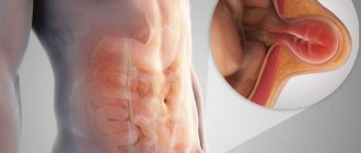
Most often the tumor is localized in the abdominal cavity, extremely rarely - in the mediastinum.
1.Burkitt's lymphoma and its types
Burkitt's lymphoma is a form of non-Hodgkin's lymphoma.
in which cancer begins in immune cells.
Burkitt's lymphoma is recognized as the fastest growing tumor in humans
, and if treatment is not started in time, it quickly leads to death. But intensive chemotherapy helps to significantly prolong the life of patients with Burkitt's lymphoma in more than half of the cases.
Burkitt's lymphoma is named after British surgeon Denis Burkitt, who first diagnosed the disease in 1956 among African children. In Africa, lymphoma affects children who also have malaria and the Epstein-Barr virus, which causes infectious mononucleosis. It is likely that malaria weakens the immune system's response to the Epstein-Barr virus, allowing it to attack infected cells and turn them cancerous.
Outside of Africa, Burkitt's lymphoma is rare. People infected with HIV, the human immunodeficiency virus, are more prone to developing this disease. According to statistics, the incidence of Burkitt's lymphoma in HIV-positive people is 1000 times higher than in those who are not infected.
Types of Burkitt's lymphoma
The World Health Organization classification identifies three types of Burkitt lymphoma:
- Endemic
(African lymphoma), which we described above; - Sporadic Burkitt's lymphoma
. This form of lymphoma occurs throughout the world. According to statistics, sporadic Burkitt lymphoma accounts for 1 to 2% of all cases of lymphoma in the adult population. - Lymphoma associated with immunodeficiency.
This type of Burkitt's lymphoma most often occurs in people with HIV/AIDS. But it can also develop in people with congenital conditions that cause immunodeficiency, as well as in patients who have had organ transplants and take immunosuppressive drugs (suppress the immune system).
A must read! Help with treatment and hospitalization!
Materials and methods
From 2003 to 2014 at the Federal State Budgetary Institution State Scientific Center M.Z. Russia, Federal Medical Research Center named after. P.A. Herzen, Medical Radiological Research Center named after. A.F. Tsyba, Orenburg Regional Clinical Hospital observed 70 patients diagnosed with LB: 45 men and 25 women aged from 15 to 62 years, median age 31 years. The stage of the disease was determined based on the S. Murphy classification. Stage I of the disease was diagnosed in 4 (5.7%) patients, stage II - in 9 (12.9%), stage III - in 25 (35.7%), stage IV - in 11 (15.7%), Burkitt's leukemia - in 21 (30%). In 23 (32.9%) patients, involvement of the BM in the tumor process was noted, in 15 (21.4%) - the central nervous system (neuroleukemia in 12, intratumor of the spinal cord in 3, intratumor of the brain in 3). B-symptoms were detected in 56 (80%) patients, increased lactate dehydrogenase (LDH) activity - in 50 (78.1%) out of 64, while 34 (56.2%) out of 64 showed an increase in LDH activity by more than 2 times compared to reference values. The median LDH level was 2398 (238–20,300) U/L. Acute renal failure (ARF) at the onset of the disease was determined in 17 (24.2%) patients, 8 chemotherapy (CT) was started against the background of therapy replacing renal function. Obstructive jaundice due to involvement of the head of the pancreas and bile ducts in the tumor process was detected in 3 patients. As the first stage of treatment, patients underwent drainage of the bile ducts (in 1 - endoscopic retrograde cholangiopancreatography, nasobiliary drainage, in 1 - minilaparotomy, transvesical drainage of the bile ducts, in 1 - laparotomy, intraoperative cholangiography, cholecystostomy). At the time of admission to the hematology clinic, 6 patients were diagnosed with gastric bleeding; in 1, in addition to gastric bleeding, multiple subdural hematomas and hemorrhages in the sclera were detected. Due to postrenal anuria caused by compression of the ureters by the pelvic tumor, nephrostomy was performed in 1 patient before admission to the clinic.
The diagnosis of LB was established in accordance with the 2008 WHO criteria based on a biopsy of the tumor, subsequent histological, immunohistochemical and cytogenetic studies. The patient examination protocol included a medical history and physical examination, a general blood test, a general urinalysis, a biochemical blood test, a coagulogram, a myelogram, an immunochemical study of blood serum and urine, computed tomography (CT) of the chest, abdominal and pelvic organs, an electrocardiogram, a biopsy lymph node - lymph node (tumor neoplasm), histological, immunohistochemical and cytogenetic studies, trepanobiopsy, lumbar puncture with cytological and biochemical studies of the cerebrospinal fluid. All patients were assessed for HIV status, the presence of HBsAg (qualitative test), and antibodies to the hepatitis C virus antigen. According to indications, fibrogastroduodenoscopy, colonoscopy with multiple biopsies of the mucous membranes of the stomach and intestines, CT scan of the soft tissues of the neck and brain, and magnetic resonance imaging of the brain were performed. brain, soft tissues of the neck, pelvis with intravenous bolus contrast, diagnostic laparoscopy or laparotomy with biopsy of the tumor formation. All women were consulted by a gynecologist; according to indications, ovarioprotection was prescribed for the period of chemotherapy. The immunohistochemical panel included antibodies to CD20, CD3, CD10, BCL2, BCL6, c-MYC, Ki-67, MUM1, cyclin D1, and in some cases, TdT. Cytogenetic studies were performed in 70 patients. A standard cytogenetic study (G-differential staining of chromosomes) was performed in 8 patients, tumor cells (LN) were examined in 4 cases, and BM cells in 4 cases. In 29 patients, a cytogenetic study was performed using fluorescent in situ
(FISH): in 9 cases, a suspension of tumor cells was used as a material, in 5 - a suspension of BM cells, in 13 - tumor prints (gastrobiopsy in 6, colon biopsy in 1, tonsil in 1, trephine biopsy in 1, lymph node in 4), in 2 — CM smears.
Histological sections of paraffin blocks were examined in 33 patients [5]. FISH study was carried out with DNA probes Vysis LSI IGH/MYC, CEP 8 Tri-color, Dual Fusion Translocation Probe (Abbott Molecular) to determine t (8;14)(q24;q32), Vysis LSI MYC Dual color, Break Apart Rearrangement Probe (Abbott Molecular) to detect rearrangement of the MYC
.
To exclude the presence of rearrangements of the BCL2
and
BCL6
genes, the Vysis LSI BCL2 Dual color, Break Apart Rearrangement Probe (Abbott Molecular) and Vysis LSI BCL6 (ABR) Dual color, Break Apart Rearrangement Probe (Abbott Molecular) probes were used, respectively.
Translocation t (8;14)(q24;q32) was detected in 56 of 63 cases, t (8;22)(q24;q11) - in 2 of 63 patients, variant translocations (presence of rearrangement of the MYC
in the absence of translocation t ( 8;14)(q24;q32)) - in 6 out of 63. In 7 cases, rearrangement of the
MYC
; FISH was not performed to detect t (8;14)(q24;q32).
In all cases, there were no rearrangements of the BCL2
and
BCL6
.
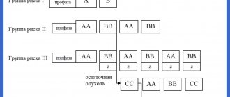
Treatment was carried out according to the LB-M-04±R protocol (4 consecutive blocks A-C-A-C) [4]. Since 2011, patients with CM lesions were treated with 6 blocks A-C-A-C-A-C with rituximab. Block A included intravenous administration of 10 mg/m2 dexamethasone on days 1–5, 1500 mg/m2 methotrexate over 12 hours on day 1, 800 mg/m2 ifosfamide on days 1–5, 1 mg/m2 vincristine on day 1, 50 mg/m2 doxorubicin on day 3, 150 mg/m2 cytarabine on days 4–5, and 100 mg/m2 etoposide on days 4–5; block C consisted of intravenous administration of 10 mg/m2 dexamethasone on days 1–5, 1500 mg/m2 methotrexate over 12 hours on day 1, 5 mg/m2 vinblastine on day 1, 2000 mg/m2 cytarabine 2 once a day on days 2-3, 150 mg/m2 etoposide on days 3-5. Rituximab was administered intravenously on day 0 of the course at a dose of 375 mg/m2. All patients received neuroleukemia prophylaxis on the 1st day of each block by intrathecal administration of 3 drugs: 30 mg of prednisolone, 30 mg of cytarabine and 15 mg of methotrexate. When neuroleukemia was detected, lumbar punctures with intrathecal administration of chemotherapy were performed 2 times a week until the cerebrospinal fluid was completely sanitized and 3 normal cerebrospinal fluid tests were obtained. As the disease progressed, the patient was removed from the protocol. In three patients with involvement of the tonsils, oropharynx and nasopharynx in the tumor process due to the threat of asphyxia, treatment was started in the intensive care unit with artificial pulmonary ventilation (ALV) [6].
29 (41%) patients had a history of surgical interventions related to the underlying disease (Table 1). The main goal of such interventions, usually carried out in general surgical hospitals, was to reduce the volume of the tumor mass or radical removal of an incidentally detected tumor. Urgent histological examination was not performed. Doctors and patients learned about the diagnosis of lymphoma after 10-14 days. During this period, in most cases, continued rapid tumor growth was observed.
Table 1. Surgical interventions in 29 patients with LB before the start of chemotherapy according to the LB-M-04 protocol
Complications of chemotherapy were assessed in accordance with CTCAE (Common Terminology Criteria for Adverse Events) 2009 [7].
Statistical data analysis.
When statistically analyzing the data using univariate and multivariate analyzes (Cox regression analysis), unfavorable prognostic factors associated with LB were discovered and assessed.
Overall survival (OS) was chosen as the main criterion for assessing unfavorable prognosis factors, and relapse-free survival (RFS) was chosen as an additional criterion. Along with the survival values, we present the standard error ( SE
), risk ratio (HR), and corresponding 95% confidence interval (CI).
The threshold level of statistical significance p
was chosen equal to 0.05. The licensed statistical package SAS 9.1 was used for calculations.
2.Symptoms of Burkitt's lymphoma
Symptoms of Burkitt's lymphoma depend on its type. Endemic (African) lymphoma usually begins with a tumor
jaw or other facial bones. It can also affect the gastrointestinal tract, breasts and ovaries, and spread to the central nervous system, causing nerve damage, weakness and paralysis.
Sporadic and immunodeficiency Burkitt lymphomas usually begin in the intestines and form large tumors in the abdomen. Later, the liver, spleen and bone marrow are often affected. The growth of cancer cells in Burkitt lymphoma can also begin in the ovaries, testicles or other organs, spreading to the brain and cerebrospinal fluid.
In addition to tumor growth, symptoms of Burkitt's lymphoma may include:
:
- Loss of appetite;
- Weight loss;
- Fatigue;
- Sweating at night;
- Unexplained fever.
Visit our Oncology page
Discussion
As noted earlier, due to the rarity of LB, the number of studies on the treatment of this disease is small, there are no randomized studies, and most of the presented studies included a small number of patients. The main international results of LB therapy are presented in Table. 8 [9-27]. These studies can be divided into 3 groups: non-intensive programs [5-14], intensive protocols (LMB, Hyper-CVAD, CODOX-M/IVAC, LMB, CALGB, NHL-BFM-83/86) without the inclusion of rituximab [13- 21] and intensive chemotherapy programs with rituximab [22–27]. As can be seen from the presented data, despite the intensification of therapy and the inclusion of rituximab in the protocols, OS and DFS did not change significantly.
Table 8. World data on LB treatment Note. n/a - no data.
The largest prospective study evaluating LB therapy in adults included 363 patients from 98 centers. Treatment was carried out according to the GMALL-B-ALL/NHL2002 protocol, which is a modified and improved version of the German group protocols B-NHL83, B-NHL86 and B-NHL90. The main features of the protocol were the inclusion of rituximab and a reduction in the dose of methotrexate to 1500 mg/m2 for patients aged 55 years or younger, to 500 mg/m2 for patients over 55 years of age. Remission was achieved in 88% of patients, 5-year OS was 80%. At the same time, in patients of the older age group (over 55 years of age), OS is significantly lower - 62% (compared to OS of patients aged 55 years or younger, which was 86%) [26]. T. Intermesoli et al. [24] conducted a study to evaluate the effectiveness of treatment of LB patients using a similar protocol GMALL-B-ALL/NHL2002. The study included 105 patients aged 17 to 78 years, median age was 47 years. 3-year OS and DFS were 67 and 75%, respectively. As can be seen, according to the results of our research group, the 5-year OS reaching 85% and the 5-year RFS reaching 95% are optimistic.
As in our study, in the work of D. Hoelzer et al. [26] showed that in the older age group there was a higher mortality rate compared to that in younger patients - 11 and 2%, respectively; Moreover, 90% of deaths are caused by infectious complications during the period of chemotherapy. Similar results are reported by T. Intermesoli et al. [24]: early treatment-related mortality was 13% and was observed predominantly in patients over 45 years of age with generalized stages of the disease (III-IV or Burkitt leukemia). In our study, treatment-related mortality was 8.6% (6 patients died), 3 patients were over 40 years of age. It is worth noting that the median age of our patients was slightly lower—31 years. J. Hong et al. [27] note that in addition to the high mortality rate in the group of older patients, it is precisely these patients who cannot be fully treated. In 25.6% of patients, the protocol program (R-Hyper-CVAD) is not followed due to its high toxicity.
J. Kelly et al. [28] analyzed data from the SEER database (Surveillance, Epidemiology and End Results) from 1973 to 2004, as well as publications according to PubMed, Web of Science, Cochrane Library from 1989 to 2007. The analysis included studies that included 10 patients or more with newly diagnosed HIV-negative LB. J. Kelly selected 13 publications (543 patients). Patients over 40 years old accounted for 42.2% ( n
=229). The OS of patients over 40 years of age in studies before 2000 was 40%, after 2000 - 60%, which is significantly lower than the OS of young patients.
Factors of unfavorable prognosis.
In a study by D. Hoelzer et al. [26] factors of unfavorable prognosis affecting OS (univariate analysis) were female gender, leukocyte levels >30·109/l, platelets <25·109/l, bone marrow lesions, involvement of the central nervous system, stages III-IV of the disease, LDH activity >250 U/L, high IPI and aaIPI. When conducting a multivariate analysis, statistically significant factors for unfavorable prognosis were age over 55 years, CM involvement, and female gender [26]. According to T. Intermesoli et al. [24], age over 60 years and ECOG score >1 point affect OS and DFS. In a study by J. Hong et al. [27], a multivariate analysis revealed that age over 60 years, ECOG somatic status score of 2 points or more, absolute lymphocyte count less than 1.2·109/l, high IPI are independent prognostic factors affecting survival. According to our data, CM, poor somatic status (ECOG score 3-4 points), Burkitt leukemia are factors of unfavorable prognosis that affect OS (according to multivariate analysis). Our results are fully consistent with the data of foreign authors.
3. Diagnosis of Burkitt's lymphoma
Because Burkitt lymphoma spreads so quickly, early diagnosis is critical to a patient's recovery.
If Burkitt's lymphoma is suspected, a biopsy
enlarged lymph nodes or other suspected foci of disease. Taking a tissue sample and further examining it under a microscope will help confirm or rule out the diagnosis of Burkitt's lymphoma.
Additional methods for diagnosing lymphoma may be:
- Computed tomography of the chest, abdomen and pelvis;
- Chest X-ray;
- PET, or positron emission tomography;
- Bone marrow biopsy;
- Cerebrospinal fluid examination;
- Blood tests to evaluate kidney and liver function;
- Testing for HIV infection.
About our clinic Chistye Prudy metro station Medintercom page!
Advantages of Top Ichilov
- The clinic’s oncologists and oncohematologists are first-class doctors and experts in the treatment of a variety of lymphomas. They have undergone many years of training and have exceptional experience.
- Diagnostic studies are carried out using the latest laboratory technology and high-precision equipment.
- Unique methods are used for treatment, ranging from highly effective chemotherapy regimens to the use of immune drugs.
- Patients of the clinic live in comfortable rooms, surrounded by an atmosphere of comfort, and a personal curator-translator is engaged in interaction with doctors and translation of medical documentation.
- 5
- 4
- 3
- 2
- 1
(12 votes, average: 4.4 out of 5)
4. Treatment of Burkitt's lymphoma
Intensive intravenous chemotherapy
, which is usually performed in a hospital, is the most effective treatment for Burkitt lymphoma.
Because Burkitt's lymphoma can spread to the fluids surrounding the brain and spinal cord, chemotherapy drugs may be injected directly into the cerebrospinal fluid. This treatment method is called intrathecal chemotherapy.
drugs may be used to treat Burkitt's lymphoma
. For example, cyclophosphamide, cytarabine, doxorubicin, etoposide, methotrexate, vincristine and others.
Intensive chemotherapy can be either an independent treatment method for Burkitt lymphoma or an integral part of complex treatment. Along with chemotherapy, the following are used:
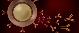
- Rituximab
is a monoclonal antibody that sticks to proteins in cancer cells and stimulates the immune system to attack the cancer cells; - Autologous stem cell transplantation
. These are stem cells that have previously been taken from the patient and stored in a stem cell bank; - Radiation therapy;
- Steroid therapy.
Burkitt's lymphoma is a fatal disease if left untreated. In children, prompt, intensive chemotherapy usually cures lymphoma and predicts long-term survival in 60-90% of cases. In adult patients, the results are very different, but on average, timely treatment means long-term survival in 70-80% of cases, which is also significant.
results
Complete remission of the disease was achieved in 62 (89%) patients (Table 2). 6 patients died from complications of therapy during the induction of remission, 2 experienced disease progression, and 3 developed a relapse of the disease (2 early, 1 2 years after completion of treatment). The 5-year OS was 85%, the 5-year DFS was 95% (Fig. 1). According to the data presented in table. 2, when considering the frequency of development of the studied outcomes (relapse, progression, death from complications of chemotherapy) in different age groups, a higher induction lethality was revealed in the group of patients over 40 years of age. The rate of complete remissions, OS and DFS were not statistically significantly different.
Table 2. Treatment results according to the LB-M-04 protocol Note. Data are presented as absolute number of patients (%) unless otherwise indicated.
Rice. 1. 5-year OS and DFS in LB for the observation period 2003—2014.
When conducting a univariate analysis of unfavorable prognosis factors, the studied parameters included gender, age, stages of the disease, involvement of the BM and the central nervous system, including combined, assessment of somatic status according to the General Condition Assessment (ECOG) scale, assessments of the International Prognostic Index (IPI) and adjusted by age International Prognostic Index (aaIPI), LDH level, B-symptoms, multiple extranodal lesions, acute renal failure, history of surgical interventions, use of rituximab in therapy. The results of univariate analysis are presented in table. 3. A statistically significant dependence of OS on the stage of the disease, the presence of BM lesions, poor somatic status and high LDH concentration was revealed. In the area of boundary values ( p
=0.05) influences on OS included male gender, high IPI and aaIPI, the presence of B-symptoms, as well as 3 extranodal lesions or more. Prognostically significant factors influencing RFS were generalized stages of the disease/Burkitt's leukemia and BM involvement. 5-year OS depending on the stage of the disease, BM involvement and assessment of somatic status according to ECOG is presented in Fig. 2.
Table 3. Results of univariate analysis of unfavorable prognostic signs in LB (total 70 patients)
Rice.
2. 5-year OS in LB depending on the stage (including the stage of ALL - Burkitt leukemia) of the disease (a), BM involvement (b) and assessment of somatic status according to ECOG (c). To assess the combined influence of unfavorable prognosis factors in patients with LB, a multivariate Cox regression analysis was performed with the inclusion of similar parameters in the study. The results of multivariate analysis are presented in table. 4. Burkitt's leukemia and BM lesions turned out to be independent prognostic factors affecting both OS and DFS. Poor physical status (ECOG score 3–4 versus 0–2) was statistically significant for OS but not for DFS.
Table 4. Results of multivariate Cox regression analysis of unfavorable prognostic features in LB
Complications of chemotherapy.
The average duration of myelotoxic agranulocytosis was 5.2 (0-29) days, the maximum duration was observed after the first course of chemotherapy - 7.2 (0-25) days, which is associated with the initial severe condition of the patients, large tumor mass, BM involvement, and often the presence OPN. Complications that developed during treatment according to the LB-M-04 protocol are presented in Table. 5. Additional confirmation of the severity of the patients’ condition is the development of tumor cytolysis syndrome (TCS) in 19 (27%) patients. SCO was diagnosed in 10 patients with Burkitt leukemia: 3 with stage IV, 5 with stage III, 1 with stage I according to S. Murphy. According to the data in Table. 4 all courses were accompanied by the development of leukopenia, mainly grade IV, thrombocytopenia grade III-IV, anemia, usually grade III-IV. The degree of leukopenia, thrombocytopenia and anemia did not differ significantly depending on the course. During the first course, severe infections and conditions requiring transfer of patients to the intensive care unit were more often observed: sepsis, pneumonia, meningitis, necrotizing enteropathy, acute respiratory failure with mechanical ventilation. Induction mortality during the first course was 5.7% (4 patients died; in all cases the cause of death was sepsis, septic shock, multiple organ failure), the second course was 3% (2 patients died: 1 from pseudomonas sepsis, 1 from traumatic subdural hematoma). There were no treatment-related deaths during subsequent courses of chemotherapy. Among the 6 patients who died from treatment-related complications, 3 were aged 40 years or older (see Table 2), which further confirms the greater toxicity of intensive chemotherapy regimens in patients over 40 years of age [8]. If we look separately at the complications that arise during treatment in patients of the older age group (Table 6), we will see that the number of severe complications (sepsis, septic shock, pneumonia, etc.) is significantly greater. In addition, the period of myelotoxic agranulocytosis was longer. Among 18 patients in the older age group, 3 died from complications of chemotherapy, 2 were withdrawn from the protocol after three courses due to high toxicity, and after two blocks, 2 were transferred to a lighter course (block B) due to prolonged cytopenia. Extension of intervals between courses (total number of courses 61) was observed in 18 (29.5%) courses, a reduction in doses of chemotherapy drugs cytarabine and methotrexate by 30-50% was noted in 11 (18%) cases.
Table 5. Complications that developed during treatment according to the LB-M-04 protocol and the need for blood transfusions Note. Here and in the table. 6, 7 data are presented as median (range) or absolute number of patients (%). ARF - acute respiratory failure; ACS - acute coronary syndrome.
Table 6. Complications that develop during chemotherapy according to the LB-M-04 protocol and their consequences in patients over 40 years of age.
The group of patients with Burkitt's leukemia is subject to separate consideration. In table For comparison, Table 7 presents the duration of myelotoxic agranulocytosis, the most severe complications that developed during treatment, and the volume of transfusion support in patients with Burkitt leukemia. As can be seen, SCO is observed in 52.4% of cases (in the general group in 27% of patients), the duration of agranulocytosis after the first course is 11.3 days (compared to 7.2 days in the general group), the most severe infectious complications: sepsis , septic shock, pneumonia, acute respiratory failure, meningitis are observed much more often - in 33.3, 9.5, 33.3, 28.6 and 14.3% of cases, respectively. For comparison, in the general group, these complications develop in 15.7, 5.7, 15.7, 8.6 and 7.1% of cases, respectively. The need and volume of transfusion support (see Table 7) is 1.5 times greater than in the general group (see Table 5). This situation is explained by the fact that patients with Burkitt’s leukemia are the most severe group. This is due to a large tumor mass, intoxication, STS, and electrolyte disturbances. Among 8 patients requiring hemodialysis, 7 had Burkitt's leukemia. The main difficulties in the treatment of patients with Burkitt's leukemia arose precisely during the first course of chemotherapy; subsequently, the duration of myelotoxic agranulocytosis, the number of infectious complications and the volume of transfusion support did not differ from those in the general group.
Table 7. Complications that develop during chemotherapy according to the LB-M-04 protocol and the need for blood transfusions in patients with Burkitt leukemia