Surgeon
Bohyan
Tigran Surenovich
Experience 36 years
Surgeon of the highest category, Doctor of Medical Sciences, member of the International Association of Surgeons, Gastroenterologists and Oncologists
Make an appointment
Lipoma (fat) is a benign formation in the form of small nodules, the appearance of which is associated with the accumulation and compaction of excess adipose tissue. The size of the lipoma may increase or remain stable. When exposed to unfavorable factors, pockets of pathogenic microflora can form inside the wen, which makes it a source of other serious diseases of internal organs. The unaesthetic appearance of formations protruding on the surface of the skin is also important. Therefore, in most cases, a decision is made to remove the tumor and its subsequent histological examination to clarify the nature of the wen - benign or malignant.
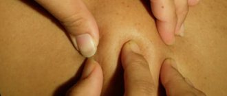
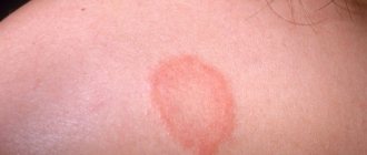
The area of localization of lipoma is the back, chest, face, limbs, mammary glands, structure of internal organs. The formation can be detected accidentally during diagnostic procedures or during palpation. The edges of the lipoma are dense and clear, the formation is mobile, and its palpation is painless. It is more difficult to detect a lipoma in the tissues of internal organs. However, it is precisely such cases that are recognized as the most dangerous, threatening disruptions in the functioning of the affected tissue area.
Services and prices
Removal of lipoma more than 3 cm
7000 ₽
Sign up
Removal of lipoma up to 3 cm
4000 ₽
Sign up
Lipoma is the most common soft tissue tumor and consists of fat cells surrounded by a thin fibrous capsule. Popularly, such a neoplasm is called a wen.
Often, a person who is attentive to his body may stumble upon such a subcutaneous formation. Such a finding can cause, at a minimum, caution, and sometimes fear of the oncological process. The unknown is always scary. If you have any concerns, you should consult your doctor. In most cases, subcutaneous neoplasms in soft tissues are benign and do not pose a threat to the owner of the wen. However, lipomas still require the supervision of a specialist who must recognize a malignant process, if one occurs.
Peculiar red flags, signs that make you wary, are rapid tumor growth, pain, and the presence of two or more similar formations on the body.
By external signs, lipoma is little distinguishable from liposarcoma, hygroma, subcutaneous cyst, hematoma, parasitic invasion, inflammation or consequences of injury. Therefore, it is important that any neoplasms be examined by a doctor.
First of all, the question of the malignancy of the tumor is decided at the appointment. Liposarcoma occurs more often in middle-aged and elderly people. This is an aggressive and fast-growing tumor that can put pressure on surrounding organs and tissues and cause pain. Situations when an existing lipoma degenerates into a malignant tumor rarely occur. However, the oncological process requires a radically different approach to diagnosis and treatment, so it is important to diagnose it as early as possible.
Lipomas may have a hereditary predisposition. This fact, combined with the spread of wen to other parts of the body, makes the doctor suspect lipomatosis. Lipomatosis accompanies a number of hereditary syndromes such as Madelung's disease and Dercum's syndrome. Diseases with a family history require a special approach to therapy.
Signs of the disease
Symptoms appear depending on the size, location and degree of damage to the organ. The formation of neoplasms in the peripheral area occurs with mild symptoms and does not cause any particular harm to a person. An unpleasant symptom occurs when the tumor grows rapidly. The compaction does not directly affect nearby organs and vessels. During the development of a lipoma, healthy cells form a pseudocapsule, which makes it difficult for the tumor to affect the organ.
Damage to large areas is dangerous because it grows into the diaphragm area, which is accompanied by severe shortness of breath and a feeling of pressure in the chest area.
Sometimes there is deformation of the pulmonary vessels with the formation of sputum mixed with blood inside. Localization in the central zone is accompanied by blockage of the bronchial ducts with the development of bronchial stenosis. This process is characterized by a severe cough with bloody sputum.
A large lipoma compresses and mechanically damages the lung tissue, which is manifested by inflammation in the bronchi and high body temperature. Lack of therapy for a long time leads to the formation of severe complications - atelectasis and internal bleeding in the bronchus.
Reasons for appearance
There are many factors that influence the formation of lipomas. Among the reasons are hereditary predisposition, impaired metabolism of fatty acids in the body, liver disease, pancreatic disease, non-compliance or violation of hygiene rules.
For a long time it was believed that soft tissue injury predisposes to the development of lipomas, but this fact was subsequently refuted in research. Thus, doctors agree that one reason that would explain all the processes has not yet been found. However, predisposition to gastrointestinal lipomas has a proven connection with a gene mutation on chromosome 12. In other cases, the reasons remain unknown.
Lipomatosis
When there are many lipomas (up to 100 or more), the disease is called lipomatosis.
Varieties:
- multiple, familial or Rocha-Lori
- multiple symmetrical or Madelunga
- painful or Dercuma
Symptoms and classification
Lipomas are distinguished by anatomical location into lipomas of the head, face and neck, lipomas of the trunk, extremities, chest (mediastinum), mammary gland, gastrointestinal tract, internal organs, retroperitoneal tissue, spermatic cord. There are also rare localizations in the myocardium, lungs, and meninges.
Another classification of adipose tissue tumors involves a clinical division:
- Lipoma surrounding nerve structures is called perineural . Due to compression of the nerves it can cause severe pain. Removal of perineural lipomas differs from subcutaneous lipomas and requires a highly qualified surgeon;
- A tumor growing in the spinal canal (usually in the lumbar region) is called lumbosacral lipoma; Mostly occurs in children and is combined with underdevelopment of spinal structures;
- Lipoma of the joint and its structures (synovium, vagina, tendons);
- Intermuscular lipomas are formed from areas of adipose tissue between muscle fibers;
- Angiomyolipoma is a tumor of fatty and muscle tissue, which in most cases grows in the kidneys and pancreas. Middle-aged and mature men are more predisposed to its formation;
- Subcutaneous lipoma is a formation of varying sizes in the subcutaneous fat tissue. In everyday life it is usually called wen.
Lipomas usually occur alone. However, some patients discover several tumors at once. Such cases are most often associated with hereditary diseases and require careful study by specialists. The most common places for lipomas to form are the neck, back and limbs.
A subcutaneous lipoma is a mobile, elastic seal in the form of a lump or ball, which does not cause pain when pressed. A neoplasm can cause pain if it grows beyond its capsule into healthy tissue or due to compression of adjacent nerves.
For example, a wen located on the head can cause headaches, and the same formation on the neck can cause hoarseness and difficulty swallowing.
Gastrointestinal lipomas differ from their subcutaneous counterparts. Small formations in the intestine do not cause symptoms and are often discovered incidentally during an instrumental examination of the gastrointestinal tract. However, if it increases in size, this happens when the tumor reaches 2 or more centimeters in diameter, the lipoma can block part of the intestinal lumen and cause intestinal obstruction, intussusception, stool problems, abdominal pain and even bleeding.
Prevention
To exclude the possibility of the appearance of benign fatty tumors, at a minimum, maintaining a healthy lifestyle is required. It is necessary to exclude bad habits and factors that can provoke changes in adipose tissue (stress, increased blood sugar). During hormone therapy or if you suspect a hormonal imbalance, you need to monitor your hormonal levels under the supervision of an endocrinologist. It is very important to periodically carefully examine and palpate the skin to prevent the appearance of neoplasms. To do this, you can involve relatives and close people. If you identify nodes, compactions, infiltrates, lumps or other formations, consult a surgeon. Don't delay.
Treatment methods
The treatment method is selected based on the location of the tumor, size and medical history of the patient. First of all, the doctor must make sure that the patient’s neoplasm is benign. To do this, the doctor carefully examines the patient, collects anamnesis and, if necessary, refers him to the necessary tests. For the differential diagnosis of lipomas, ultrasound examination of soft tissues, computed tomography, magnetic resonance imaging are used and, if a malignant tumor is suspected, a tumor biopsy is taken.
The most common type of tumor from adipose tissue is a subcutaneous lipoma, which does not cause dysfunction of organs, systems and does not threaten the patient’s life, and removal is performed only for cosmetic purposes.
Also, indications for surgical treatment of lipoma are large tumor sizes, 5 cm or more, and the presence of symptoms caused by the tumor.
There is no effective conservative treatment for lipomas. It is also worth noting that traditional methods such as heating and applying ice do not affect tumors from adipose tissue. However, they can cause serious complications if the neoplasm is of a different nature. For example, when atheroma is heated (atheroma is an accumulation of sebaceous secretion that clogs the duct of the sebaceous gland and causes inflammation), inflammation may spread and infect healthy tissues that are nearby.
Among the surgical methods for treating lipomas are:
- lipoma excision
- liposuction
- laser removal
- endoscopic method (for gastrointestinal lipomas)
Diagnosis and treatment of lipoma at the JSC Meditsina clinic in Moscow
The clinic of JSC "Medicine" in the Central Administrative District of Moscow is ready to offer professional assistance to patients with suspected single or multiple cases of lipoma development. The center’s specialists have high-quality equipment and extensive practical experience in using various methods of removing a tumor focus. Each patient is guaranteed attentive attention, referral to a full course of examinations and an individual approach to developing lipoma treatment tactics. Appointments are available on the clinic’s website and by calling the numbers provided.
Advantages and disadvantages of various methods
- Lipoma excision.
Excision of a lipoma is the simplest and most affordable way to remove a wen. The operation is performed under local anesthesia. The surgeon injects the lipoma with an anesthetic and removes the tumor along with the capsule through an incision in the skin. Removal of the fibrous capsule is an undeniable advantage of this method. This prevents the re-formation of lipoma in the old place, which means it reduces the risk of relapse to a minimum. This method also allows one to examine the histological structure of the tumor. In general, the operation lasts no more than half an hour.
- Liposuction.
Liposuction allows the tumor to be removed through a small hole. A special device is inserted into the cavity of the wen, which destroys the lipoma. Many doctors and patients love this method for its minimal invasiveness and good cosmetic results. However, the disadvantage is the inability to remove the fibrous capsule of the wen, which creates the possibility of future relapse of the tumor.
- Laser removal.
Laser techniques are used to eliminate tumors larger than 3 cm. The method is gentle, carries minimal risks of bleeding and infection, and does not leave wounds or scars.
Diagnosis of the disease
To confirm or refute the diagnosis, you need to undergo a detailed examination of the body. Diagnostics includes the following activities:
- an x-ray of the chest area with respiratory organs is prescribed, which helps to examine the tumor and determine the location;
- fluorography examines the extent of lung damage and the exact number of tumors;
- pulmonology and tracheobronchoscopy provide a detailed study of the structure of the affected area with signs of compression of the ducts;
- computed tomography (CT) is considered an effective procedure for diagnosing pathology - it provides complete information about the structure of the formation, shape, size and degree of growth into the depths of the organ;
- X-ray of the pulmonary arteries using a contrast agent allows you to identify disturbances in the functioning of the system.
After receiving the test results, the doctor can assess the patient’s condition and the extent of damage to the body. Treatment depends on the medical and physical conditions of the patient.
Preparation for the procedure
In case of subcutaneous lipoma removal, no special preparation is required. The mini-surgery is performed on an outpatient basis, meaning it does not require hospitalization. The surgeon performs all necessary manipulations under local anesthesia. Thus, the procedure is painless for the patient.
Giant subcutaneous lipomas, as well as neoplasms of the intestines, internal organs, and peritoneum require more serious and thorough preparation. Operations of this type are carried out with hospitalization of the patient. Before the intervention, samples are taken and, if necessary, additional studies are done. Operations performed under general anesthesia require restriction of water and food on the eve of the operation.
Reviews:
Alexander
I have hereditary lipomatosis. My father also has the same problem. About 5 years ago, lipomas appeared and began to increase in size on the arms, legs and abdomen. At first I tried not to pay attention to them, but others notice them. He began to hide them. I had to wear only long sleeves and only trousers, even in the summer, and this is really annoying. But most importantly, new formations appeared. They hurt when pressed. On my back they even began to interfere with my sleep. Sheer discomfort. I turned to Alexander Shadzhievich, he explained everything and at the same time removed 11 wen in one visit using the radio wave method. I liked that there was no pain during removal, only minor discomfort at the beginning of anesthesia. The syringe and needle were small. The operation went almost unnoticed. After the operation there was no pain either. I did the dressing myself. Very simple. The doctor recommended comfortable small bandages that do not come off. I was very afraid that the scars after the operation would be large, but I managed to remove lipomas from small incisions. Small scars remain. The entire operation took about 1.5 hours. Previously, I applied to other places, but they offered to remove 2-3 formations at a time and the price was higher. The clinic at the place of residence refused to remove it at all. It is very convenient that many formations were removed at once, most of them. The main thing is that the largest ones were removed. It became easier. There are a few very small, unnoticeable lipomas left; if they grow, I’ll remove them later too. Thank you doctor for your attentive attitude, I am very pleased.
Valery
Diagnosis criteria
Lipoma is not diagnosed by clinical manifestations. The pulmonologist, having noted the clinical picture, will definitely refer the patient to a plain chest radiograph (CHX). The X-ray results clearly show the approximate location of the tumor.
Having suspected a lipoma, doctors will send the patient for further diagnostics. Usually, when it is detected, a bronchoscopy with a biopsy of tumor tissue is performed. Bronchoscopy allows you to determine the exact location of the lipoma, and the results of a biopsy will tell you about the nature of the tumor.
Doctors will make the final diagnosis after the results of the biopsy. Without tissue examination, a pulmonologist or oncologist cannot confirm or refute the diagnosis. Before this, the detected tumor is called a neoplasm.
Bronchoscopy with biopsy reveals centrally located tumors. They are found in large bronchi. If the tumor is located on the periphery, it does not manifest itself for a long time. It is discovered accidentally during routine preventive examinations.
Complications of unoperated lipoma
A lipoma that is not removed in time will grow and begin to compress the lung. After some time, complications will follow. Some can be fatal:
- Impaired outflow of mucus from the bronchial tree due to compression of the bronchi by a tumor.
- Difficulty in ventilation of the lungs: development of infections inside the lungs (bronchitis, pneumonia, tuberculosis) or oxygen starvation due to insufficient breathing.
- stenosis , which leads to oxygen starvation or suffocation, requires emergency assistance.
- Pulmonary hemorrhage, a condition that can lead to death, requires emergency surgical treatment.
Prevention of the development of complications is a regular x-ray or fluorographic examination of the chest.
Localization of wen
Where do children usually develop wen? They can also occur on internal organs, but this is rare. Tumors grow under the skin, in muscle tissue, and in places above the bone tissue. Although lipoma is called a wen, it itself, as a rule, is located where there is almost no fat. Most often, a child has only one wen. However, if more than one of them is detected, then a diagnosis of lipomatosis is made.
Most often, formations are localized on:
- head and neck;
- face and back;
- shoulders and legs.