A person lives for himself, lives, does not complain about his health, goes in for sports and does not know that trouble is approaching, which can develop into trouble. And in the most real way. And it all starts with the fact that one day he either manages to feel the lump on his back, or one of his close people notices it. Some people will react to this news quite calmly; just think, a small bump has popped up. It doesn’t hurt and doesn’t bother you at all, and it’s invisible under clothes.
The more cautious public still goes to see a surgeon, who will make a diagnosis and initially reassure that the lipoma is just a wen, and not a malignant tumor, as one might think. But he will add that it still won’t hurt to keep an eye on the lipoma, and it’s even better to remove it, since it can quickly grow, become inflamed and fester. And, most importantly, any neoplasm may well become a malignant tumor. As for lipoma, its sad future in this regard is degeneration into liposarcoma.
Types of lipomas
There are several main classifications of lipomas that are actively used in medical practice. Depending on the type of tissue that is involved in the pathological process, the following types of neoplasms are distinguished:
- perineural - localized around nerve trunks;
- intermuscular – located between the muscles of the body;
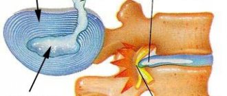
- lumbosacral - grow near the vertebrae or in the spinal canal;
- soft tissues - located on the surface of the skin, less often subcutaneous;
- joints - are located in the synovial membrane or vagina of the joints.
The formation can appear in almost any part of the body and internal organs. Depending on the location of the compaction , the following types of lipomas are most often diagnosed:
- mammary gland - forms in the glandular tissue and deforms the shape of the breast as it grows;
- breasts - a soft and mobile formation that appears in the subcutaneous fatty tissues;
- head - a frequently occurring pathology, which is mainly formed as a result of insufficient hygiene;
- back - one of the most common neoplasms, characterized by extremely slow development;
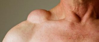
- neck is a hereditary disease that, during development, can impair the airways, cause weakness and angina.
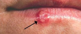
There are also other, less common places where pathology forms, which include the brain, limbs, skin, peritoneum, eyes, lips and face.
Also, these compactions are divided into two large groups: single and multiple. The first represent a single formation in any part of the body. The latter, accordingly, are characterized by multiple manifestations in different areas of the body and are much less common.
How are atheromas and lipomas treated?
Atheromas are treated surgically; the type of operation will depend on the size of the formation. Small sebaceous cysts can be removed with a laser. Most often, atheromas are large in size and are removed with a scalpel. The “bump” on the skin is surrounded by two incisions, then the cyst is peeled out and removed along with a small piece of skin. Stitches are placed on the wound.
Atheroma cannot be cured by “sucking out” the contents with a needle and syringe. It is imperative to remove the walls of the stretched sebaceous gland - if they remain, they will begin to produce sebum again, and the cyst will grow again.
If atheroma suppurates, surgical treatment is performed, and the doctor may prescribe a course of antibiotics. Lipomas do not need to be treated. The operation is performed if:
- education is large and growing rapidly;
- the lipoma is in an inconvenient place and is constantly in the way;
- bothered by soreness;
- The patient himself insists on removing the lipoma.
Wen is removed in the classic way, using a scalpel. Relapses are possible, but extremely rare. Typically, atheromas and lipomas are removed on an outpatient basis; hospitalization is not necessary. Anesthesia is also not needed - local anesthesia is sufficient. The operation lasts on average 15–20 minutes.
Make an appointment by phone +7 (495) 120-08-07.
Causes of lipomas
Experts identify a set of prerequisites that can provoke the formation of lipomas. Among the most common:
- genetic factors, hereditary predisposition;
- disruption of metabolic processes in the body and fatty tissues;
- insufficient level of personal hygiene;
- malfunctions of the thyroid and pancreas;
- chronic pathologies that significantly affect the body’s immune forces (diabetes, hepatitis, HIV, etc.);
- dependence on alcohol, tobacco, drugs;
- poor nutrition;
- obesity, excess fatty tissue;
- injuries, damage;
- sedentary lifestyle, lack of physical activity.
Despite the fact that lipoma is a benign formation, there is always a risk of developing its malignant form. This probability is especially high for people who are included in the relevant risk groups: they have precancerous diseases (polyps, dysplasia), have already suffered from oncological pathologies, have a hereditary predisposition to the formation of tumors, and are under the constant influence of carcinogens, radiation and other harmful environmental factors.
Services and prices
Removal of lipoma more than 3 cm
7000 ₽
Sign up
Removal of lipoma up to 3 cm
4000 ₽
Sign up
Lipoma is the most common soft tissue tumor and consists of fat cells surrounded by a thin fibrous capsule. Popularly, such a neoplasm is called a wen.
Often, a person who is attentive to his body may stumble upon such a subcutaneous formation. Such a finding can cause, at a minimum, caution, and sometimes fear of the oncological process. The unknown is always scary. If you have any concerns, you should consult your doctor. In most cases, subcutaneous neoplasms in soft tissues are benign and do not pose a threat to the owner of the wen. However, lipomas still require the supervision of a specialist who must recognize a malignant process, if one occurs.
Peculiar red flags, signs that make you wary, are rapid tumor growth, pain, and the presence of two or more similar formations on the body.
By external signs, lipoma is little distinguishable from liposarcoma, hygroma, subcutaneous cyst, hematoma, parasitic invasion, inflammation or consequences of injury. Therefore, it is important that any neoplasms be examined by a doctor.
First of all, the question of the malignancy of the tumor is decided at the appointment. Liposarcoma occurs more often in middle-aged and elderly people. This is an aggressive and fast-growing tumor that can put pressure on surrounding organs and tissues and cause pain. Situations when an existing lipoma degenerates into a malignant tumor rarely occur. However, the oncological process requires a radically different approach to diagnosis and treatment, so it is important to diagnose it as early as possible.
Lipomas may have a hereditary predisposition. This fact, combined with the spread of wen to other parts of the body, makes the doctor suspect lipomatosis. Lipomatosis accompanies a number of hereditary syndromes such as Madelung's disease and Dercum's syndrome. Diseases with a family history require a special approach to therapy.
Symptoms of lipomas
The clinical picture of the pathology is quite sparse and is characterized by:
- the presence of a palpable formation, which is distinguished by its soft consistency, mobility, and elasticity;
- the appearance of pain due to compression of nerve trunks or growth in internal organs;
- stability of the compact size or its increase with weight loss;
- swelling of the limbs and disruption of their function (if the formation compresses nerves and blood vessels).
If a malignant process develops, general malaise and headaches appear, blood pressure rises and other characteristic symptoms of intoxication of the body appear.
In this case, depending on the exact location of the compaction, other, more pronounced symptoms and signs may occur:
- neoplasms in the esophagus cause nausea and cough;
- seals on the trachea and bronchi cause a painful dry cough that does not subside after taking antitussive drugs;
- a fatty tumor on cartilage and tendons causes pain in the joints and impedes movement;
- formation in the mammary gland provokes pain in this area;
- compaction in the kidney area causes increased blood pressure, colic and lower back pain;
- a formation in the head causes neurological symptoms – headaches, dizziness;
- lipoma of the neck is accompanied by hoarseness, hoarseness, difficulty swallowing;
- a neoplasm in the heart area causes the development of cardiac pathologies: arrhythmia, heart failure, etc.
What is atheroma?
Atheroma is a cyst of the sebaceous gland that occurs when its duct is blocked. Sebum cannot come out and accumulates inside, it stretches the gland, and it turns into a cystic cavity. This can happen for a variety of reasons, often due to trauma to the skin.
Atheromas most often occur where there is a lot of hair and sebaceous glands: atheromas on the scalp, face, neck, back, genitals.
In its manifestations, atheroma resembles a lipoma: it is also a “bump” that is located under the skin, does not cause pain and easily moves when pressed. But there is one characteristic sign: usually there is a hole in the skin in the area of atheroma through which contents with an unpleasant odor are periodically released. Inside the atheroma there is a substance that resembles cottage cheese or paste in consistency.
Atheroma is a benign formation. It's not cancer. The only thing that is dangerous about it is the possibility of suppuration. An infected and inflamed “bump” swells, turns red, and becomes painful. Sometimes it opens and pus comes out along with fat.
Atheromas can degenerate into malignant tumors, but this happens very rarely.
Diagnosis of lipomas
Since the development of a neoplasm in the body practically does not cause symptoms, the patient may not be aware of its presence for a long time. Therefore, in most cases it is diagnosed accidentally during a preventive examination or treatment of other pathologies.
If the localization of the formation allows the doctor to palpate, the specialist can determine the lipoma even during a standard physical examination. However, the placement of a node does not always allow it to be detected by superficial palpation. At the same time, quite a few dangerous diseases have symptoms similar to lipoma. Therefore, diagnostics are carried out not only to identify a neoplasm, but also to exclude other diseases and clarify the benign quality of the process.
A comprehensive examination includes a number of laboratory and instrumental examinations, the need for which is determined individually by the attending physician in each medical case:
- Blood tests. The most accessible method of primary assessment of the body’s condition. The results of the study allow us to identify the presence of pathological changes, inflammation, viruses or bacteria.
- X-ray examination. Depending on the location of the compaction, an X-ray of the chest, abdominal cavity, and extremities is prescribed. Allows you to diagnose a formation, identify its exact location, and also analyze the condition of bone tissue and structures.
- Ultrasonography. Scanning soft tissues and organs allows you to determine the size of the node, identify the clarity of its contours, and analyze the contents. Not the most informative examination method for suspected lipoma, since even in the presence of a capsule, the compaction is often difficult to visualize using ultrasound waves.
- CT scan. Allows you to establish an accurate diagnosis and distinguish lipoma from malignant neoplasms if there is suspicion.
- Magnetic resonance imaging. It is prescribed, if necessary, to evaluate the signs of compaction and distinguish it from malignant liposarcoma. Using this method, the diagnosis is established with maximum accuracy.
- Biopsy. Tissue collection from the compaction and their further cytological and histological analysis. Allows you to exclude the possible oncological nature of the pathology.
The examination also reveals the reasons that caused the formation of a compaction in the body. If other chronic diseases are a prerequisite for the development of pathology, additional diagnostics are also carried out for an accurate diagnosis and further effective treatment.
Lipoma treatment
The only medical treatment for lipoma is surgical removal. Clinic specialists prescribe surgery if the tumor:
- grows rapidly, involving surrounding tissues and organs in the pathological process;
- affects appearance, causes aesthetic defects;
- causes pain;
- disrupts the functioning of internal organs.
Only surgical intervention can avoid future complications and prevent the transformation of pathology into malignancy.
Depending on the characteristics of the tumor and taking into account the individual characteristics of the body, SM-Clinic doctors choose one of the most effective methods for removing the lump:
- endoscopic method - the advantage is a small incision, but relapses are possible;
- excision of the lipoma – the likelihood of relapse is almost completely absent;
- liposuction is a gentle method with a good cosmetic effect and very frequent recurrence.
SM-Clinic specialists initially conduct a comprehensive examination, after which they establish an accurate diagnosis and provide consultation regarding surgical excision of the tumor. Before removing a lipoma, specialists must provide detailed information about the possible risks of the operation, as well as the consequences of non-intervention. Interventions are performed exclusively by experienced surgeons with many years of experience.
Doctors warn that, despite the apparent harmlessness of the pathology and its benign course, it is not worth delaying its treatment. It is important to remember that along with the increase in neoplasm, the risk of developing malignant processes and the occurrence of concomitant pathologies of internal organs significantly increases.
Sources:
- Congenital lipomas of the brain and spinal cord: clinical and MRI diagnostics. Bein B.N., Syrchin E.F., Yakushev K.B. Medical almanac, 2013
- Accidental detection of cardiac lipoma. Alekseeva I.V., Gordova V.S. Bulletin of the Baltic Federal University. I. Kant. Series: Natural and medical sciences, 2021.
- A rare congenital anomaly: a cerebral lipoma connecting to a subcutaneous lipoma through a defect in the frontal bone. Miloserdov M.A., Korneva Yu.S., Gelt T.D., Rudenko Y.A. Difficult patient, 2021.
The information in this article is provided for reference purposes and does not replace advice from a qualified professional. Don't self-medicate! At the first signs of illness, you should consult a doctor.
Consequences of lipoma removal surgery
Complications after removal of a wen are extremely rare. And yet they happen. For example, bleeding. This is least common if the tumor was removed with a scalpel. After removal of large lipomas, the cavity remaining in their place can be filled with serous fluid and a small amount of blood. Hyperthermia for several days accompanied by the appearance of edema at the intervention site. Then you should visit a doctor, as inflammation may occur. You should consult a doctor if the wound begins to fester, which may be due to infection. You should also be wary of loss of sensitivity at the site of the former tumor.
Nothing like this will happen if the operation is performed by an experienced surgeon, and the patient does not skimp on the recommendations prescribed to him.
Author: Ekaterina Solovyova
Please note that the information presented on the site is for informational and educational purposes only and is not intended for self-diagnosis and self-medication. The selection and prescription of medications, treatment methods, as well as monitoring their use can only be carried out by the attending physician. Be sure to consult a specialist.