The inner ear is located deep in the temporal bone and is responsible for perceiving sound signals, maintaining balance and identifying the body in space. Symptoms of inner ear diseases are not always specific, and it is difficult to make an accurate diagnosis.
To understand whether the inner ear or another organ is the source of the malaise, a thorough examination of the patient should be performed.
The best method for making an accurate diagnosis is an MRI of the ear.
Advantages of the method:
- high image quality, the possibility of detailed study of the structure of the organ;
- health safety, no radiation exposure;
- no damaging manipulations are performed during the procedure;
- Due to layer-by-layer scanning, there is no effect of overlapping images on each other.
MRI clearly shows the soft tissues located in the bony labyrinth of the temporal bone pyramid. The inner ear consists of the vestibule (a small cavity bordering the middle ear), from which the cochlea extends anteriorly, and the semicircular canals of the vestibular apparatus extend posteriorly. With the help of the inner ear, a person not only perceives sounds, but also orients himself in space, instantly determining the bottom and top with any movements of the head and torso.
What can you see on magnetic resonance imaging of the ears?
What does a magnetic resonance imaging scan of the ears show:
- ear structure;
- anatomical features;
- foci of inflammation, tumors;
- condition of the vestibulocochlear (VIII pair of cranial nerves) and facial (VII pair) nerves;
- neighboring structures of the head - eyes, temporal bones, cerebellopontine angle, blood vessels.
MRI of the inner ear allows you to detect minor changes, the initial forms of diseases. Such information makes it possible to carry out timely treatment and stop the disease at the very beginning of its development.
MRI of the ear with contrast allows you to look at the inner ear in more detail and assess the extent of its damage. The method is based on the ability of the metal gadolinium, which is part of contrast agents, to give an enhanced signal from the tissues in which it accumulates. The inflammatory focus has a stronger blood supply than the healthy ear, so it is clearly visible. MRI of the ear with contrast clearly shows areas of insufficient blood flow and tumors.
When is an MRI of the inner ear prescribed?
The examination is carried out if there are symptoms that indicate damage to the ears or surrounding tissues, so not only the ear, but also the brain is often checked. MRI of the ear allows you to confirm or exclude its participation in the formation of symptoms and conduct an accurate diagnosis.
Symptoms for which an MRI of the inner ear is indicated:
- progressive hearing loss, partial or complete loss;
- congestion, tinnitus;
- headache;
- dizziness at rest and when changing position, accompanied by nausea, vomiting, headache;
- distortion of sound perception;
- unstable balance at rest and when walking;
- head injury;
- signs of damage to the facial nerve - asymmetrical facial expressions, immobility and weakness of one half of the face;
- elevated body temperature of unknown origin;
- suspected tumor of the ears or surrounding structures.
The symptoms that form the basis for indications for MRI of the ear may be similar in diseases of the brain, blood vessels, or the ears themselves. An MRI will show the cause of the ailment with high accuracy and allow you to begin the correct treatment. The examination allows us to identify the initial manifestations of pathology, which increases the success of treatment and allows you to preserve your hearing.
An important indication for MRI of the inner ear is preparation for surgery. Depending on what the image shows, the surgeon decides on how to access the inner ear and draws up a surgical plan. In the postoperative period, the MRI method is most optimal for monitoring the stages of healing and the condition of the ears and implant.
Research methods
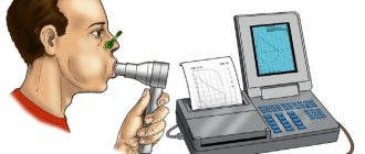
Modern methods of studying the function of V. at. are very complex and consist in determining the state of both of its functions - auditory and vestibular. When studying auditory function, an adequate stimulus is used - sound of varying frequency and intensity in the form of pure tones, noise and speech signals. Tuning forks, audiometers (tone and speech), whispered and loud speech are used as a sound source. This set of studies makes it possible to determine the state of the function of the sound-conducting system, the receptor apparatus of the auditory system, as well as the conductive and central sections of the auditory analyzer (see Audiometry).
When studying the function of the vestibular apparatus, the presence of balance disorders, spontaneous vestibular reactions (see), as well as the occurrence of motor and autonomic reflexes in response to various irritations of the vestibular apparatus (rotational, caloric, galvanic, pressor and other tests) are determined. The use of special equipment makes it possible to objectively assess the state of the function of the vestibular apparatus with a high degree of accuracy (see Vestibulometry).
What does an MRI scan of the ears show?
Good quality images allow you to thoroughly study the structure of the anatomical structures of the area under study, note pathologies and developmental anomalies, and determine the stage and extent of the lesion.
MRI of the inner ear helps in diagnosing the following diseases and pathological conditions:
- neuritis of the vestibulocochlear (auditory), facial nerve;
- inflammation of the inner ear (labyrinthitis) in acute or chronic form;
- acoustic neuroma;
- congenital and acquired deformations of ear structures;
- Meniere's disease;
- meningioma of the cerebellopontine angle;
- otosclerosis of the bone labyrinth;
- vascular pathologies of the temporal region;
- complications of purulent labyrinthitis - meningitis, encephalitis, abscess;
- traumatic injuries to the ears and the area next to them.
Since the examination does not expose the body to radiation, it can be performed an unlimited number of times. Treatment of diseases is much more effective if it is possible to control the body’s reaction and, if necessary, make adjustments or change tactics.
Neuroma of the vestibulocochlear nerve
The most common benign tumor of the cerebellopontine angle, its development may be due to a hereditary predisposition. Acoustic neuroma develops from Schwann cells surrounding the nerve fiber and is an irregularly shaped formation surrounded by a capsule. The disease practically does not become malignant, but as it increases in size, the tumor affects the inner ear and brain, causing clinical manifestations.
The disease is characterized by decreased hearing, impaired balance, tinnitus, headache, and numbness of the face. Less commonly observed are pain in the ears, paresis of the facial nerve, double vision, and involuntary frequent eye movements (nystagmus). Most often, symptoms are observed on one side.
In most cases, acoustic neuroma grows slowly, and symptoms do not always appear clearly. MRI of the auditory canal allows you to detect a tumor and begin its treatment, preserve hearing and avoid compression of brain structures and intracranial hypertension.
Labyrinthitis (internal otitis)
Inflammation of the inner ear occurs as a result of infection entering from the middle ear or from other sources through the circulatory system. Infection can get into the ears due to injury to the eardrum or temporal bone. By nature, labyrinthitis can be serous or purulent, acute or chronic.
With the development of internal otitis, hearing decreases (impairments can be irreversible, especially with purulent labyrinthitis), attacks of dizziness with nausea and vomiting, nystagmus occur, balance and coordination of movements are impaired. Internal otitis is dangerous due to complications when the infection spreads to the brain and meninges with the formation of an abscess and meningitis.
Severe forms of internal otitis may lead to removal of the labyrinth on the affected side and complete hearing loss. If symptoms of damage to the auditory analyzer appear, an MRI of the ear should be performed - this will identify otitis media and stop its spread.
Meniere's disease
The disease is characterized by attacks of dizziness in combination with tinnitus and progressive hearing loss. The symptoms are caused by increased pressure in the inner ear due to an increased amount of endolymph, the fluid that fills the labyrinth cavity. The causes of Meniere's disease are unknown.
The disease is characterized by three main symptoms:
- sudden dizziness, the attack lasts 4-5 hours, accompanied by nausea and vomiting;
- unilateral low-frequency noise in the ear, intensifies during dizziness;
- periodic unilateral hearing loss.
An MRI of the inner ear in Meniere's disease is performed primarily to exclude other diseases that may present with similar symptoms.
Sensorineural hearing loss
The disease occurs when the ears are affected or is a consequence of other diseases. In this case, there is a loss of the sound-perceiving function of the nervous apparatus of the ear with a gradual decrease in hearing acuity until it is completely lost.
The causes of hearing loss may be the following:
- infectious diseases - influenza, measles, tick-borne encephalitis, mumps, meningitis, syphilis;
- acute intoxication with ototoxic substances at home and at work;
- taking certain antibiotics, anti-inflammatory drugs, diuretics, antidepressants;
- cerebrovascular accident due to arterial hypertension, atherosclerosis;
- genetic predisposition;
- long-term exposure to industrial noise exceeding maximum permissible levels.
MRI of the ear canal will allow us to establish the true cause of hearing loss and make a differential diagnosis. An examination is necessary to exclude or confirm ear diseases as a cause of hearing loss.
Histology
The histological structure of the wall of the membranous labyrinth is relatively simple. The wall of the vestibular section of the membranous labyrinth is lined with flat single-layer epithelium. This epithelium in the area of ampullary ridges turns into cubic and cylindrical, located on the basement membrane, to which the connective tissue is adjacent to the outside. The network of tense perilymphatic bridges consists of connective tissue fibers that penetrate on one side into the Endoste, lining the walls of the bone labyrinth, and on the other into the connective tissue membrane of the membranous walls. Blood vessels pass through these bridges.
In their structure, the terminal nervous apparatus of the vestibular department is similar to each other. The receptor apparatus maculae sacculi et utriculi, located in the form of somewhat elevated spots, is also called the otolith apparatus (see). On these spots, the epithelial cover consists of supporting cells - sustentites (cellulae sustentantes), which are not related to the transmission of irritation, and hair (sensory epithelial) cells (cellulae pilosae), braided with nerve fibers and not reaching the basement membrane with their lower ends. Cell processes, intertwining with each other at the top, form a thin fibrous network located parallel to the upper surface of the epithelium and with its ends passing directly into the otolithic membrane (membrana statoconiorum). The latter consists of fibers, grains and numerous hexagonal crystals formed by impregnation of the protein skeleton with calcium and magnesium bicarbonate salts - otoliths, or statoconia. The space between the upper surface of the epithelium and the otolithic membrane is filled with a network of hairs and impregnated with a liquid mass.
Hair cells are divided into two types based on their ultramicroscopic structure. Cells of the first type have a rounded, wide base, adjacent to which are nerve endings that form a cup-shaped case around it. On their outer surface there is a cuticle. 60-80 immobile hairs (steriocilia) approx. long emerge from it. 40 microns and one mobile kinocilium, the edges contain 9 peripheral and 2 central fibrils, starting from the basal bodies. The kinocilium is always located polar to the bundle of steriocilia. In the cytoplasm of cells lie mitochondria and membranes of the cytoplasmic reticulum, forming cisterns. Ribosomes lie on the surface of the membranes. Cells of the second type are cylindrical in shape and differ little in structure from cells of the first type, but are less well equipped with nerve endings.
Ultramicroscopic structure of the receptor epithelium of the ampullar comb: I - hair cell of the second type; II - supporting cell; III - hair cell of the first type; 1 - hairs of hair cells; 2 - granules in the supporting cell; 3 - microvilli of the supporting cell; 4 - nerve endings that look like a bowl; 5 and 7 - pulpy nerve fibers; b — nucleus of the supporting cell; 8 - basement membrane; 9 - intracellular mesh apparatus; 10 - hair cell mitochondria.
The nervous apparatus of the ampullae of the semicircular canals is somewhat different from that of the vestibule sacs. Crista ampullaris, in comparison with the macula, rises strongly above its base in the form of a narrow truncated cone protruding into the lumen of the ampulla. The cone is covered with hair (sensory-epithelial) cells, above which there are jelly-like formations - cupula, as if planted on the hairs of the epithelium. In the scallops, the sensory hairs extend straight up from their cells and only deviate slightly at the edges and penetrate into the cupula covering them, distributing them quite evenly. There is no otolithic membrane. The fine structure of the hair cells of the ampullar combs (Fig.) and their innervation are almost the same as the cells of the spots.
The histological structure of the walls of the membranous canal of the cochlea is quite complex. The vestibular membrane is most simply structured, consisting of connective tissue covered with single-layer squamous epithelium facing the endolymph, endothelium facing the perilymph. The outer wall of the ductus cochlearis is fused with the spiral ligament. Capillaries penetrate into it from the vascular network embedded in the spiral ligament, which form a significant thickening - the stria vascularis. The most complex structure is the lower wall with the nervous epithelium - the spiral organ - a receptor for auditory stimuli (see Organ of Corti).
Contraindications for magnetic resonance imaging
Obtaining MRI images is possible due to the interaction of a high-power electromagnetic field with the structures of the body. If you place objects that have ferromagnetic properties in a magnetic field, they will react in a certain way. The special magnetic susceptibility of ferromagnets forms the basis of contraindications for MRI.
The examination is prohibited for people with implants containing a magnet and prostheses made of iron, cobalt, nickel, and steel. In a magnetic field, objects implanted into the body are displaced and cause pain and internal injuries. Implants can fail and distort the image.
MRI of the ear is not performed on patients who have inside the body:
- pacemaker;
- insulin pump;
- cochlear implant;
- hemostatic vascular clips;
- metal fragments after injury.
Modern prosthetics and implants can be made from MRI-compatible materials. If there are contraindications, it is necessary to study their characteristics to determine the feasibility of the procedure.
There are relative contraindications that, under certain conditions, allow examination. These include pregnancy in the first trimester, serious condition of the patient, age under 7 years, claustrophobia, mental illness, epilepsy.
MRI of the ear with contrast is a safe procedure, but it is contraindicated in people with severe renal failure and at any stage of pregnancy. Breastfeeding mothers can be administered a contrast agent, provided that feeding is stopped for 1-2 days - until the drug is completely eliminated from the body.
Hearing loss
Hearing loss can have several causes: traumatic brain injury, complications after previous infectious diseases, aging of the body, inflammatory processes of the middle ear, genetic predisposition, autoimmune reactions, work related to working in conditions of increased noise. It is quite difficult to track the process of decrease in sound perception at an early stage. It becomes clear that there are certain problems only when hearing defects become too obvious.
Most people with hearing loss do not seek help for an average of 5-7 years. But it's not right. At first, if your hearing is impaired, you have to constantly ask again, turn on the TV or radio at full volume, which quite annoys those around you. With further hearing loss, and no longer able to understand speech, these people try to avoid communication - which leads to limited social skills and loss of incentive to live. Moreover, the lack of new information leads to involution of brain tissue. With hearing loss, there is no constant stimulation of the brain neurons responsible for hearing, and they “atrophy” over time.
Remember paralyzed people who cannot move without a wheelchair. After some time, the muscles of their legs become thinner, and destruction processes begin in the bones. The same thing happens with the part of the brain responsible for hearing. Our body lives according to the laws of nature. If something in the body is not used, then local blood circulation decreases, metabolic processes slow down, and self-destruction mechanisms are subsequently activated.
In 90% of cases, the cause of hearing loss is the death of the hair cells of the cochlea, and, unfortunately, if this has already happened, they are no longer restored. This means that the brain will not receive all the sound information. It's like a piano without several keys: you won't be able to play the entire composition, even if only 6-7 keys are missing - the melody will be unrecognizable.
To prevent hearing loss from becoming irreversible, it is necessary to diagnose hearing loss as early as possible. In modern medicine, there are specialists who deal with hearing problems - audiologists.
Preparing and conducting a study using a magnetic resonance imaging scanner
At the appointment, it is advisable to take the results of all previous tests and studies related to the current state of health, as well as advisory opinions and extracts from medical records. No special preparations are required.
If an MRI of the ear with contrast is performed, a renal function test should be performed first to rule out renal failure. Women should be aware of the possibility of pregnancy - contrast agents have a negative effect on the developing fetus. A contrast agent should not be administered if an allergic reaction to its components has previously been documented. The examination is carried out on an empty stomach.
Metal objects and magnetic devices must not be brought into the room where the research is being conducted. The patient changes into light disposable clothing, having previously removed jewelry, watches, belts, removable dentures, clothing with metal parts and leaving a mobile phone, money, and cards outside the office. The subject is positioned on a table that slides into the body of the tomograph. Most devices are not designed for body weights exceeding 130 kg. When performing an MRI of the ear, a special coil is placed on the area under study to read the signal. The procedure lasts about 20 minutes.
If an MRI of the ear canal is performed using a contrast agent, the drug is administered intravenously after the first scan. After a few seconds, you can rescan. The contrast agent rarely causes side effects or allergic reactions. You may feel warmth or tingling at the injection site.
The examination can be stopped at any time at the request of the patient if he becomes ill during the procedure. People suffering from mild claustrophobia are recommended to take sedatives half an hour before the test. In some cases, a loved one may be present during the procedure.
Physiology
In V. u. receptors of the auditory and statokinetic analyzers are located.
The receptor (sound-perceiving) apparatus of the auditory analyzer (see) is located in the cochlea and is represented by hair (sensory-epithelial) cells of the spiral (Corti) organ. The cochlea and the receptor apparatus of the auditory analyzer contained in it are called the cochlear apparatus. Sound vibrations arising in the air are transmitted through the external auditory canal, the eardrum and the chain of auditory ossicles to the vestibular window of the labyrinth, causing wave-like movements of the peri-lymph, which, spreading, are transmitted to the spiral organ (see Hearing). These fluid movements are possible due to the presence of the membrane of the cochlear window, the edges with each push of the stapes and the corresponding movement of the perilymph protrude towards the tympanic cavity. Transfer of vibrations from the environment to liquid media V. at. occurs directly through the bones of the skull (bone sound conduction). In the receptor cells of the spiral organ, the physical energy of sound vibrations is converted into the energy of nervous excitation - nerve impulses arriving through the conductive section of the auditory analyzer to its cortical section. With the help of electrophysiology, research has established that with sound stimulation, electrical potentials arise in the cochlea - cochlear currents, which in frequency and shape of vibrations correspond to the sound vibrations entering the ear. Cochlear currents, after amplification, can be transformed again into sound vibrations using a telephone and exactly repeat the sound entering the ear. This phenomenon, called the cochlear microphone effect, or the Weaver-Bray phenomenon, reflects the function of the receptor apparatus of the cochlea. The cochlear current curve recorded on an oscilloscope (cochleogram) also allows one to judge the integrity of the cochlear receptor apparatus.
The receptor apparatus of the statokinetic analyzer (see Vestibular analyzer), located in the semicircular canals and sacs of the vestibule, is called the vestibular apparatus. Receptors located in the semicircular canals perceive angular accelerations that occur when turning the head or rotational movements of the whole body, and receptors in the vestibule respond to linear accelerations.
What does the result of magnetic resonance imaging of the ears show?
Preparing a report can take from one hour to a day. MRI images of the inner ear and surrounding brain structures are analyzed by a radiologist. The patient receives a description of everything that the MRI shows and a conclusion. The photographs can be received electronically and the most significant images can be additionally printed on film.
A healthy MRI of the ear is characterized by:
- absence of foci of increased or decreased intensity;
- the ventricles of the brain are not displaced;
- the internal auditory canals on both sides are of a normal shape, not expanded;
- the course of the nerves (vestibulocochlear, facial) is not disturbed, the diameter is within normal limits;
- the configuration of the cochlea and semicircular canals is typical;
- the cerebellopontine angles on both sides are unremarkable;
- The pyramids of the temporal bones are not changed.
The administration of a contrast agent should not be accompanied by pathological accumulation of the drug in any areas of the inner ear.
What research shows in some diseases:
- meningioma looks like a volumetric formation of irregular shape with clear, uneven edges, the structure of the formation is heterogeneous, cysts may appear inside, there is an area of edema around it, the ventricles of the brain are deformed by the tumor;
- neuroma of the vestibulocochlear nerve is characterized by the appearance of a space-occupying formation that extends into the internal auditory canal, the cavity of the fourth ventricle of the brain is deformed;
- Meniere's disease may appear on magnetic resonance imaging as the absence of the endolymphatic duct.
Depending on what the study shows, the patient may need additional consultation or an appointment with a specialist doctor. This could be an oncologist, surgeon, neurologist, otorhinolaryngologist or audiologist - a doctor who treats hearing disorders and specific lesions of the auditory analyzer.
Anatomy
Rice.
2. Section of the cochlea: 1 - ganglion spirale cochleae;
2 - scala vestibuli; 3 - ductus cochlearis; 4 - scala tympani; 5 —pars cochlearis n. vestibulocochlearis; 6 - modiolus; 7 - organon spirale. Rice.
3. Cross section of the cochlea coil: 1 - membrana vestibularis; 2 - ductus cochlearis; 3 - membrana tectoria; 4 - stria vascularis; 5—cellulae phalangae ext.; 6 - membrana basilaris with a spiral organ located on it; 7 - cellula pilaris ext.; 8—cellula pilaris int.; 9 - pars cochlearis n. vestibulocochlearis; 10 - scala tympani; 11 - lamina spiralis secundaria; 12 - ganglion spirale. Rice. 4. The right bony labyrinth (opened) and the membranous labyrinth contained within it: 1 - saccus endolymphaticus; 2 -ductus endolymphaticus; 3 - ductus semicircularis post.; 4 - crus membranaceum commune; 5 - n. ampullaris post.; 6 - ductus reuniens; 7 - v. labyrinthi; 8 - n. sacculars; 9 - ductus cochlearis; 10 - a. labyrinthi; 11 - pars cochlearis n. vestibulocochlearis; 12 - pars vestibularis n. acustici; 13 - sacculus; 14 - ductus utriculosaccularis; 15 - utriculus; 16 - ductus semicircularis ant.; 17 - dyctus semicircularis lat.
The inner ear, or ear labyrinth (tsvetn. fig. 2-4), is located in the thickness of the stony part (pars petrosa) of the temporal bone and consists of a system of bone canals communicating with each other - the bone labyrinth (labyrinthus osseus), in which the the membranous labyrinth (labyrinthus membranaceus) is strengthened. The outlines of the bony labyrinth almost completely repeat the outlines of the membranous labyrinth, being, as it were, its capsule. The membranous labyrinth is a closed system of canals in which the terminal devices of the vestibulocochlear nerve are located (n. vestibulocochlear is). The space between the bony and membranous labyrinth, called the perilymphatic labyrinth, is filled with a fluid - perilymph, the composition of which is similar to the composition of the cerebrospinal fluid. The membranous labyrinth is, as it were, immersed in the perilymph, it is movably strengthened in its bone case with the help of a number of connective tissue strands and is filled with a liquid - endolymph, which is somewhat different in composition from the perilymph. The perilymphatic space (spatium perilymphaticum) is connected to the subarachnoid narrow bony canal called the cochlear aqueduct (aquaeductus cochleae, s. ductus perilymphaticus). The endolymphatic space does not have such communication with the subarachnoid space. From the endolymphatic space, a very narrow passage - the vestibular aqueduct (aquaeductus vestibuli, s. ductus endolymphaticus) - leads into a small closed reservoir - the endolymphatic sac (saccus endolymphaticus), embedded in the thickness of the dura mater on the posterior surface of the pyramid.
The bony labyrinth consists of three sections: the vestibule (vestibulum), semicircular canals (canales semicirculares ossei) and cochlea (cochlea).
The vestibule forms the central part of the labyrinth. Posteriorly and outwardly it passes into the semicircular canals, and anteriorly and inwardly into the cochlea. The inner wall of the vestibule cavity faces the posterior cranial fossa and forms the bottom of the internal auditory canal. Its surface is divided by a small bone ridge into two parts, one of which, the anterior-inferior one, is called a spherical recess (recessus sphaericus), and the other is an elliptical recess (recessus ellipticus). In the spherical recess there is a membranous spherical sac - sacculus, in the elliptical one - an elliptical sac - the utricle (utriculus), into which the semicircular canals flow at their ends. In the middle wall of both recesses there are groups of small holes that form flat elevations on the surface in the form of lattice spots - maculae cribrosae. They are intended for branches of the vestibular part (pars vestibularis) of the nerve. The outer wall of the vestibule faces the tympanic cavity and is mostly occupied by the window of the vestibule (fenestra vestibuli). The semicircular canals are located in three planes almost perpendicular to each other. One of the ends of each canal is expanded and is called the ampullary leg (crus osseum ampullare), the other - a simple leg (crus osseum simplex). According to their location in the bone, they are distinguished: upper - frontal, or anterior (canalis semicircularis ant.), posterior - sagittal (canalis semicircularis post.) and lateral - horizontal (canalis semicircularis lat.) canals. Both ends of each semicircular canal lead into the vestibule, only two simple legs of the posterior and superior canals are connected to each other, forming a common leg (crus osseum commune), and communicate with the vestibule through one common opening.
The bony cochlea is a convoluted canal extending from the vestibule; it spirals around its horizontal axis 2/2 times and gradually narrows towards the apex. The central bony core is called modiolus. A narrow bone plate spirals around the rod, to which a connective tissue membrane called the basement membrane (membrana basilaris) is firmly attached, forming its direct continuation. In addition, a thin connective tissue membrane, the vestibular membrane (membrana vestibularis), also called Reissner's membrane, extends from the bony spiral plate (lamina spiralis ossea) at an acute angle upward and laterally. The space formed between the basal and vestibular membranes is called the cochlear duct (ductus cochlearis), it is filled with endolymph. Above and below it are the perilymphatic spaces, forming two floors. The lower floor is called the scala tympani (scala tympani), the upper floor is called the staircase vestibule (scala vestibuli). The scalae at the top of the cochlea are connected to each other by a cochlear opening called the helicotrema. The cochlear shaft is pierced by longitudinal canaliculi (canales longitudinales modioli) for the passage of nerve fibers. Along the periphery of the rod, its winding canal (canalis spiralis cochleae) stretches spirally; nerve cells are placed in it, which form the spiral ganglion of the cochlea - ganglion spirale cochleae. The internal auditory canal (meatus acusticus internus) leads into the bone labyrinth from the skull, into which the vestibulocochlear and facial nerves pass. The cranial opening of the canal is located on the posterior surface of the pyramid, and the internal one ends with a bone plate, the edges are called the bottom of the internal auditory canal (fundus meatus acustici interni) and forms part of the medial wall of the vestibule and cochlea.
The membranous labyrinth consists of two vestibular sacs, three semicircular canals, the cochlear duct, the aqueducts of the vestibule and the cochlea. All these sections of the membranous labyrinth represent a system of formations communicating with each other. The elliptical sac is located in the upper part of the vestibule; it is connected to the medial wall of the vestibule by connective tissue bundles and fibers of the n. utricularis passing through the superior ethmoidal spot. Accordingly, the inner surface of the lower wall of the sac has an elevation formed by the sensitive epithelium, and is called the spot of the elliptical sac (macula utriculi). Three ampullary and two simple legs of the semicircular canals lead into the elliptical sac. The sacculus has the shape of a flat-convex lens and in the lower part it passes into the duct (ductus reuniens), connecting it with the cochlear duct (ductus cochlearis). The sacculus and utriculus communicate with each other by the endolymphatic duct (ductus endolymphaticus).
In the wall of the membranous labyrinth, the fibers of the vestibulocochlear nerve terminate in certain places. Three of them are located in ampoules and are called ampullary combs (cristae ampullares), two are in sacs and are called spots (maculae sacculi et utriculi), the last is the entire terminal nervous apparatus of the cochlea, known as the organ of Corti (organon spirale) .
The membranous cochlea is a spirally convoluted duct of triangular cross-section.
Arteries
the inner ear originates from the labyrinthine artery (a. labyrinthi), the edges depart from the basilar artery (a. basilaris). The venous blood of the labyrinth collects in the plexus lying in the internal auditory canal. Venous blood flows from the vestibule and semicircular canals. arr. through the vein passing through the aqueduct of the vestibule into the transverse sinus of the dura mater. The veins of the cochlea carry blood to the inferior petrosal sinus. The inner ear receives innervation from the VIII pair of cranial nerves, each of which, upon entering the internal auditory canal, splits into 3 branches: upper, middle and lower. The upper and middle branches form the nerve of the vestibule - n. vestibularis, the lower corresponds to the nerve of the cochlea - n. cochlearis (see vestibulocochlear nerve).
Where can I get an MRI of the ears?
Choosing a clinical diagnostic center for magnetic resonance imaging of the ears is not easy to do. Our website contains information about 18 clinics in Moscow where you can undergo an examination of the inner ear at the best prices. Any patient can independently compare and choose a medical center or call by phone to receive a free consultation and make an appointment at a convenient time at any of the proposed clinics.
On the website you can find the following information:
- contact details of the clinic (address, metro);
- detailed price list for services;
- type of equipment used (closed or open tomograph);
- operating hours of the institution;
- availability of free places for registration;
- special offers and promotions;
- possibility of undergoing tomography for children;
- patient reviews.
The average price for magnetic resonance imaging of the ears is 4,400 rubles. The price range is between 3,600-10,500 rubles. When you make an appointment through our website for your first appointment, you can get a discount of up to 1,000 rubles.
Literature
- Jackler RK Congenital malformations of the inner ear: a classification based on embryogenesis//RK Jackler, WM Luxford, WF House/ Laryngoscope. – 1987. – Vol. 97, no. 1. – P. 1 – 14.
- Jackler RK The large vestibular aqueduct syndrome//RK Jackler, A. De La Cruz/ Laryngoscope. – 1989. – Vol. 99, No. 10. – P. 1238 – 1243.
- Marangos N. Dysplasien des Innenohres und inneren Gehörganges//N. Marangos/HNO. – 2002. – Vol. 50, no. 9. — P. 866 – 881.
- Sennaroglu L. A new classification for cochleovestibular malformations//L. Sennaroglu, I. Saatci/Laryngoscope. – 2002. – Vol. 112, No. 12. – P. 2230 – 2241.
- Siebenmann F. Grundzüge der Anatomie und Pathogenese der Taubstummheit// F. Siebenmann/Wiesbaden: JF Bergmann; 1904. – 76s.
- Stellenwert der MRT bei Verdacht auf Innenohrmissbildung//S. Kösling, S. Jüttemann, B. Amaya et al. / Fortschr Röntgenstr. – 2003. – Vol. 175, No. 11. – S. 1639 – 1646.
- Terrahe K. Missbildungen des Innen- und Mittelohres als Folge der halidomidembryopathie: Ergebnisse von Röntgenschichtuntersuchungen//K. Terrahe/Fortschr Röntgenstr. – 1965. – Vol. 102, No. 1. – P. 14.
The head of the department for the development and implementation of high-tech treatment methods is Doctor of Medical Sciences, Professor Anikin Igor Anatolyevich. Clinic phone number
Commission for the selection of patients for the provision of high-tech medical care Secretary of the commission - candidate of medical sciences Vladislav Evgenievich Kuzovkov, phone
Schedule of consultation appointments with clinic staff


