Urolithiasis
Urolithiasis
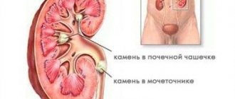
(urolithiasis) is a disease characterized by the formation of stones in the urinary organs. Stones can be found in all organs of the urinary system, however, they most often occur in the kidneys and bladder. Also a serious problem are stones in the ureter, where they enter after descending from the kidneys. Urolithiasis can develop at any age, but most cases of its initial detection occur between the ages of 20-55 years.
Causes of urolithiasis
The appearance of stones indicates metabolic disorders, as a result of which insoluble salts begin to accumulate in the body. It is believed that the tendency to urolithiasis is congenital, but stones will not appear without the influence of one of the additional factors, including:
- hot climate, provoking active sweating;
- composition of drinking water (hard water promotes stone formation);
- addiction to spicy, salty and sour foods, excess calcium and animal protein in food;
- lack of vitamins (A and group B), ultraviolet radiation (life “without the sun”);
- severe dehydration, for example as a result of infectious diseases;
- sedentary lifestyle, including forced inactivity as a result of injuries;
- chronic diseases of the stomach and intestines (gastritis, colitis, peptic ulcer);
- diseases of the kidneys and other organs of the genitourinary system (pyelonephritis, cystitis, prostate adenoma, prostatitis);
- metabolic diseases (for example, gout).
Medicines and substances that increase risk
- Acetazolamide
- Efilrin
- Phosphate-binding anatcides
- Uricosuric substances
- Calcium/Vitamin D
- Guaifenesin
- Topiramate
- Triamterene
- Vitamin C
In modern medicine there is no remedy that cures urolithiasis. After pain relief and stone removal, the only goal is to prevent relapse
The process of stone formation can be controlled. Proper nutrition and plenty of water in the diet will reduce the likelihood of recurrence.
Symptoms of urolithiasis
The main symptom is pain, the localization of which depends on the organ in which the stones are located. The hard body of the stone injures the organ, especially when moving.
Symptoms of kidney stones
Kidney stones manifest themselves as pain in the lumbar region on one or both sides (depending on whether there are stones in one or both kidneys). As a rule, the pain is dull, aching in nature. Pain sensations change with movement or change in body position. Blood may be found in the urine after an attack of severe pain, walking or physical activity. Stones may also pass (stones are carried away by a stream of urine).
If the kidney stone is small, there may be no obvious symptoms.
Symptoms of stones in the ureters
Stones in the ureter cause pain, the location of which changes as the stone moves: the pain moves from the lower back to the groin, lower abdomen or genitals.
The stone can completely block the lumen of the ureter. In this case, urine accumulates in the kidney, causing severe pain ( renal colic
). The pain can last for hours (up to several days), and only stops when the stone passes from the ureter into the bladder. In case of renal colic, emergency medical care is required (you must call 03).
A stone in the lower ureter may cause frequent urination.
Symptoms of bladder stones
The presence of stones in the bladder is manifested by pain in the lower abdomen, radiating to the perineum or genitals, primarily during movement or urination. Sudden movements can also trigger the urge to urinate (a symptom of frequent urination). During urination, the stream of urine may be interrupted and resumed only when the stone moves as a result of a change in body position.
Life story...
As an introduction, here is a real incident that happened several years ago...
The ambulance was flying through the city at night, cutting through the darkness with its headlights. An ordinary challenge that doesn’t stand out in any way from a series of hundreds of others. Male, forty-two years old, acute pain in the lumbar region. The doctor looked at the call sheet, weighing a possible diagnosis in his head. The car ran its headlights along the wall of the house, turning into the yard. In the faint light of the lantern one could see a well-groomed courtyard with a children's swing and a flowerbed. The ambulance stopped at the right entrance. Grabbing his suitcase, the doctor hurriedly ran up the steps and dialed the number on the intercom. The elevator took him to the sixth floor, where a pretty, fragile woman was already waiting for him, chillyly wrapped in a coat thrown over her shoulders.
The doctor entered the apartment, hastily washed his hands and, wiping them with a towel as he went, walked into the room. The man was lying on the sofa, the rumpled sheets indicating that he had apparently recently had a seizure. Now, apparently, the pain had subsided and he, gradually coming to his senses, squinted, trying to see the doctor’s face. The doctor said hello, sat down on the edge of the sofa and, asking the usual questions about complaints, threw back the blanket. The patient's entire abdomen, thighs and back were covered with small bruises. The doctor looked at the man in confusion:
- Who are you? “Wife,” answered the patient. - Is she hitting you? – the doctor asked in complete confusion, glancing sideways at the pretty woman standing next to him. - It stings. - Does it sting? For what?? The patient sighed and weakly waved his hand. “Doctor, he has a kidney stone,” the woman explained. “We were told that if you pinch it, it can come out on its own...
Methods for diagnosing urolithiasis
Diagnosis of urolithiasis
A stone is, in fact, a foreign object, and its existence in the organs of the urinary system can lead to serious consequences. Stones in the kidneys and ureter over time lead to the development of pyelonephritis in acute or chronic form; and in the absence of proper treatment, relapses of pyelonephritis and impaired urine flow can cause kidney death. Bladder stones can cause inflammation of the mucous membrane (cystitis).
Therefore, if you notice symptoms of urolithiasis, you should definitely consult a doctor. To confirm the diagnosis, an ultrasound or x-ray examination, urine and blood tests are usually performed.
General blood analysis
A general blood test allows you to identify the inflammatory process, which in a significant number of cases is present in urolithiasis. However, if there is no inflammation, the test results may remain within normal limits.
More information about the diagnostic method
General urine analysis
In case of urolithiasis, a general urine test may show an admixture of blood (the result of stones injuring the organs of the urinary system). Also, urolithiasis is characterized by the presence of salts and protein in the urine. With the development of inflammation, leukocytes and bacteria are detected in the urine.
To obtain a more complete picture, in addition to the clinical analysis, tests according to Nechiporenko and Zimnitsky can be prescribed.
More information about the diagnostic method
Blood chemistry
A biochemical blood test for urolithiasis makes it possible to evaluate metabolic disorders that cause the stone formation process. Attention is paid to the level of electrolytes, creatinine, calcium and phosphorus, uric acid, and parathyroid hormone.
More information about the diagnostic method
Biochemical urine analysis
Biochemical urine analysis is used to determine the composition of stones (this is necessary to decide on treatment tactics). Biochemical analysis indicators make it possible to judge not only metabolic disorders in the body, but also the functional state of the kidneys. For this purpose, daily urine output is collected (daily urine analysis).
More information about the diagnostic method
X-ray of the abdominal cavity
When complaining of abdominal pain, a plain radiography of the abdominal organs is usually performed. This study allows us to discover the cause of complaints. In particular, X-rays will allow you to detect the shadows of stones in the projection of the urinary system. However, there are stones that are not x-ray positive; they cannot be detected using this method.
More information about the diagnostic method
Ultrasound examinations
Ultrasound of the urinary system can reveal the presence of stones, their location, number and size. Ultrasound can detect urate and cystine stones that are not visible on x-rays. If an ultrasound is performed based on the results of a plain radiography, it may be aimed at examining a specific organ - the kidneys or bladder. It is usually not possible to detect stones in the ureters using ultrasound (with the exception of the uppermost and lowermost sections). Ultrasound of the kidneys also allows you to determine the size of the kidneys, the condition of the pelvis and calyces, and possible concomitant pathologies.
More information about the diagnostic method
Excretory urography
The excretory urography method involves preliminary intravenous administration of a radiocontrast agent. Using excretory urography, evaluate renal function, the structure of the abdominal system and the patency of the ureters. Used to definitively confirm the diagnosis.
More information about the diagnostic method
Computed tomography (CT)
Computed tomography of the kidneys is the most informative diagnostic method. Allows you to see almost all types of stones. However, compared to x-rays and ultrasound, this is a more expensive test.
More information about the diagnostic method
Sign up for diagnostics To accurately diagnose the disease, make an appointment with specialists from the Family Doctor network.
How does a person with a stone in the ureter feel?
— The stone moves, tearing the mucous membrane and small vessels. It's just eyes on the forehead. The pain from renal colic is considered the most severe. It’s not even pulpitis when the tooth suddenly hurts.
Hospital, operation.
CC0
A person basically cannot tolerate colic. There are quite a lot of nerve endings that become irritated and cause terrible pain. As a rule, it comes to nausea and vomiting, blood pressure drops, dizziness, and the person simply loses consciousness from painful shock.
These sensations are not easy to remove; you have to use several strong medications. Even with medications, some people do not relieve pain, so they go straight to the operating table. There are two options - either open surgery or endoscopic surgery, when the stones are crushed with a laser.
Treatment methods for urolithiasis
Treatment of urolithiasis is mainly surgical - an operation is performed to remove the stones. If the stones are formed by uric acid salts, their dissolution is possible. In this case, a course of special medications is prescribed.
However, if the conditions that caused the stone formation remain the same, then the stones may appear again after they are removed. To avoid this, a course of preventive treatment is carried out, including adherence to a special diet, medication, maintaining the necessary water balance, and physical therapy.
By contacting JSC “Family Doctor” with complaints about a disease of the urinary organs, you will receive advice from qualified urologists. Their experience, as well as equipping the Family Doctor clinics with modern equipment, including its own laboratory, will allow you to quickly and accurately establish a diagnosis and prescribe effective treatment. Stone removal is carried out in the Hospital.
Removing Stones
Currently, stone removal is usually done using minimally invasive methods. Laparoscopy and open surgery should be resorted to only in extreme cases. First of all, methods such as pyelolithotripsy are used (stones are destroyed as a result of mechanical, ultrasound or laser action using instruments placed along the natural urinary tract) and percutaneous nephrolitholapaxy (removal is performed through an incision in the lumbar region. This is how large and coral-shaped stones are removed).
Make an appointment Do not self-medicate. Contact our specialists who will correctly diagnose and prescribe treatment.
What types of stones are there?
- There are hard and soft. If the stone is soft, it cannot be operated on. The X-ray machine does not see it. On an x-ray, all solid structures appear white. So if the stone is soft, it will simply merge with the intestines into one dark spot.
There is the only drug in the world for elimination - blemarene. However, you will have to drink it for six months, and the stone will dissolve.
Stones. Pebbles.
CC0.
Hard stones - oxalates, phosphates, carbonates and others - can be seen in the photographs. There are salts of oxalic acetic acid, there are a lot of them in cabbage. An ultrasound will show all the stones.
Some just crumble into dust right in your hands, while others are very hard. Some are small and smooth, like pebbles, and some are spiky, like hedgehogs. Such a stone has a small base from which dozens of sharp needles protrude.
There are coral-shaped ones. They grow naturally like branched corals and often occupy the entire cavity of the kidney, all the cups and the pelvis.
It's very scary. Previously, they could only be removed together with the kidney, but in the last decade they have learned to split them and remove them through a small side incision.
The stones grow like a pearl in a shell: there is a small sediment, and more and more deposits grow on it from all sides.
Doctors. Medicine. Operation.
CC0
How often should you do an ultrasound?
- Once a year, be sure to get examined for everything. Because there are a lot of hidden diseases. According to statistics, the first complaints appear 5-10 years after the disease actually began to develop.
If you don't have any pain, it doesn't mean you're healthy. If you take all the tests for everything, then doctors will find at least 20-25 diseases in everyone.
Kidneys.
CC0.
Regardless of whether the body is prone to forming stones or not, an ultrasound of the kidneys should also be done once a year. It is better to prevent the disease.
Should I drink more water?
- You need a lot of fluid - 30 ml per kilogram of body weight per day. That is, if you weigh 100 kg, then you need to drink three liters. This means not only clean water, but also compotes, tea, soup - anything that contains liquid.
In general, you need to drink so much that your urine is always light yellow or clear, like water itself.
If it is dark or deep yellow, it means it is concentrated, and this is very bad. The more a person drinks, the more often they urinate and the better the kidneys work, so that all the salts can be released without the formation of stones.
Without enough fluid, urine circulates all the time and salts precipitate.
It is difficult, however, to talk about the quality of the water that is sold in stores: is there a well stated 300 meters long, or is it poured from the tap?
Kidneys.
CC0.