Kidney ultrasound is a modern, accurate and highly informative diagnostic method that helps to identify pathology in the functioning of the urinary system and kidney disease. The method is based on the ability of ultrasonic waves sent by the transducer of the device to be reflected from organ tissues with different acoustic resistance and return again to the transducer. As a result, a 2 D image is obtained on the device’s monitor in real time. The study visualizes the functioning of the genitourinary system in dynamics, so the doctor conducting the examination can detect even minor deviations from the norm.
How is the ultrasound procedure performed?
Ultrasound waves provide data on the condition of internal organs. The results of the study help the attending physician confirm, clarify or refute the diagnosis and evaluate the effectiveness of the chosen treatment method.
An ultrasound of the kidneys is performed as follows:
- The patient lies on the couch on his side, then on his stomach;
- A special gel is applied to the lower back;
- The sensor touches the skin and slides over its surface;
- From the sensor, ultrasonic waves travel to the internal organs, and the reflected signal is displayed on the computer screen;
- The doctor takes the necessary measurements and prints out the examination results.
The procedure lasts from 15 to 20 minutes.
Carrying out the procedure
In our medical center, the ultrasound procedure is performed in a specially equipped room. First, you need to undress to the waist or expose the abdomen and lower back to provide the doctor with access to the area being examined. After this, the patient takes a lying position on a medical couch. An acoustic gel is applied to the area under study, which ensures the closest possible contact of the transducer with the skin, increases ultrasound absorption, and increases the ability of the transducer to slide. During the examination, the doctor can move the sensor over the skin to better visualize a particular organ. The doctor may ask the patient to change his body position: stand up, turn sideways, or lie on his stomach. After the procedure is completed, the remaining gel is removed with a sanitary napkin.
The sonologist enters the results of the study into a protocol,, if necessary, prints out a two-dimensional image of the organ, deciphers the study data, and draws up a conclusion. All stages of the examination take no more than half an hour.
In what cases is an ultrasound of the kidneys required?
Various disorders can occur in the urinary system - bladder, kidneys and adrenal glands:
- neoplasms: stones, polyps and cysts;
- inflammatory processes: cystitis and pyelonephritis;
- congenital features and injuries.
Symptoms may include: lower back pain, change in urine color, pain when urinating, swelling, abnormal urine tests.
Ultrasound of the urinary system and kidneys is almost always included in the preoperative examination.
What kidney diseases can be detected by ultrasound examination?
Ultrasound can detect almost all kidney diseases. Let's take a closer look at them.
Kidney abscess
This type of disease can occur in three stages:
- Acute inflammation.
- Purulent melting.
- Chronic condition.
In the acute stage, a rounded focal formation with reduced echogenicity is recorded, no structural changes are observed.
Depending on the location, the inflamed area may move slightly out of the kidney area or, on the contrary, be pressed into the pelvis.
If the abscess is small, then the size of the kidney remains normal. With large lesions, when there is low echogenicity, the organ will be enlarged.
With purulent melting, the formation appears on the ultrasound image with a fuzzy contour, low echogenicity and the presence of pinpoint inclusions.
The chronic stage is characterized by the presence of an echogenic space enclosed in a capsule. Its content appears with low and high echogenicity, which indicates the presence of liquid and dense pus.
Chronic pyelonephritis
With this disease, the kidney usually maintains its size, but sometimes there are cases of its reduction. The tissue filling the organ gives different echogenicity, has scars and calcifications. Sometimes small liquid formations are recorded on the surface of the kidney (their size is usually no more than 2 cm).
Pyonephrosis
This disease is understood as the last purulent-destructive stage of the inflammatory process. It can manifest itself in urolithiasis and secondary pyelonephritis.
According to the ultrasound picture, the kidney has an uneven contour with bulges and a different level of echographic signals, which indicates the presence of pus in the area being examined.
Purulent total melting is similar to hydropyonephrosis, when individual cavities of the kidney are filled with pus and urine. The picture will resemble parapelvic cysts. The plane of the parenchyma is displayed as a thick strip, pushed to the periphery and having increased echogenicity.
Glomerulonephritis
The disease is characterized by structural changes in both kidneys during the course of the disease in chronic and acute forms. Echogenicity does not provide clear criteria. However, in the classic version, the parenchymal area has diffuse swelling, uniform expansion and a low echographic signal.
Ultrasound scans are also similar to nephrosclerosis and polyenephritis, so doctors prioritize analysis of the clinical picture when diagnosing the disease.
Specific inflammations
This problem appears when a secondary infection occurs against the background of the main disease. This may include tuberculosis, sepsis, malaria, syphilis, bacterial endocarditis, diabetes mellitus, anaerobic infection and others.
In the presence of sepsis, the kidney enlarges due to the expansion of the parenchyma area, shows low echogenicity and foci of necrosis.
The presence of anaerobic infection leads to an increase in the size of the organ and the presence of gas bubbles in the perinephric tissue. Round foci of necrosis reveal low strength of echographic impulses.
Ultrasound can detect actinomycosis. In this case, small patches of pus can create large lesions.
Perifocal inflammation leads to deformation of the kidney and changes in the perinephric tissue. The echographic picture resembles an ordinary abscess, the distinctive feature of which is its sluggish fluidity.
With tuberculosis, unilateral organ damage is observed with the appearance of low-echoic small granulomas. After healing, such lesions form petrification. But if the disease continues to progress, the cavity is filled with weakly echogenic pus, which leads to the formation of pyonephrosis.
Echinococcosis of the kidneys
A rather rare disease is characterized by the appearance of alveolar tumors and cysts localized inside and outside the organ.
Echinococcal damage after the death of the parasitic source manifests itself in the form of thickening of the cyst wall, calcification and a high, heterogeneous echographic signal.
Alveolar echinococcus is rare and resembles a tumor with indistinct edges, the oval shape of which becomes irregular over time. After eliminating the problem, the formation calcifies.
Immunological tests help distinguish alveolar echinococcus from a malignant formation.
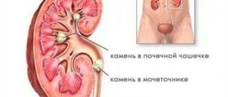
Urolithiasis disease
This pathology is one of the most common, since its development depends on the region of residence, the quality of food consumed, water and hereditary factors.
Echolocation allows you to completely detect kidney damage from urolithiasis, to see the accumulation of fine sand and large deposits, regardless of their actual chemical composition.
To carry out diagnostics and improve visualization, a water load is used (before the procedure, the patient is asked to drink 4 glasses of water).
In the ultrasound picture, stones show high echogenicity and clear contours. Their density is determined by the strength of the echo signal.
A significant drawback of diagnostics is the inability to establish the integrity of the stone, whether it is glued together from several parts, and also to determine the exact location. Today this problem is solved by intraoperative echography.
Stones in the ureter
Ultrasound examination of the ureters for the presence of stones in them has some difficulties due to the specific structure of the organ. Informative results of the study can be obtained provided that the ureters are significantly expanded in an area of 5 cm from the pelvis.
The best research results are obtained when ultrasound is performed through the abdominal area and in a lateral decubitus position.
Ureteral stones have a clear contour and low echographic positivity. Acoustic shadow may only be present in oxalates measuring 8-10 mm.
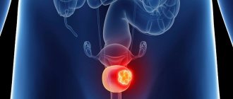
Tumors of the renal parenchyma
Both benign and malignant tumors can develop in the kidneys. On the echogram they appear with a clear outline, oval/round shape and weak echogenicity. Cystic and vascularized tumors have good visualization.
Cancer is easily detected by ultrasound. In 97% of cases it is clearly visible. But, unfortunately, it is discovered already in advanced stages. Such a tumor is heterogeneous, foci of necrosis merge into cysts, and their calcification is observed. The pelvicalyceal area can be filled with a tumor. In such cases, the kidney ceases to differentiate into separate zones.
Nephroblastoma
Most often this tumor occurs in children. Children usually do not complain about anything (complaints can only arise when the cancer grows to a large size). With an echogram, the oncological formation (its second name is Wilms tumor) has an oval shape. When the size of nephroblastoma reaches large parameters, multiple echogenic septa may be visible on ultrasound, which resemble the picture of polycystic disease.
Tumors of the renal pelvis and ureters
If we talk about benign tumors, the most common are angiomas and papillomas. When the size of the tumor does not exceed 2 cm, then due to stagnation of urine in the dilated pelvis, the papilloma is considered as a round formation with clear contours. This type of tumor often has a thin stalk that connects the formation to the wall of the renal pelvis.
The angioma has a similar rounded shape and contours that are also clearly visible on ultrasound.
If we talk about malignant tumors, then most often papillary cancer is found on ultrasound. It characterizes itself as a round, weakly echogenic formation, the contours of which are intermittent. Increasing in size, papillary cancer seriously deforms the kidney.
Metastases
Malignant neoplasms metastasize to the lymphatic system or hematogenously. Therefore, more than half of people with cancer have metastases. Metastases first affect the lungs, then the bones, liver, and brain. The kidneys are affected in advanced cases and this process will be noticeable on ultrasound.
On ultrasound, metastases will have the same appearance as malignant neoplasms.
Venous lesions
Patients are often diagnosed with such abnormalities as varicoceles, which are cystic dilations of the veins of the spermatic cord. Such a deviation may occur due to compression or germination of the tumor. Thrombosis of the left renal vein or inferior vena cava can also be a provocateur. Varicocele of the renal veins is understood as expansion along the walls of the pelvis. If there is a chronic form of venous damage, the kidney noticeably decreases in size. At the same time, echogenicity becomes high.
Acute renal failure
This disease usually occurs due to injury or dysfunction of one or more kidneys. Acute renal failure can be caused by:
- prerenal cause (bleeding, trauma, surgery);
- post-renal cause (problems with the patency of the kidneys or a single ureter);
- arenal cause (implies traumatic crushing of both kidneys or only one paired organ).
Regardless of what cause caused acute renal failure, the ultrasound picture is always almost the same. The kidneys or one kidney are enlarged in size, the parenchyma area has a pronounced expansion, the pyramids are enlarged, their echogenicity is weak in comparison with the structure of the parenchyma.
Chronic renal failure
This pathology occurs for various reasons. Its provocateurs can be:
- developmental anomalies of the ureters and kidneys;
- bilateral renal artery stenosis;
- nephrosclerosis;
- nephritis of various etiologies;
- glomerulonephritis;
- diseases associated with collagen formation.
When chronic renal failure occurs, the echographic picture corresponds to one or another ailment that provokes chronic renal failure. In the case of chronic renal failure, the kidneys are smaller than the permissible norm, the capsule has the same echogenicity as the tortuous contours (in this case we can also talk about nephrosclerosis), the parenchyma zone in this case is somewhat narrowed.
How to prepare for a kidney ultrasound? Nutrition
Preparing for a kidney ultrasound involves following a diet. Three days before the examination, you must avoid foods that increase gas formation and possible inflammation. You cannot eat spicy, salty, sweet or fried foods. Cabbage, legumes and other foods that promote gas formation should also be excluded.
What you should not eat or drink before a kidney ultrasound:
- legumes;
- dried fruits;
- nuts;
- fatty meats and fish;
- sauerkraut, pickled tomatoes;
- sauces with mayonnaise;
- hot spices;
- bakery products;
- fast food;
- sausages;
- sweets;
- alcoholic drinks;
- carbonated drinks;
- mushrooms and dishes based on them;
- strong tea and coffee.
It is recommended to eat porridge - buckwheat and rice. You need to eat small portions 5-6 times a day. The last meal should be 8 hours before diagnosis. 30 minutes after it, you should take an absorbent, for example, activated carbon at the rate of 1 tablet per 10 kg of weight. Reducing gas formation in the intestines improves the visualization of the kidneys on ultrasound.
Nuances of kidney diagnostics
Kidney ultrasound can detect many diseases and pathologies even in the early stages. There are no contraindications for the procedure, except for significant damage to the skin in the area where the gel needs to be applied. Difficulties in performing an ultrasound examination may occur in cases where there is significant obesity, gases are present in the intestines, or an examination has recently been carried out using a barium contrast agent. Over the past 35 years, no negative consequences from the ultrasound procedure have been identified, therefore it is considered completely safe.
Medication preparation
Patients who regularly take diuretics, on the day of the ultrasound, with the obligatory agreement of the doctor, should skip taking them until the examination is completed.
Medicines to normalize blood pressure and sugar levels should be taken as usual. They do not reduce the information content of the study.
In the acute phase of urolithiasis, before an ultrasound scan of the kidneys, you should take analgesics and antispasmodics prescribed by your doctor to reduce pain.
In medical, competent specialists perform ultrasound using modern expert-class equipment. We do everything to ensure that our patients receive quality services in a comfortable environment.
Call the phone number listed on the website, or leave a request in the feedback form. We will answer your questions and conduct all necessary examinations within one business day. Let's take care of your health together!
When is an ultrasound scan of the adrenal glands prescribed?
An ultrasound may be prescribed if adrenal disease is suspected. Symptoms of such diseases are:
- muscle weakness;
- weight change (loss or, conversely, excess weight gain);
- chronic hypertension (high blood pressure) or hypotension (low blood pressure);
- sexual dysfunction, decreased potency, menstrual irregularities, excess hair growth.
Ultrasound of the adrenal glands can reveal:
- cysts;
- tumors;
- hematomas;
- foci of inflammation;
- increase in the size of the adrenal glands (hyperplasia).
What documents do you need to bring with you to a bladder ultrasound?
The patient, entering the room for scanning using the described clinical method, must have the following papers with him:
- Information from previous checks.
- A document identifying a person.
- Medical card or medical history.
- Referral for examination prescribed by the treating specialist.
- A clean sheet or household towel.
- Disposable wipes (will allow you to remove residual gel from the body).
- Cash to pay for the procedure.
- A 1-1.5 liter bottle of plain water will help fill the MP if necessary.
Indicators and standards
During an ultrasound of the kidneys, the following is assessed:
- Their number and location. Normally, a person has two kidneys; they are localized retroperitoneally, approximately at the level of the XI-XII thoracic and III-IV lumbar vertebrae. The left kidney is often located slightly higher than the right. There are pathologies such as fusion of the kidneys, the absence of one kidney, or localization in an atypical location.
- Shape and dimensions. The kidneys are normally bean-shaped, but occasionally horseshoe-shaped, L-shaped and others are found. The weight of one bud is about 150-200 grams, the length ranges from 100 to 130 mm, and the width is on average 50 mm. The ultrasound diagnostician also takes into account such important parameters as the thickness of the parenchymal layer (about 20 mm) and the thickness of the capsule (normally no more than 1.5 mm).
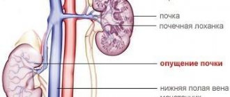
- Mobility. Normally, this parameter does not exceed 1-2 cm, which is important when diagnosing a wandering kidney.
- The structure of the collecting apparatus. This device performs the main functions of the kidneys - the formation and filtration of urine, and suffers from a variety of kidney diseases, in particular inflammatory ones.
- Structure. In many kidney diseases, changes in the structure of the kidneys are observed, for example, after suffering from inflammatory diseases, cysts, tumors. Normally, the structure is heterogeneous: the pyramids look like dark areas (low density, hypoechoic), the parenchyma is much lighter (it is hyperechoic, that is, it has a high density). The kidney capsule has an increased density; on the ultrasound machine monitor it looks the lightest.
- Ureters. Ultrasound evaluates their number, location, size and function. Normally, a person has two ureters, each of them has a length of 25 to 30 cm, a thickness of 5-9 mm. With pathology, you can detect an increase or decrease in the number of ureters, incorrect location or torsion, the formation of stagnation of urine and the formation of stones. On the monitor of an ultrasound diagnostic device, the ureter looks like a monochromatic dark hollow formation with light walls.