Urolithiasis
Urolithiasis
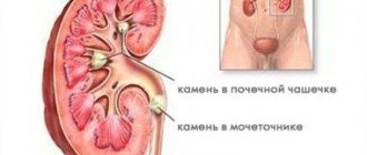
(urolithiasis) is a disease characterized by the formation of stones in the urinary organs. Stones can be found in all organs of the urinary system, however, they most often occur in the kidneys and bladder. Also a serious problem are stones in the ureter, where they enter after descending from the kidneys. Urolithiasis can develop at any age, but most cases of its initial detection occur between the ages of 20-55 years.
What is urolithiasis
Urolithiasis is a common disease characterized by the formation of stones in the organs of the urinary system (kidneys or bladder).
The largest number of hospitalizations of patients with urological diseases is due to urolithiasis. The formation of stones occurs in the kidneys, much less often in the bladder. Subsequently, stones can migrate and be localized at any level of the urinary system: ureter, bladder, urethra. In addition to renal colic, the most serious complication is the development of obstructive pyelonephritis (acute inflammation in the kidney due to blockage of the ureter with a stone).
Visual Research
Imaging studies confirm the clinical diagnosis of urolithiasis. Determining the size of the sediment, its location, and the degree of obstruction of urine flow are important in determining the correct procedure.
- Overview image of the abdominal cavity
. About 90% of urinary tract stones are visible on the overview image. The strongest shadow on the radiograph is produced by calcium oxalate stones, weaker struvite stones and even weaker cystine and gout stones. Uric acid deposits are colorless. - Urography.
Allows you to accurately determine the location of the deposit, and also provides additional information about kidney function and the degree of difficulty in the outflow of urine. In the case of ureteral stones, a photo should be taken after urination to show the obstruction covered by a full bladder. In case of kidney dysfunction caused by impaired urine flow, be sure to take a late photo approximately 2-3 hours after contrast injection. - Ureteropyelography
. It is carried out in the absence of renal function on urography and in the absence of hydronephrosis detected by ultrasound of the abdominal cavity. Additionally, a search is carried out for a non-obscuring stone in the ureter in the survey photograph. - Ultrasound of the abdominal cavity and
. Widely available non-invasive testing. Ultrasound should be performed regularly along with survey photography. Depending on the type of device, it allows you to visualize deposits with a diameter of 4-5 mm. In addition, ultrasound of the bladder and kidneys allows us to assess the degree of stagnation of urine, the condition of the renal parenchyma and the presence of non-obscuring stones. Because ultrasound examinations are harmless, they can be performed on patients with allergies to contrast agents, children and pregnant women.
Abdominal ultrasound
Causes of development and risk factors
Urolithiasis is a multifactorial disease. Stones form when the content of crystal-forming substances such as calcium, oxalates, and uric acid increases in the urine. At the same time, there is a decrease in substances that prevent stone formation.
The causes leading to the formation of stones in the urinary system are divided into exogenous (external) and endogenous (internal).
The following can be classified as exogenous:
- long-term use of hard water;
- living in climatic zones with a lack of ultraviolet rays;
- excessive consumption of sour, salty, spicy foods, as well as foods high in oxalates (legumes, beer, berries, coffee, chocolate, spinach, potatoes);
- not drinking enough water during the day;
- taking medications containing calcium, vitamin D preparations, ascorbic acid;
- concomitant diseases such as obesity, diabetes mellitus, urinary tract infections, chronic kidney disease, arterial hypertension increase the risk of developing urolithiasis;
- sedentary lifestyle.
The endogenous ones include the following:
- impaired renal function as a result of chronic diseases;
- genetic predisposition to the formation of stones (the presence of urolithiasis in blood relatives increases the risk of developing this pathology.);
- infectious diseases accompanied by dehydration;
- severe illnesses in which the patient is immobile for a long time;
- pathologies of the gastrointestinal tract associated with disruption of the processes of digestion and absorption;
- metabolic disorders (hyperparathyroidism, gout);
- congenital anomalies of the structure of the kidneys and urinary tract.
Causes
There are many causes of urolithiasis, the main ones are as follows:
- drinking water with plenty of minerals. It is also called hard water. Water supersaturated with calcium salts most strongly contributes to the development of the disease;
- a menu rich in high-protein foods, as well as too sour and spicy dishes;
- deficiencies of individual vitamins - most often belonging to group B and vitamin A;
- physical inactivity – that is, an insufficiently active lifestyle also contributes to urolithiasis;
- some bad habits. Thus, the development of stones is promoted by excessive alcohol consumption;
- taking certain medications not according to instructions, in excessive quantities;
- structural features of the urinary system. We are talking about anomalies such as a narrowed lumen of the urinary tract;
- various inflammations, including urethritis, cystitis, etc.;
- metabolic disorders;
- severe and regular dehydration;
- increased acidity of the body itself.
Many of these causes of urolithiasis can act in combination, in a complex manner - it all depends on the situation.
Symptoms of urolithiasis
Stones localized in the kidneys usually do not cause any inconvenience and are asymptomatic. If stones obstruct the outflow of urine, reflux develops (backflow of urine into the kidney), which leads to the development of edema of the kidney parenchyma, severe pain, which can be manifested by the following symptoms
- acute, severe, constant or paroxysmal pain in the lumbar region on the affected side, sometimes accompanied by nausea and/or vomiting;
- pain radiating down the abdomen and into the groin area on the affected side;
- burning or stinging sensation when urinating;
- occasional blood in the urine.
Patients suffering from urolithiasis also describe the following symptoms:
- change in the color, character, smell of urine;
- increased urge to urinate;
- nausea and vomiting;
- increase in body temperature (with infection).
Complications
Urolithiasis, if the patient is not treated in time, can lead to more serious consequences:
- penetration, development and spread of infections in the urinary tract;
- pyelonephritis and other kidney diseases;
- nephrosclerosis. These are transformations of kidney tissue that are associated with degenerative processes;
- problems with the intestines - in particular, paresis;
- accumulation of purulent formations;
- renal failure and bacterial shock.
If the stone is not removed, then as the disease develops the kidney may even die - this is extremely dangerous and risky, so the disease requires effective and thorough treatment.
Diagnosis of urolithiasis
Diagnosis of urolithiasis begins with a general medical examination, collecting an anamnesis about the patient’s health condition and complaints, laboratory and instrumental diagnostics are performed.
- Clinical and biochemical blood test. Shifts in these tests make it possible to identify the inflammatory process (increased blood leukocytes), the phenomenon of renal failure (increased creatinine and urea);
- A general urine test in the presence of an infectious agent in the urinary system reflects inflammatory changes (presence of leukocytes, nitrite-forming bacteria);
- A survey X-ray of the abdominal organs allows you to visualize stones in the kidneys, ureters and bladder;
- Excretory urography. It is performed on the same principle as a survey photograph, only with the use of an X-ray contrast agent;
- Ultrasound of the kidneys and bladder with the orifices of the ureters;
- Computed tomography of the retroperitoneal organs with contrast enhancement. It is the most effective diagnostic method, allowing a detailed assessment of the location, number of stones and their density, as well as obtaining additional information about the anatomical and functional state of the organs of the urinary system. The data obtained helps in choosing the optimal method of stone removal.
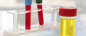
Laboratory tests
Urinalysis and culture should be performed routinely. In most cases, micro- or macroscopic hematuria is present.
Examination of urine sediment makes it possible to identify crystals and thus indirectly determine the chemical composition of the sediment.
Blood urea and creatinine levels are also measured to rule out kidney failure. A markedly elevated white blood cell count with accompanying fever indicates acute infection.
Urine sediment examination
Treatment of urolithiasis
When renal colic develops, it is necessary to relieve pain. For this purpose, the doctor prescribes non-steroidal anti-inflammatory drugs, less often narcotic analgesics.
Surgical treatment is performed in the case of a large stone, the inability to relieve pain with medications, recurrent attacks of renal colic, inflammatory processes in the organs of the urinary system, or impaired urine outflow.
- Draining the kidney using a special internal catheter-stent or performing percutaneous puncture nephrostomy (creating an artificial path for urine outflow) often precedes other surgical interventions.
- External shock wave lithotripsy is a non-invasive treatment method aimed at fragmenting stones under X-ray or ultrasound guidance with high-intensity pulses focused on the stone.
- Ureteroscopy is a minimally invasive endoscopic method for removing stones from the ureters. The destruction of the stone occurs using laser, pneumatic or ultrasonic energy.
- Percutaneous nephrolithotripsy is an invasive endoscopic removal of kidney stones. This treatment method is used for stones larger than 1.5 cm. Laser, ultrasonic or pneumatic energy is used to destroy the stone. Fragments of destroyed stone are evacuated with tongs.
Sources
- Tizelius H.G. et al.: Recommendations for urolithiasis, 2000.
- Preminger GM: Treatment of urolithiasis: pathogenesis and diagnosis, 1995.
- Dretler S.P. Treatment of ureteral stones, 1995.
- Macfarlane MT: Urolithiasis, 1997.
- Drach G.V.: Urinary lithiasis. Campbell Urology, 1992.
ONLINE REGISTRATION at the DIANA clinic
You can sign up by calling the toll-free phone number 8-800-707-15-60 or filling out the contact form. In this case, we will contact you ourselves.
If you find an error, please select a piece of text and press Ctrl+Enter
Prevention of urolithiasis
Important in the treatment of urolithiasis and the prevention of repeated attacks of renal colic is the elimination of risk factors, changes in lifestyle and eating habits. After stone removal, patients are advised to undergo regular follow-up with a urologist. Correction of metabolic disorders should be carried out with the participation of endocrinologists, gastroenterologists, and nutritionists.
You can prevent the development of urolithiasis by following a number of recommendations:
- follow a diet: minimize the consumption of spicy, salty and sour foods, control the amount of protein foods and vitamins;
- give up bad habits: smoking, drinking alcohol;
- drink enough water (2-3 liters of fluid per day);
- avoid stress and nervous tension;
- regularly undergo a medical examination, including a general urine test and ultrasound of the kidneys and bladder, and also promptly treat diseases of the urinary system;
- get rid of excess weight;
- move more;
- don't get too cold.
Epidemiology of the disease - how common are stones in the urinary system?
The content of the article
Urolithiasis occurs in approximately 2% of the population. Most often, stones are discovered after 30 years. Men suffer from this disease three times more often than women.
People who have an episode of renal colic have about a 15% chance of having another episode within 3 years, and a 30-50% chance of having another episode within the next 15 years. As a rule, urolithiasis is a long-term disease, with an average period of 9 years between attacks.
Urolithiasis disease
Genesis of formal type
The basis of the stone is its matrix, which has a protein structure. The matrix is formed as a result of metabolic disorders, infection of the body, various inflammatory diseases, and atypical blood circulation in the kidneys. Insoluble compounds (calculi) are deposited in the components of the urinary system, which accumulate and increase over time. The kidneys and other organs that remove urine from the body are in conditions of insufficient blood flow, which reduces their nutrition and leads to malfunctions.