In this article:
- What is mechanical ventilation?
- Indications for artificial ventilation of the lungs
- Invasive ventilation
- Who needs invasive ventilation and when?
- How does an invasive ventilator work?
- Features of equipment for invasive ventilation
- NIV - what is it?
- When is non-invasive ventilation used?
- Advantages of NIV
- Non-invasive ventilation in CPAP and BIPAP modes
- CPAP and BIPAP machines to help patients with COVID-19
- Long-term use of non-invasive ventilation: benefit or harm
Artificial pulmonary ventilation (ALV) is used to help patients with acute or chronic respiratory failure, when the patient cannot independently inhale the amount of oxygen necessary for the full functioning of the body and exhale carbon dioxide. The need for mechanical ventilation arises in the absence of natural breathing or in case of its serious disturbances, as well as during surgical operations under general anesthesia.
Emergency assistance from the medical team: what is the algorithm of action?
To provide emergency care in case of sudden cardiac arrest, a special cardiology team arrives on site, whose task is to carry out advanced resuscitation measures and immediately transport the patient to the hospital. It works according to a protocol that includes the following sequence of actions:
- Checking vital signs and making a diagnosis. For this purpose, a wider arsenal of equipment is used, including an electrocardiograph. It is necessary to exclude other causes of clinical death, such as bleeding or blockage.
Resumption of conductivity of the upper respiratory tract. To ensure maximum oxygen supply, they are intubated.- Resuscitation measures are carried out according to the same algorithm as indicated above, but for mechanical ventilation they use breathing masks, an Ambu bag or a ventilator.
- In the presence of atrial fibrillation or ventricular fibrillation on the ECG, the question of using defibrillation is raised.
- Drug support is provided by intravenous or intracardiac administration of drugs such as Adrenaline (1 ml 0.1% in 19 ml NaCl 0.9%) and Cordarone (in the presence of arrhythmias, 300 mg IV).
What is artificial ventilation?
In the process of mechanical ventilation, respiratory support is provided by forcibly pumping air or a gas mixture into the patient’s lungs and removing them out. The procedure can be carried out in several ways:
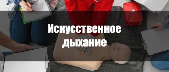
- without any devices, providing passive inhalation and exhalation by rhythmic pressing on the costal area and mouth-to-mouth breathing;
- using a resuscitation bag (such as an Ambu bag);
- using a device for NIV or invasive ventilation - if possible and necessary.
Modern ventilators provide complete respiratory support for the patient
Preparing for artificial respiration
Before performing expiratory artificial respiration, the patient should be examined. Such resuscitation measures are contraindicated for facial injuries, tuberculosis, poliomelitis and trichlorethylene poisoning. In the first case, the reason is obvious, and in the last three, performing expiratory artificial respiration puts the person performing resuscitation at risk.
Before starting expiratory artificial respiration, the victim is quickly freed from clothing squeezing the throat and chest. The collar is unbuttoned, the tie is undone, and the trouser belt can be unfastened. The victim is placed supine on his back on a horizontal surface. The head is tilted back as much as possible, the palm of one hand is placed under the back of the head, and the other palm is pressed on the forehead until the chin is in line with the neck. This condition is necessary for successful resuscitation, since with this position of the head the mouth opens and the tongue moves away from the entrance to the larynx, as a result of which air begins to flow freely into the lungs. In order for the head to remain in this position, a cushion of folded clothing is placed under the shoulder blades.
Causes of respiratory arrest
There are many different causes of breathing problems. Common causes include certain health conditions and sudden medical emergencies.
Breathing problems can be caused by:
- anemia (low red blood cell count);
- suffocation;
- chronic obstructive pulmonary disease (COPD), emphysema or chronic bronchitis;
- heart failure;
- lung cancer or cancer that has spread to the lungs;
- respiratory infections, including pneumonia, acute bronchitis, whooping cough and others;
- pericardial effusion (fluid surrounding the heart and preventing it from functioning properly);
- pleural effusion (fluid surrounding the lungs and compressing them);
- blood clot in the lungs (thromboembolism);
- collapsed lung (pneumothorax);
- myocardial infarction;
- injury to the neck, chest wall or lungs;
- severe allergic reaction;
- drowning, in which fluid accumulates in the lungs.
Artificial respiration from mouth to nose
This method of artificial ventilation is carried out if it is not possible to properly unclench the patient’s jaws or there is an injury to the lips or oral area.
The rescuer places one hand on the victim’s forehead and the other on his chin. At the same time, he simultaneously throws back his head and presses his upper jaw to the lower. With the fingers of the hand that supports the chin, the rescuer must press the lower lip so that the victim’s mouth is completely closed. Taking a deep breath, the rescuer covers the victim’s nose with his lips and forcefully blows air through the nostrils, while watching the movement of the chest.
After artificial inspiration is completed, you need to free the patient's nose and mouth. In some cases, the soft palate may prevent air from escaping through the nostrils, so when the mouth is closed, there may be no exhalation at all. When exhaling, the head must be kept tilted back. The duration of artificial exhalation is about two seconds. During this time, the rescuer himself must take several exhalations and inhalations “for himself.”
IVL AND NIVL - what is the difference?
There are two options for delivering air into the patient's airway:
- invasive pulmonary ventilation (IVL);
- non-invasive pulmonary ventilation (NIV).
To perform invasive ventilation, an endotracheal tube is inserted into the trachea. It can be administered through the nose (nasotracheal intubation) and/or mouth. If long-term mechanical ventilation is needed and the patient is unconscious or in serious condition, a tracheostomy is performed - a surgical operation to dissect the anterior wall of the trachea to insert a tracheostomy tube directly into its lumen.
After completing the above-described manipulations, the ventilator is connected. The advantage of invasive mechanical ventilation is its effectiveness, because the air mixture is delivered directly to the lungs without loss.
With bulbar disorders, the patient loses the separation of the digestive and respiratory tracts, and this is also taken into account when determining the indications for tracheostomy. Through a tracheostomy, the patient's sputum is also removed.
Invasive respiratory support is usually performed in the following cases:
- intolerance to NIV in the patient or lack of effect from the therapy;
- increased salivation, excessive sputum;
- the need for urgent intubation during emergency hospitalization,
- patient coma, disturbances of consciousness;
- presence of facial burns and injuries.
Although invasive ventilation is highly effective, it is used only when it is impossible to help the patient in a more gentle way, i.e. without invasive intervention. After all, a person connected to a ventilator can neither speak nor eat. Intubation is not only inconvenient, but also painful. Therefore, a patient on mechanical ventilation is usually put into a medically induced coma. The procedure is performed only in a hospital setting under the supervision of specialists; it may have complications and side effects. In connection with the above, whenever possible, preference is given to non-invasive ventilation.
The principle of non-invasive ventilation
Rules for checking a ventilator
For safe operation and to prevent iatrogenicity, it is necessary to check the breathing apparatus before each patient connection. The algorithm for testing breathing equipment consists of 1) checking the ability to perform mechanical ventilation, providing inhalation and exhalation, and 2) the functionality of the control, alarm and safety system. Testing the device for its ability to perform mechanical ventilation is carried out in two stages - a) testing the ability to provide sufficient inspiration and b) testing the ability to provide exhalation. This can be done in two ways. The first method is applicable for “old” ventilators. To test the ability to take a breath, i.e. to develop sufficient pressure in the breathing circuit, close the hole on the tee for connecting the patient with your finger and observe the pressure gauge readings. At the same time, the depressurization valve is closed or the response value is set to maximum. With the established average parameters (UP TO 500-600 ml), the device must create a pressure in the circuit of at least 50-60 cm of water column. If the device creates such pressure and there is no audible leak of the gas mixture, then proceed to the second stage. If the device does not develop sufficient pressure, then it is necessary to check the tightness of the connections of hoses, adapters, humidifier, etc. If there are no leaks and the device does not develop sufficient pressure during inspiration, then it is considered faulty and is not allowed for use! The next step is to check the device for its ability to allow the patient to exhale. To do this, the doctor himself must try to exhale calmly through a gauze pad into the device through the tee for connecting the patient. This is done during the exhalation phase. If exhalation is carried out without difficulty, then the device can be considered in good working order in this regard. The second method is applicable to any ventilators. To implement it, you need a rubber bag with a capacity of 1.5-2 liters - a lung simulator. It joins the tee instead of the patient. The ventilation parameters are set close to the maximum - UP TO 900-1000 ml, the depressurization valve is closed or the response value is set to the maximum, PEEP (PEER) is removed. Observe the bag and the pressure gauge - the bag should inflate as much as possible during inhalation, creating a resistance of about 30 cm of water column. and collapse during exhalation. To be more sure, you can try squeezing the bag while inhaling and getting more pressure. Squeezing the bag during exhalation will confirm the ability to exhale unimpeded. Checking the functionality of the control, alarm and security systems. Carry out when the depressurization valve opens (RO-5, RO-6) or set its response value to the usual parameters - 30-40 cm of water column. When a finger closes the hole on the patient connection tee or squeezes the simulator bag during inspiration, the pressure in the circuit should not exceed the set limit and the alarm system should be activated. When the circuit depressurizes - the bag is removed, the alarm system should also trigger. The importance of such a routine check should be emphasized before each connection of the device, before each anesthesia. Practice shows that any, even the most advanced, device is capable of breaking down, and if this breakdown is not detected before the start of operation, then its detection during mechanical ventilation is usually accompanied by significant disturbances in external respiration and blood circulation, and sometimes barotrauma. These complications are fatal and iatrogenic! The most common cause of malfunction of breathing equipment is sticking of rubber membranes exposed to condensation. When drying - outside of operation, condensate can stick or deform the membrane valves. Only half an hour to an hour is enough for this - just the break time between anesthesia.
Removal from the device
Of course, in most cases the device is not turned off immediately with one button. It is necessary to remove the patient from mechanical ventilation correctly. Lung support is gradually removed, and the patient begins to breathe better on his own. This process is complex, doctors need to determine whether everything is under control. Usually they are disconnected from mechanical ventilation if compliance is close to normal, there are no signs of heart failure, and sepsis is absent or subsiding (if there was one).
When using the device for a short time, a long shutdown is not required; it is maintained until the end of the anesthetic effect. In other cases, when mechanical ventilation has been prolonged, parameters begin to be reduced, primarily those that can lead to serious side effects.
Bibliography
1. Satishur O.E. Mechanical ventilation. M.: Med.lit., -2006 2. Belebezev G.I., Kozyar V.V. Physiology and pathophysiology of artificial ventilation. Kyiv, -2003 3. Butylin Yu. P., Butylin V. Yu., Butylin D. Yu. Intensive care of emergency conditions. Kyiv. 2003 4. Vaiman V.A., Avakov V.E. Critical and emergency conditions in medicine. 5. Grippi M.A., Pathophysiology of the lungs. Saint Petersburg. 2001 6. Zilber A.P., Etudes of critical medicine, volume II, Respiratory medicine. Petrozavodsk. 1996 7. Kassil V.L., Leskin G.S., Vyzhigina M.A., Respiratory support. Moscow "Medicine". 1997 8. Malysheva V.D., Intensive care. Moscow "Medicine". 2003 9. Shurygin I. A., Breath monitoring in anesthesiology and intensive care. Saint Petersburg. 2003
Preparing to provide assistance
Before performing artificial respiration, the following manipulations must be performed:
- Remove the victim from tight clothing.
- Place him on a horizontal surface on his back.
- Tilt the person's head back, placing your palm under the back of the head, and with the other press on the forehead, ensuring that the chin is level with the neck. Place folded clothing under your shoulder blades to secure this position.
- Check with your fingers to see if there is anything foreign in your mouth. If necessary, delete. Also remove dentures.
Assistance in case of mass accidents[edit | edit code]
The main attention should be paid to the clear organization of rescue, for which an experienced swimmer or someone on shore must lead the overall management of relief efforts.
In the absence of a sufficient amount of rescue equipment, various floating objects (logs, boards, benches, etc.) can be used, which rescuers push to the scene of the incident. When providing swimming assistance to a group of drowning people, you should first save children and the elderly. It should be borne in mind that swimming into the middle of a group of victims is dangerous for rescuers, and it is necessary to save only those from the edge, encouraging and giving advice to others. If these basic rules are followed, the rescue of a group of people in distress will be successful and will ensure the saving of many lives.
Poisoning
Poisoning is a disorder of the body's functioning that occurs due to the ingestion of a poison or toxin. Depending on the type of toxin, poisoning is distinguished:
- carbon monoxide
- pesticides
- alcohol
- medications
- food and others
First aid measures depend on the nature of the poisoning. The most common food poisoning is accompanied by nausea, vomiting, diarrhea and stomach pain. In this case, the victim is recommended to take 3-5 grams of activated carbon every 15 minutes for an hour, drink plenty of water, refrain from eating and be sure to consult a doctor.
In addition, accidental or intentional drug poisoning, as well as alcohol intoxication, .
In these cases, first aid consists of the following steps:
Read also: How to make baby sling beads with your own hands
- Rinse the victim's stomach.
To do this, make him drink several glasses of salted water (for 1 liter - 10 g of salt and 5 g of soda). After 2–3 glasses, induce vomiting in the victim. Repeat these steps until the vomit is clear. Gastric lavage is only possible if the victim is conscious. - Dissolve 10–20 tablets of activated carbon in a glass of water and give it to the victim to drink.
- Wait for the specialists to arrive.
NIV, advantages and disadvantages
During non-invasive ventilation, the patient should:
- Be conscious.
- Be able to follow doctors' instructions.
There must also be a clear prospect of stabilization of the patient's condition within a few hours or days after the start of this type of respiratory support.
Absolute contraindications for NIV:
- coma;
- heart failure;
- respiratory arrest;
- other condition requiring immediate intubation of the patient.
The NIV technique is the simplest and most comfortable for the patient, because this method can help a patient with acute or chronic respiratory failure without resorting to endotracheal intubation or tracheostomy.
Advantages of NIV compared to invasive ventilation:
- lower cost;
- better tolerability (no need for sedatives);
- Ease of use;
- better accessibility outside the intensive care unit (eg, at home);
- the ability to interrupt therapy, which makes it easier to refuse it in the future;
- no need for airway intubation skills.
Disadvantages of NIV compared to invasive ventilation:
- difficulties in communicating with patients if they are not ready (or cannot, due to mental characteristics) interact with the doctor;
- impossibility of use in patients with limited physical capabilities (for example, if they cannot remove the mask when vomiting);
- an obstacle to the effective removal of secretions (sputum), difficult access for its suction;
- cannot be performed on patients with a reduced level of consciousness;
- the mask-face interface is not easy to manage - it is necessary to take into account the anatomical features of the patient’s face, which is not always possible;
- oxygen leakage from the mask causes discomfort and reduces the effectiveness of therapy;
- forced mechanical breaths may be impossible or dangerous.
The ability to use the NIV technique at home is its important advantage
Of course, when prescribing NIV, a positive effect is expected to be achieved, and it most likely will not be long in coming. However, it is worth mentioning possible complications. These include:
- skin irritation in places where the mask fits tightly (with prolonged use);
- discomfort when exhaling due to the constant supply of air;
- dry mucous membranes and nasal congestion;
- night awakenings from possible signals from the device.
There are different categories of ventilators, divided according to the principle of their operation.


