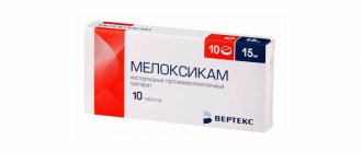
Alfadol-Ca caps 0.25 mcg+500 mg x100
Alfadol-Ca caps 0.25 mcg+500 mg x100 ATX code: A12AX (Calcium preparations in combination with other drugs)
Active substances
alfacalcidol Rec.INN WHO registered calcium carbonate Ph.Eur. European Pharmacopoeia
Dosage form
Alfadol-Sa
Capsulereg. No.: P N013997/01 dated 02/09/09 - Indefinitely Re-registration date: 10/22/15
Release form, packaging and composition of the drug Alfadol-Sa
Capsules 1 capsule.
alfacalcidol 0.25 mcg
calcium carbonate 500 mg,
which corresponds to a calcium content of 200 mg Clinical-pharmacological group: Drug that regulates calcium and phosphorus metabolism Pharmaco-therapeutic group: Calcium-phosphorus metabolism regulator
pharmachologic effect
A combined drug that regulates the metabolism of calcium and phosphorus. Replenishes the lack of calcium and vitamin D3 in the body.
The main therapeutic effects of alfacalcidol are an increase in the concentration of 1,25-hydroxycolecalciferol in the blood and, as a consequence, an increase in the absorption of calcium and phosphate, an improvement in mineralization and a decrease in bone resorption, a normalization of the content of parathyroid hormone in plasma, and a decrease in bone and muscle pain.
Calcium is involved in the formation of bone tissue, blood clotting, transmission of nerve impulses, contraction of skeletal muscles, and regulation of heart function.
Rationale for the combination of alfacalcidol and calcium
Alfacalcidol enhances the absorption of calcium from the gastrointestinal tract and causes a therapeutic effect in osteoporosis. The elemental calcium content in calcium carbonate is 40%, i.e. 500 mg of calcium carbonate contains 200 mg of elemental calcium, which corresponds to the minimum physiological requirement.
Pharmacokinetics
Absorption is high, TCmax is 8-18 hours. In the blood it binds to specific alpha globulins.
Metabolized in the liver to form the active metabolite calcitriol (1,25-dihydroxycolecalciferol), a smaller part is metabolized in bone tissue. Unlike natural vitamin D, it is not metabolized in the kidneys, which allows it to be prescribed for vitamin D deficiency in patients with renal failure (the effect does not depend on hydroxylation in the kidneys).
It is excreted by the kidneys and bile in approximately the same amount.
Indications for the drug Alfadol-Sa
osteoporosis (menopausal, senile, steroid, idiopathic and others) and its complications (bone fractures), renal osteodystrophy, hypoparathyroidism and hyperparathyroidism (with bone damage), chronic renal failure. Open list of ICD-10 codes
ICD-10 code Indication
E55 Vitamin D deficiency
E58 Nutritional calcium deficiency
M80.0 Postmenopausal osteoporosis with pathological fracture
M80.1 Osteoporosis with pathological fracture after spayectomy
M80.4 Drug-induced osteoporosis with pathological fracture
M80.5 Idiopathic osteoporosis with pathological fracture
M81.0 Postmenopausal osteoporosis
M81.1 Osteoporosis after spayectomy
M81.4 Drug-induced osteoporosis
M81.8 Other osteoporosis (senile osteoprosis)
M82 Osteoporosis in diseases classified elsewhere
Dosage regimen
For osteoporosis - 1-2 capsules per day.
For osteodystrophy and chronic renal failure - 2 capsules per day.
For hypoparathyroidism and hyperparathyroidism (with bone damage) - 1-2 capsules per day.
Chronic renal failure - 1-2 capsules per day.
The duration of taking the drug is determined by the doctor.
Side effect
From the side of metabolic processes: hypercalcemia. In patients with severe renal impairment, hyperphosphatemia may develop.
From the digestive system: constipation, diarrhea.
From the urinary system: nephrocalcinosis.
Allergic reactions: itching, rash, urticaria.
Contraindications for use
increased sensitivity to vitamin D3, urolithiasis (formation of calcium stones), increased calcium levels in the blood (in persons with increased function of the parathyroid glands, hypervitaminosis D, osteochondrosis, metastases of tumors in the bones), hyperphosphatemia (except in cases where the latter develops against the background hypoparathyroidism), hypermagnesemia, Zollinger syndrome, peanut or soy intolerance, children under 18 years of age.
Take with caution to persons suffering from renal failure, as well as patients on hemodialysis. In case of long-term treatment, it is necessary to check the amount of calcium excreted by the kidneys.
Use during pregnancy and breastfeeding
No studies have been conducted, so the drug is contraindicated during pregnancy and breastfeeding.
During pregnancy, the drug is prescribed only for absolute indications and only if the potential benefit of its use outweighs the possible risk to the fetus. Hypercalcemia during pregnancy may affect fetal development. The daily dose should not exceed 1500 mg of calcium carbonate.
Use for impaired renal function Take with caution to persons suffering from renal failure, as well as patients on hemodialysis. In case of long-term treatment, it is necessary to check the amount of calcium excreted by the kidneys.
Use in children Contraindicated in children under 18 years of age.
special instructions
Alfacalcidol is an active derivative of vitamin D3. During treatment, therapeutic doses of vitamin D3 and its derivatives should not be prescribed due to the possibility of additive effects and an increased risk of hypercalcemia.
The drug enhances the intestinal absorption of calcium and phosphate, the concentration of which must be controlled, especially in patients with renal failure. Monitor concentrations of calcium, phosphate, alkaline phosphatase, magnesium and creatinine.
If hypercalcemia or hypercalciuria occurs, they can be quickly eliminated by discontinuing the drug until the plasma calcium concentration normalizes. The drug should be used with caution in patients with hypercalciuria, in particular in those with urolithiasis.
Impact on the ability to drive vehicles and operate machinery
The drug does not affect driving or engaging in other potentially hazardous activities that require increased concentration and speed of psychomotor reactions.
Overdose
If hypercalcemia develops, you should stop taking the drug.
Hypercalcemia manifests itself as malaise, fatigue, weakness, dizziness, drowsiness, headache, nausea, dry mouth, constipation, diarrhea, heartburn, vomiting, abdominal pain, gastrointestinal disorders, muscle pain, bone pain, joint pain, itching
Treatment: the drug should be discontinued. A severe form of hypercalcemia requires supportive treatment measures - hydration with the introduction of infusion saline solutions, in some cases - the appointment of “loop” diuretics and corticosteroids.
In cases of acute overdose, it is necessary to carry out preliminary treatment with gastric lavage and/or the introduction of mineral oil, which helps to reduce absorption and increase excretion of the drug through the intestines.
Drug interactions
Alfacalcidol/digitalis glycosides
Hypercalcemia in patients taking cardiac glycosides may exacerbate cardiac arrhythmia, and therefore their combined use should be avoided.
Alfacalcidol/barbiturates/anticonvulsants that are inducers of microsomal liver enzymes
Patients systematically receiving barbiturates or other anticonvulsants require increased doses of alfacalcidol to achieve a therapeutic effect.
Alfacalcidol/drugs affecting absorption
The absorption of alfacalcidol may be impaired by concomitantly taken mineral oils (with long-term use), cholestyramine, colestipol, sucralfate, as well as large quantities of aluminum-containing antacids.
When using alfacalcidol together with antacids and laxatives containing magnesium in patients on hemodialysis, the possibility of developing hypermagnesemia should be taken into account.
Alfacalcidol/thiazide diuretics
The interaction of alfacalcidol with thiazide diuretics increases the risk of developing hypercalcemia.
Calcium carbonate may interfere with the absorption of concomitantly taken tetracycline and ciprofloxacin.
Storage conditions for the drug Alfadol-Sa
In a dry place, protected from light, at a temperature not exceeding 25°C. Keep out of the reach of children.
Shelf life of the drug Alfadol-Sa Shelf life - 2 years.
Terms of sale
On prescription.
The use of alfacalcidol in the treatment of osteoporosis
Osteoporosis (OP), a systemic skeletal disease from the group of metabolic osteopathies, is characterized by a decrease in bone mass and disruption of the microarchitecture of bone tissue, which leads to a decrease in bone strength and, as a consequence, an increased risk of fractures [1]. The goal of drug therapy for AP is to reduce the incidence and risk of fractures, increase bone mineral density (BMD) and improve the quality of life of patients [1].
Among the drugs used for the treatment and prevention of systemic AP, antiresorptive drugs occupy an important place, which include vitamin D and its active metabolites - alfacalcidol and calcitriol [2, 3]. Postmenopausal AP is characterized by increased peri- and postmenopausal bone loss, which can occur with normal or initially low peak bone mass. The leading pathogenetic mechanism of AP is estrogen deficiency and the associated decrease in the activity of the renal enzyme 1α-hydroxylase, accompanied by a decrease in the synthesis of calcitriol in the kidneys [4]. This results in calcium (Ca) leaching from bone tissue, concomitant parathyroid hormone (PTH) suppression, Ca malabsorption, and vitamin D receptor (VDR) deficiency, primarily in the classic target tissues (intestine, bone, kidney, and parathyroid glands). It is important that in postmenopausal AP, a decrease in calcitriol activity is always secondary to estrogen deficiency [5, 6]. The rate of bone formation in this type of AP, as a rule, remains within the normal range or is slightly reduced [7–11].
The main mechanism for the development of senile (involutive) AP is a decrease in the synthesis of calcitriol as a result of deficiency of renal 1α-hydroxylase, as well as a deficiency and decrease in the affinity of receptors for calcitriol in target organs: the gastrointestinal tract (GIT), bones and parathyroid glands. Increased Ca malabsorption and, accordingly, its leaching from the bone, as well as a decrease in the expression of genes responsible for the synthesis of matrix proteins produced by osteoblasts, ultimately have a negative effect on the mass and quality of bone tissue [6, 7, 12, 13].
Deficiency of sex hormones (estrogens/testosterone), as well as somatopause, accompanied by a decrease in insulin-like growth factors (IGF) and their binding proteins (IGFBP-4↑, IGFBP-3/5↓), in senile AP have an additional effect on the reduction of cofactors 1α-hydroxylase [14–16].
As a result, a decrease in the synthesis, reception and activity of D-hormone stimulates the synthesis of PTH with the development of parathyroid hyperplasia and tertiary hyperparathyroidism [15, 17]. Moreover, an increase in PTH levels in patients over 70 years of age with osteoporosis is accompanied by an increase in endocortical resorption, especially in the area of the proximal femur, intracortical porosity, and underlies the patients’ susceptibility to fractures. Moreover, bone resorption induced by PTH is not accompanied by an adequate increase in bone formation [9, 18–20].
Thus, restoring calcitriol levels is a key area of prevention and treatment for AP and requires the mandatory use of vitamin D or its active forms (calcitriol and alfacalcidol).
Both regular vitamin D3 and the D-hormone prodrug alfacalcidol (1α,25(OH)D3) act through a common biologically active metabolite, calcitriol (1α,25(OH)2D3; D-hormone). Moreover, in the body, alfacalcidol (Alpha D3-Teva®) is converted into calcitriol, bypassing endogenous regulation and without the participation of the renal enzyme 1α-hydroxylase [21].
As already noted, the effect of alfacalcidol (via the active metabolite calcitriol) in maintaining calcium and bone homeostasis is through interaction with nuclear VDR in target organs, primarily the intestine, bones, kidneys and parathyroid glands [14, 22, 23]. In conditions of vitamin D3 deficiency, the main effects of physiological and pharmacological concentrations of calcitriol (1α,25(OH)2D3) are thought to be:
- increasing the level of Ca in the blood plasma by stimulating its absorption in the intestine and reabsorption in the distal renal tubules;
- a decrease in the content of PTH in the blood plasma, which occurs in two ways: firstly, due to direct inhibition of PTH gene transcription due to binding to the VDR of the parathyroid glands, and secondly, as a result of indirect inhibition of PTH secretion due to an increase in the concentration of Ca in the blood plasma [14 –23];
- decrease in resorption and increase in bone tissue formation due to a decrease in PTH content and an effect on Ca and phosphate homeostasis [24–26].
Data on the effects of calcitriol from randomized prospective clinical trials vary widely. Some studies have shown a significant effect of calcitriol (1α,25(OH)2D3) on the increase in BMD in postmenopausal osteoporosis [21, 23, 26], while others have not obtained similar effects [27], which may be due to the use of lower doses of the drug. Additionally, a 3-year prospective multicenter study of 622 women with vertebral compression fractures found that treatment with calcitriol (1α,25(OH)2D3) resulted in a reduction in the incidence of new vertebral fractures [28].
A feature of vitamin D preparations is their good tolerability. Supplementation of regular vitamin D and Ca in elderly patients with natural vitamin D deficiency and concomitant low levels of 25OHD3 substrate is in most cases sufficient to overcome clinical or even subclinical osteomalacia. According to some studies, in patients with natural vitamin D deficiency and low Ca intake, vitamin supplements reduce the severity of osteoporosis and the incidence of non-vertebral fractures [17, 29]. Meanwhile, the use of regular vitamin D in physiological daily doses of 400–3000 IU (or 15 μg of 25OHD3) is not always effective in the treatment of osteoporosis [21, 30]. Moreover, in elderly people, correction of disturbances in the synthesis, reception and activity of calcitriol requires the administration of vitamin D in doses that significantly exceed not only physiological, but also pharmacological, which can lead to vitamin D intoxication due to its long-term retention in soft tissues.
A study using the D-hormone precursor alfacalcidol, compared with native vitamin D3 in combination with calcium supplements, revealed an increase in BMD and a decrease in the incidence of vertebral fractures [21, 24, 31].
Bone effects and safety of the use of 1 mcg/day alfacalcidol (Alpha D3-Teva®) and a combination of vitamin D 880 IU/day and Ca carbonate 1000 mg/day, in the treatment of white patients with postmenopausal AP and the absence of vitamin D deficiency in the blood plasma, evaluated in a multicenter randomized comparative study. After 12 months from the start of treatment, in the group of patients taking alfacalcidol, there was an increase in BMD of the lumbar spine (from the initial level) by 2.33%, and after 18 months - by 2.87% (p < 0.001); in the group taking vitamin D and calcium - only by 0.7%. Moreover, the statistical differences between the groups were significant (p = 0.018; 0.005).
The main mechanisms of action of alfacalcidol in AP are the normalization of reduced calcitriol synthesis and, accordingly, the correction of Ca malabsorption by stimulating the expression of estrogen receptors in bone cells, mediated by calcitriol [14–26]. Although calcitriol synthesized from alfacalcidol is not directly involved in the regulation of mineralization, but rather increases Ca levels, this does not exclude its effects on the organic matrix of bone or on bone growth factors. On the contrary, the effects of alfacalcidol can also include an increase in the secretion of calcitonin and the normalization of disconnected processes of bone tissue remodeling by increasing the level of transforming growth factor beta (TGF-β) and osteoprotegerin (OPG), which, in turn, determines the inhibition of postmenopausal production of bone-resorbing cytokines tissue, especially tumor necrosis factor alpha (TNF-α), and an increase in the release of TRF-β, which ensures restoration of weakened osteoclast apoptosis and slowdown of resorption processes [9, 32–34].
Along with this, data have now been accumulated that can be used to justify the advantages of using alfacalcidol over vitamin D in osteoporosis, also in the context of bone effects, but not associated only with stimulated calcium absorption and a reduced content of endogenous PTH as the only necessary conditions for the anabolic action of these drugs.
Studies evaluating the relationship between the ability of alfacalcidol and vitamin D3 to increase calcium levels and have a protective effect on bones in osteoporosis caused by estrogen deficiency have shown that both drugs increase bone mineral density. Moreover, the increase in MIC is accompanied by a slight (within the normal range) increase in the Ca content in the blood plasma and directly depends on the dose of the drugs. However, at a fixed plasma calcium concentration, alfacalcidol increases BMD more effectively than vitamin D3, and higher doses of vitamin D3 are required to achieve comparable levels of BMD.
Similar patterns can be traced in terms of an increase in bone strength when taking both drugs. Of course, the effect depends on the rate of increase in the concentration of Ca in the blood. However, at the same plasma Ca level, alfacalcidol is more effective than vitamin D3 in increasing bone strength, which is reduced in estrogen deficiency. Moreover, the study showed that the effect of vitamin D3 on bone strength reached a plateau at a dose of 200 μg/kg, and a dose of 400 μg/kg did not lead to a corresponding increase in BMD at all [35].
When comparing the effect of drugs depending on the excretion of Ca in the urine, unidirectional trends are also observed: at the same level of Ca in the urine, alfacalcidol is more effective than vitamin D3 in increasing bone mass and strength, which is reduced with estrogen deficiency.
A comparison of the bone effects of alfacalcidol and vitamin D3 at a well-defined plasma calcium concentration of less than 10 mg/dl (that is, at dosages that do not cause hypercalcemia) shows that bone strength increases with alfacalcidol, but does not change with vitamin D3. Obviously, in order to cause an increase in BMD comparable to alfacalcidol, large doses of vitamin D3 may be required, and this is fraught with the development of hypercalcemia.
An assessment of the effect of drugs on the urinary excretion of deoxypyridinoline (a marker of bone resorption) against the background of a decrease in estrogen concentrations shows that both alfacalcidol and vitamin D3 dose-dependently reduce the content of deoxypyridinoline in the urine, but alfacalcidol inhibits bone resorption more effectively than vitamin D [21, 35] . A comparison of the same effects of the drugs, but with a given low Ca content in plasma, shows that the administration of alfacalcidol leads to a decrease in the excretion of deoxypyridinoline in the urine, while the administration of vitamin D3 in doses that maintain the concentration of Ca in the blood plasma below 10 mg/dl, is not accompanied by significant suppression of deoxypyridinoline excretion.
Thus, both alfacalcidol and vitamin D3 increase BMD and bone strength while increasing plasma and urinary Ca levels. It is reasonable to conclude that the bone effects of these drugs are directly dependent on their calcium effects. However, correlation of the bone and calcium effects of both drugs obtained at a given Ca level suggests that alfacalcidol increases bone mass and improves bone quality more effectively than vitamin D3. It is obvious that the implementation of the protective effect of alfacalcidol on bones occurs partly independently of its normalizing effect on calcium balance. Of course, the mechanisms of the protective effect of alfacalcidol on bone remain not fully understood, however, there is convincing evidence of its suppression of bone resorption caused by estrogen deficiency.
Since, as studies show, the protective effect of alfacalcidol on bones is not due solely to the normalization of calcium balance, it is possible that suppression of endogenous PTH is also not the only necessary condition for the development of bone effects of the drug.
This is confirmed by the results of a study in which the effect of alfacalcidol on bones and calcium metabolism was assessed in an experiment on animals that had undergone parathyroidectomy [35]. Postoperative hypocalcemia and hyperphosphatemia were reversed by continuous infusion of human parathyroid hormone (hPTH) (amino acid sequences 1–34 responsible for the calcemic effects of PTH). Thus, against the background of a fixed PTH level and relative normocalcemia, the dose of alfacalcidol was titrated upward, but, and this was the main condition, it was not accompanied by the development of hypercalcemia. Then, at the end of the study, the animals were killed and the bones were examined. The study showed that over a 2-week period, alfacalcidol dose-dependently increased proximal tibial BMD and trabecular bone volume. The bone surface of alfacalcidol-treated animals was lined with large cuboidal cells resembling active osteoblasts [29, 35].
The main conclusion that the study allowed us to draw is that the protective effect of alfacalcidol on bones does not depend on the level of PTH and is partly carried out independently of its effect on Ca absorption and the resulting suppression of PTH secretion.
Thus, the phenomenon of stimulation of osteoclastic bone resorption observed in vitro (as a result of induction of the formation of osteoclastogenesis-supporting molecules (RANKL/TRANCE) in bone marrow stromal cells), while taking large doses of 1α,25(OH)2D3, contradicts the data obtained in vivo , indicating that in doses that do not lead to the development of hypercalcemia, the active metabolite of vitamin D3 increases bone mass, at least by suppressing bone resorption [14. 15, 36–38]. Given that the protective effects of alfacalcidol on bone are observed at constant PTH levels, it is reasonable to assume that the active metabolite of vitamin D3 inhibits bone resorption independent of PTH suppression. Further studies are certainly needed to shed light on the mechanisms by which the active metabolite of vitamin D3 inhibits bone resorption [34, 35].
Another target organ for the effects of calcitriol [14–17] is the muscular system. Activation of VDR by alfacalcidol on the membrane of muscle cells, where they regulate the transport of calcium and phosphate, as well as in the cell nucleus, where they participate in the production of energy for muscle contraction, contributes to the improvement of motor activity, optimization of motor coordination and, as a result, preventing the risk of falls in elderly patients. In addition, alfacalcidol regulates the expression of nerve growth factor (NGF), and also promotes a dose-dependent increase in the effect of IGF-1, one of the most significant factors of muscle activation [15, 27]. It is obvious that some pathogenetic factors of age-related sarcopenia can be balanced by alfacalcidol therapy.
Clinical data have been obtained on the effectiveness of alfacalcidol in terms of increasing muscle strength [24] and reducing the incidence of vertebral and femoral neck fractures by 50–70%, reducing the intensity of back pain compared to native vitamin D [33].
An estimate of the relative risk of falls in postmenopausal women receiving vitamin D showed a small beneficial effect of cholecalciferol on falls risk reduction of 0.92 (95% CI 0.75–1.12). However, no differences were observed with those patients who did not receive vitamin D supplementation. A randomized, placebo-controlled clinical trial involving 9,440 home-dwelling older women and men over the age of 75 years found that annual intramuscular administration of 300,000 IU vitamin D for 3 years failed to reduce falls and thus the risk of hip fractures and hip fractures. non-vertebral fractures. Daily oral supplementation of native vitamin D (800 IU) and/or calcium (1 g) in a group of 5292 women (85%) and men aged 70 years and older with established AP and follow-up at 24 and 62 months was also not confirmed reducing the risk of falls and fractures of the vertebrae and femoral neck [9, 22, 29, 39, 40, 41].
Along with this, a comparative meta-analysis of the effectiveness of two treatment regimens - alfacalcidol and native vitamin D (14 studies with a total number of patients 21,268) showed a statistically significant reduction in the absolute risk of falls by 3.5 times on therapy with active metabolites compared with vitamin D preparations 0, 79 (95% CI 0.64–0.96) vs. 0.94 (95% CI 0.87–1.01) (p = 0.049). Moreover, the number of patients who needed to be treated to prevent 1 fall was 12 for alfacalcidol, and 52 for vitamin D [41].
Thus, alfacalcidol (Alpha D3-Teva®) not only effectively increases BMD, improves the quality of bone tissue, but also improves neuromuscular conduction and contractility of motor muscles, as well as coordination of movements, which ultimately reduces the tendency to fall, and therefore, and risk of fractures.
Literature
- Institute for Clinical Systems Improvement (ICSI) Health Care Guideline: Diagnosis and Treatment of Osteoporosis. 3rd edition, July 2004. www.icsi.org
- Bouillon R., Okamura WH, Norman AW Structure-function relationships in the vitamin D endocrine system // Endocr. Rev. 1995; 16: 200–257.
- Ph., Cooper C. Osteoporosis // Lancet. 2006. Vol. 367. P. 2010–2018.
- Kuchuk NO, van Schoor NM, Pluijm SM, Chines A., Lips P. Vitamin D status, parathyroid function, bone turnover, and BMD in postmenopausal women with osteoporosis: global perspective // J. Bone Miner. Res. 2009; 24: 693–701.
- Lips P. Epidemiology and predictors of fractures associated with osteoporosis // Am. J. Med. 1997; 103:3–11.
- Lips P. Vitamin D deficiency and osteoporosis: the role of vitamin D deficiency and treatment with vitamin D and analogues in the prevention of osteoporosis - related fractures // Eur. J. Clin. Ivest. 1996; 26:436–442.
- Massart F., Reginster JY, Brandi ML Genetics of menopause-associated diseases // Maturitas. 2001; 40 (2): 103–116.
- Runge M., Schacht E. Multifactorial pathogenesis of falls as a basis for multifactorial interventions // J. Musculoskel Neuronal Interact. 2005.
- Dukas L., Schacht E., Bischoff HA Better functional mobility in community living elderly is related to D–hormone serum levels and to a daily calcium intake // J. Nutrition Health and Aging. 2005; 9: 347–351.
- Rodan GA, Martin TJ Role of osteoblasts in hormonal control of bone resorption - a hypothesis // Calcif. Tissue Int. 1981; 33:349–351.
- Papadimitropoulos E., Wells G., Shea B. et al. Osteoporosis Methodology Group and The Osteoporosis Research Advisory Group Meta-analyses of therapies for postmenopausal osteoporosis VIII: Meta-analysis of the efficacy of vitamin D treatment in preventing osteoporosis in postmenopausal women // Endocr. Rev. 2002; 23(4):560–569.
- McDonnell DP, Mangelsdorf DJ, Pike JW, Haussler MR, O'Malley BW Molecular cloning of complementary DNA encoding the avian receptor for vitamin D // Science. 1987; 235:1214–1217.
- Miyamoto H., Suzuki T., Miyauchi Y., Iwasaki R., Kobayashi T., Sato Y. et al. Osteoclast stimulatory transmembrane protein and dendritic cell-specific transmembrane protein cooperatively modulate cell-cell fusion to form osteoclasts and foreign body giant cells // J. Bone Miner. Res. 2012; 27:1289–1297.
- Holick MF Vitamin D. Deficiency // N. Engl. J. Med. 2007; 357(3):266–281.
- Haussler MR, Whitfield GK, Kaneko I. et al. Molecular mechanisms of vitamin D action // Calcif. Tissue Int. // 2013; 92(2):77–98.
- Adams JS, Hewison M. Update in vitamin D // J. Clin. Endocrinol. Metab. 2010; 95(2):471–478.
- Larsen ER, Mosekilde L., Foldspang A. Vitamin D and calcium supplementation prevents osteoporotic fractures in elderly community living residents: A pragmatic population–based 3–year intervention study // J. Bone Miner. Res. 2004; 19: 270–278.
- Pfeifer M., Begerow B., Minne HW et al. Effects of a short-term vitamin D and calcium supplementation on body sway and secondary hyperparathyroidism in elderly women // J. Bone Miner. Res. 2000; 15: 1113–1118.
- Pike JW, Lee SM, Meyer MB Regulation of gene expression by 1,25-dihydroxyvitamin D3 in bone cells: exploiting new approaches and defining new mechanisms // 2014; 3:482.
- Calvo MS, Whiting SJ, Barton CN Vitamin D intake: A global perspective of current status // J. Nutr. 2005; 135:310–316.
- Shiraishi A., Higashi S., Ohkawa H., Kubodera N., Hirasawa T., Ezawa I., Ikeda K., Ogata E. The advantage of alfacalcidol over vitamin D in the treatment of osteoporosis // Calcif. Tissue Int. 1999; 65 (4): 311–316.
- Richy F., Dukas L., Schacht E Differential effects of D-Hormone analogs and native vitamin D on the risk of falls: a comparative meta-analysis // Calcific. Tissue International. 2008; epub ahead of print.
- Takeda S., Yoshizawa T., Nagai Y., Yamato H., Fukumoto S., Sekine K. et al.. Stimulation of osteoclast formation by 1,25-dihydroxyvitamin D requires its binding to vitamin D receptor (VDR) in osteoblastic cells: studies using VDR knockout mice. Endocrinology 1999; 140:1005–1008.
- Richy F., Ethgen O., Bruyere O. et al. Efficacy of alphacalcidol and calcitrilol in primary and corticosteroid–induced osteoporosis: a meta–analysis of their effects on bone mineral density and fracture rate // Osteoporos. Int. 2004; 15 (4): 301–310.
- Chambers TJ, Owens JM, Hattersley G., Jat PS, Noble MD Generation of osteoclast-inductive and osteoclastogenic cell lines from the H-2 KbtsA58 transgenic mouse // Proc. Natl. Acad. Sci. USA 1993; 90:5578–5582.
- O'Donnell S., Moher D., Thomas K., Hanley DA, Cranney A. Systematic review of the benefits and harms of calcitriol and alfacalcidol for fractures and falls // J. Bone Miner. Metab. 2008; 26(6):531–542.
- Shikari M., Kushida K., Yamazaki K., Nagai T., Inoue T., Orimo H. Effects of 2 years' treatment of osteoporosis with 1 alpha-hydroxy vitamin D3 on bone mineral density and incidence of fracture: a placebo- controlled, double-blind prospective study//Endocr. J. 1996; 43 (2): 211–220.
- Nuti R., Bianchi G., Brandi M.L. et al. Superiority of alfacalcidol compared to vitamin D plus calcium in lumbar bone mineral density in postmenopausal osteoporosis // Rheumatol Int. 2006; 26 (5): 445–453.
- Ringe JD, Schacht E. Potential of alfacalcidol for reducing increased risk of falls and fractures // Rheumatol. Int. 2009; 29 (10): 1177–1185.
- Bischoff-Ferrari HA, Dawson-Hughes B, Willett WC et al. Effect of vitamin D on falls: A meta-analysis // JAMA. 2004; 291: 1999–2006.
- Jackson C., Gaugris S., Sen SS, Hosking D. The effect of cholecalciferol (vitamin D3) on the risk of fall and fracture: a meta-analysis // QJ Med. 2007; 100: 185–192.
- Suda T., Takahashi N., Martin TJ Modulation of osteoclast differentiation // Endocr. Rev. 1992; 13: 66–80.
- Yasuda H., Shima N., Nakagawa N., Mochizuki S.I., Yano K., Fujise N. et al. Identity of osteoclastogenesis inhibitory factor (OCIF) and osteoprotegerin (OPG): a mechanism by which OPG/OCIF inhibits osteoclastogenesis in vitro//Endocrinology 1998; 139:1329–1337.
- Yasuda H., Shima N., Nakagawa N., Yamaguchi K., Kinosaki M., Mochizuki S. et al. Osteoclast differentiation factor is a ligand for osteoprotegerin/osteoclastogenesis-inhibitory factor and is identical to TRANCE/RANKL // Proc. Natl. Acad. Sci. USA. 1998; 95:3597–3602.
- Uchiyama Y., Higuchi Y., Takeda S. et al. ED-71, a vitamin D analog, is a more potent inhibitor of bone resorption than alfacalcidol in an estrogen-deficient rat model of osteoporosis // Bone. 2002; 30(4):582–588.
- Nakamura I., Takahashi N., Jimi E., Udagawa N., Suda T. Regulation of osteoclast function // Mod. Rheumatol. 2012; 22: 167–177.
- Takahashi N. Mechanism of inhibitory action of Eldecalcitol, an active vitamin D analog, on bone resorption in vivo // J. Steroid Biochem. Mol. Biol. 2013; 136:171–174.
- Harada S., Mizoguchi T., Kobayashi Y. et al. Daily administration of eldecalcitol (ED-71), an active vitamin D analog, increases bone mineral density by suppressing RANKL expression in mouse trabecular bone // J. Bone Miner. Res. 2012; 27(2):461–473.
- Bishoff HA, Stahelin HB, Urscheler N., Ehrsam R., Vontheim R., Perrig-Chiello P., Tyndall A., Theiler R. Muscle strength in the elderly: its relation to Vitamin D metabolites // Arch. Phys. Med.Rehabil. 1999; 80:54–58.
- Orimo H., Nakamura T., Hosoi T. et al. Japanese 2011 guidelines for prevention and treatment of osteoporosis - executive summary // Arch. Osteoporos. 2012; 7 (1–2): 3–20.
- Dukas L., Bischoff HA, Lindpaintner LS et al. Alfacalcidol reduces the number of fallers in a community-dwelling elderly population with a minimum calcium intake of more than 500 mg daily // J. Am. Geriatr. Soc. 2004; 52(2):230–236.
M. I. Shupina1, Candidate of Medical Sciences G. I. Nechaeva, Doctor of Medical Sciences, Professor
State Budgetary Educational Institution of Higher Professional Education Omsk State Medical Academy of the Ministry of Health of the Russian Federation, Omsk
1 Contact information


