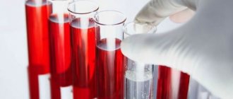
If your joints are swollen and painful at night, your rheumatologist will suggest you check your rheumatology profile. This examination will help make an accurate diagnosis, monitor the dynamics of the disease and prescribe the correct treatment.
If rheumatic disease is suspected, the following tests are used:
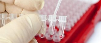
- blood test for uric acid levels;
- blood test for antinuclear antibodies;
- blood test for rheumatoid factor;
- blood test for ACCP (antibodies to cyclic citrulline-containing peptide);
- blood test for C-reactive protein.
Blood test for uric acid levels
Uric acid is the final breakdown product of purines. Every day a person receives purines through food, mainly meat products. Then, with the help of certain enzymes, purines are processed to form uric acid.
In normal physiological quantities, uric acid is needed by the body; it binds free radicals and protects healthy cells from oxidation. In addition, just like caffeine, it stimulates brain cells. However, high levels of uric acid have harmful consequences, in particular, they can lead to gout and some other diseases.
Testing uric acid levels makes it possible to diagnose uric acid metabolism disorders and related diseases.
1
Rheumatological examination
2 Rheumatological examination
3 Rheumatological examination
When to conduct an examination:
- with the first attack of acute arthritis in the joints of the lower extremities, which arose without obvious reasons;
- with recurrent attacks of acute arthritis in the joints of the lower extremities;
- if you have relatives in your family who suffer from gout;
- for diabetes mellitus, metabolic syndrome;
- with urolithiasis;
- after chemotherapy and/or radiation therapy for malignant tumors (and especially leukemia);
- with renal failure (the kidneys excrete uric acid);
- as part of a general rheumatological examination necessary to determine the cause of joint inflammation;
- with prolonged fasting, fasting;
- with a tendency to excessive consumption of alcoholic beverages.
Uric acid level
The level of uric acid is determined in the blood and urine.
Uric acid in the blood is called urecemia , and in urine - uricosuria . An increased level of uric acid is hyperuricemia , a decreased level of uric acid is hypouricemia . Only hyperuricemia and hyperuricosuria are of pathological significance.
The concentration of uric acid in the blood depends on the following factors:
- the amount of purines entering the body with food;
- synthesis of purines by body cells;
- the formation of purines due to the breakdown of body cells due to disease;
- the function of the kidneys, which excrete uric acid in the urine.
Under normal conditions, our body maintains normal uric acid levels. An increase in its concentration is one way or another associated with metabolic disorders.
Normal levels of uric acid in the blood
Men and women may have different concentrations of uric acid in the blood. The norm may depend not only on the gender, but also on the age of the person:
- in newborns and children under 15 years of age – 140-340 µmol/l;
- in men under 65 years old – 220-420 µmol/l;
- in women under 65 years of age – 40-340 µmol/l;
- in women over 65 years of age – up to 500 µmol/l.
If excess of the norm occurs for a long time, then crystals of uric acid salt (urate) are deposited in joints and tissues, causing various diseases.
Hyperuricemia has its own symptoms, but can also be asymptomatic.
Reasons for increased uric acid levels:
- taking certain medications, such as diuretics;
- pregnancy;
- intense loads in athletes and people engaged in heavy physical labor;
- prolonged fasting or eating foods containing large amounts of purines;
- some diseases (for example, endocrine), consequences of chemotherapy and radiation;
- impaired metabolism of uric acid in the body due to a deficiency of certain enzymes;
- insufficient excretion of uric acid by the kidneys.
How to reduce uric acid levels
Those who suffer from gout know how much trouble an increased concentration of uric acid can cause. Treatment of this disease must be comprehensive and must include taking medications that reduce the concentration of uric acid in the blood (xanthine oxidase inhibitors). It is recommended to drink more fluids and reduce the consumption of foods rich in purines.
It is also important to gradually lose excess weight, since obesity is usually associated with increased uric acid. The diet should be designed so that the amount of foods rich in purines is limited (red meat, liver, seafood, legumes). It is very important to give up alcohol. It is necessary to limit the consumption of grapes, tomatoes, turnips, radishes, eggplants, sorrel - they increase the level of uric acid in the blood. But watermelon, on the contrary, removes uric acid from the body. It is useful to consume foods that alkalize urine (lemon, alkaline mineral waters).
Purine metabolism is a complex biochemical process in which many enzyme systems take part. Most of the uric acid in the body is formed during the metabolism of nucleic acids, but other routes of its synthesis have been described. In all variants, the most important intermediate is inosinic acid, which subsequently undergoes hydrolysis. The resulting hypoxanthine is converted into xanthine and uric acid under the influence of the enzyme xanthine oxidase. From a biochemical point of view, disorders of purine metabolism represent various types of imbalance between the enzyme systems responsible for the synthesis and transport of uric acid and its precursors.
The content of purines in the body consists of their intake from food and endogenous synthesis. It is believed that the body of an adult healthy person contains about 1000 mg of uric acid. With gout, this figure can increase several times. The content of uric acid in the body is not a rigid and main clinical indicator of the state of purine metabolism. The range of normal values of blood uric acid varies widely, and in men it is in the range of 200-360 µmol/l, in women – 160-340 µmol/day. In healthy people, approximately 750 mg, or 2/3 of the total volume of uric acid, is excreted and synthesized again per day. Of this amount, about 80%, or 600 mg, is excreted by the kidneys, the remaining 20% is excreted through the gastrointestinal tract. According to calculations by P.M. Klimenko et al. (2010), normal uric acid clearance is 5.4-9.0 ml/min [1].
The metabolism of urate in the kidneys is a complex and multi-step process. Filtration of plasma urate occurs in the glomeruli. The urates that enter the ultrafiltrate are almost completely reabsorbed in the proximal tubule and then secreted. Some of the secreted urate is reabsorbed. The process of active secretion of urates is very sensitive to various chemical agents. It is believed that renal secretion of urate is increased by orotic acid, losartan, estrogens, and tetracycline breakdown products. Renal excretion of urate is reduced by ethambutol, thiazides and thiazide-like diuretics, and to a lesser extent by furosemide and acetazolamide [2]. The tendency of thiazides and indapamide to reduce renal excretion of urate and increase its serum concentration makes these drugs contraindicated in gout and hyperuricemia.
When purine metabolism is disrupted, overproduction of uric acid occurs, which is manifested by an increase in its concentration in the blood (hyperuricemia) and urine (hyperuricuria).
CLINICAL VARIANTS OF PURINE METABOLISM DISORDERS
Purine metabolism disorders themselves are not nosological entities, just as various congenital and acquired fermentopathies without clinical manifestations are not diseases. However, diseases associated with disorders of purine metabolism are relatively common in real urological practice, which makes this aspect relevant. Most urologists are well acquainted with the features of urate nephrolithiasis, and at the same time, in most cases, they have absolutely no idea about the existence of other, sometimes more serious, diseases caused by disorders of purine metabolism. Meanwhile, they all occur with varying frequency in urological hospitals, as well as during consultative work.
The most clinically significant consequence of disorders of purine metabolism is an increase in the level of uric acid in the blood - hyperuricemia, which is the main pathogenetic factor of various pathological conditions. Depending on the etiology, hyperuricemia is divided into primary and secondary to any disease.
The clinical consequence of primary hyperuricemia is gout in the broad sense of the term. This includes classic acute microcrystalline arthritis, as well as other types of damage to the musculoskeletal system, and various types of gouty nephropathy, one of which is urate nephrolithiasis, and tophi of various locations, and complications of all these conditions.
It is quite obvious that for a urologist, diseases of the genitourinary system caused by hyperuricemia are of greater interest. In nephrological and general therapeutic practice, the concept of “gouty kidney” was introduced several decades ago to determine kidney damage due to hyperuricemia, which in modern medicine has been transformed into “gouty nephropathy”. Considering the experimentally proven damaging effect of hyperuricuria on renal structures, the term “urate nephropathy” was also proposed. These concepts are generalizing and unite several processes that are quite different in their pathogenesis: acute uric acid nephropathy, urate nephrolithiasis and chronic tubulointerstitial nephritis. Some authors also note the possibility of immune complex glomerulonephritis due to impaired purine metabolism [3].
In urological practice, patients with urate nephrolithiasis are most often encountered. Up to 80% of such patients had an episode of arthritis at least once in their lives, and not necessarily in the classical localization - the first metatarsophalangeal joint. Recently, atypical variants of arthritis, for example, driving, have become increasingly common. In addition, the widespread and uncontrolled use of non-steroidal anti-inflammatory drugs often blurs the clinical picture, increasing the proportion of arthritis with less activity of the inflammatory process. It may be noted that the combination of arthritis and urate nephrolithiasis is not mandatory, but rather characteristic.
The clinical picture of a kidney or ureteral calculus is well known to all urologists, so there is no point in describing it in detail again. However, it should be noted that in the most severe, “malignant” course, along with the formation of urate stones in the lumen of the urinary tract, deposition of uric acid crystals in the renal interstitium is also possible, which is called nephrocalcinosis. Unlike nephrolithiasis, nephrocalcinosis in gout is always bilateral. Nephrocalcinosis does not have any specific symptoms. Clinical manifestations are reduced to the progression of renal failure due to the progression of nephrosclerosis. Nephrocalcinosis is detected in most cases by ultrasound scanning.
Tubulointerstitial nephritis is a characteristic and common variant of gouty nephropathy. However, due to the less vivid clinical picture, it is known mainly to nephrologists and rheumatologists.
In the initial stages of tubulointerstitial nephritis, the pathological process mainly affects the tubules and renal interstitium, so the leading symptom is a violation of the concentration function of the kidneys - polyuria with low urine density (hyposthenuria). Proteinuria does not exceed 1 g/day or is completely absent - it is associated with impaired protein reabsorption by the tubules. Gouty interstitial nephritis is characterized by persistent uraturia, as well as persistent or episodic microhematuria, especially after a respiratory viral infection. In an immunohistochemical study of renal biopsy specimens, some patients with a clinical picture of gouty tubulointerstitial nephritis showed fluorescence of the C3 fraction of complement and IgG, which is characteristic of immune complex glomerulonephritis. This made it possible to identify chronic glomerulonephritis as a separate variant of gouty nephropathy [3].
As gouty tubulointerstitial nephritis progresses, arterial hypertension and nephrosclerosis develop.
Acute uric acid nephropathy (acute gouty kidney) is based on obstruction of the renal tubules by uric acid crystals, which leads to acute renal failure. The disease begins with oliguria. Some patients simultaneously complain of renal colic-type pain and gross hematuria, which may be explained by the migration of large urate crystals through the ureter. High uraturia, which is not typical for acute renal failure of other etiologies, is pathognomonic. The assumption of acute uric acid nephropathy is based on a combination of three clinical signs - highly active arthritis with characteristic localization, a sharp decrease in diuresis and brick-brown urine. The diagnosis is all the more likely if there is a history of hypohydration of any origin - from visiting a bathhouse to inadequate infusion therapy and overdose of diuretics, as well as consumption of significant amounts of meat products and/or alcohol. In the natural course of the disease, oliguria almost always progresses to anuria with a detailed clinical picture of acute renal failure.
The problem of acute uric acid nephropathy is closely related to secondary hyperuricemia. The reasons for increased levels of uric acid in the blood serum are quite numerous and varied. Among them: chronic renal failure, regardless of etiology, obesity, especially high degrees, poorly compensated type 2 diabetes mellitus, dyslipidemia, acromegaly, hypothyroidism, hypoparathyroidism, toxicosis of pregnancy, myeloproliferative diseases, chronic lead intoxication, chronic alcoholism, severe forms of psoriasis, chemotherapy radiation therapy. In most cases, the severity of hyperuricemia in these diseases is small, less often – moderate. Thus, disorders of purine metabolism rarely significantly affect the clinical picture of the disease. However, their presence affects the development of complications and also increases the relative risk of cardiovascular and overall mortality.
The most striking and clinically significant variant of secondary hyperuricemia is “tumor lysis syndrome” (“tumor disintegration syndrome”), which develops during chemotherapy and radiotherapy for lymphoproliferative diseases, less often for tumors of other localizations. A key component of this syndrome, along with hyperphosphatemia and hyperkalemia, is the overproduction of uric acid, leading to the development of acute uric acid nephropathy, often in intact kidneys [4].
DRUG THERAPY FOR DISEASES CAUSED BY DISORDERS OF PURINE METABOLISM
Therapy for purine metabolism disorders consists of several components. A diet limiting foods rich in purines is required. Specific drugs are allopurinol and citrate.
Allopurinol is indicated for gouty tubulointerstitial nephritis, acute uric acid nephropathy, urate nephrolithiasis in combination with hyperuricemia, as well as chemotherapy for malignant neoplasms to prevent the development of secondary hyperuricemia and acute renal failure. The minimum effective dosage is 200 mg/day, the average therapeutic dosage is 300-400 mg/day. Chemotherapy for malignant neoplasms requires high, close to maximum dosages of allopurinol – 600-900 mg/day [2].
Citrate therapy is an integral part of the drug treatment of gouty nephropathy. The effect of citric acid salts on the process of crystal formation in urine is multifaceted. The solubility of uric acid varies significantly depending on the reaction of the medium. In an acidic environment, urates have very poor solubility and easily pass into the solid phase - they crystallize. With a neutral or alkaline reaction, the solubility of these salts increases. The main effect of citrates is the ability to reduce the acidity of urine, which prevents the crystallization of urates and creates conditions for the dissolution of already formed crystals. This is the basis of litholytic therapy. However, with an alkaline reaction of the environment, the solubility of phosphates decreases. The layering of a phosphate film on the urate stone makes the process of further litholysis practically hopeless. This dictates the need for careful monitoring of the urine reaction throughout the course of treatment. In modern conditions, the empirical use of plant materials rich in citric acid and its salts has been replaced by drugs that include chemically pure citrate and a set of test strips for monitoring urine reactions.
In addition to alkalization with citrate, potassium and sodium ions are needed to convert uric acid into a soluble keto form. It is their quantity that determines the “ionic strength of the solution.” The ratio of these ions to each other is also important, because it has been shown that an excess of sodium ions provokes the formation of a calcium “crust” on the stone.
Studies in the 80-90s demonstrated the effectiveness of litholysis of urate stones using citrates in monotherapy on the order of 75-80% [5, 6]. Currently, as a result of improving the technique, the effectiveness of litholysis has been increased to 85-90%, depending on the characteristics of the chemical composition of the stones [1, 7, 8].
In recent years, studies have appeared indicating the advisability of including citrate preparations in multicomponent treatment regimens. In particular, with urate stones of the ureter, especially in its distal third, combination therapy, including citrate and tamsulosin, led to spontaneous passage of 84.8% of stones, which significantly differs from the groups of patients receiving monotherapy with these drugs (68.8% and 58. 8%, respectively), as well as from patients receiving placebo (26.1%) [9].
There is convincing evidence for the effectiveness of the combination of allopurinol and citrate in gouty interstitial nephritis. A twelve-week course of combination therapy, including potassium sodium hydrogen citrate 3 g/day and allopurinol 100-200 mg/day, led to an increase in glomerular filtration rate by an average of 15 ml/min compared with the control group of allopurinol monotherapy. The clearance of uric acid also increased significantly and there was a greater decrease in its level in the blood. Let us note the low dosage of allopurinol. It can be assumed that there is a possible potentiation of the effects of allopurinol and potassium sodium hydrogen citrate. An additional positive consequence should be a reduction in the frequency of side effects of allopurinol, which is a significant limiting factor in the drug treatment of gouty nephropathy. Particularly significant was the increase in creatinine clearance in the initial stages of chronic renal failure. [10]
A more striking effect of citrate on renal function was noted in the treatment of chronic interstitial nephritis caused by hyperuricemia in obese patients [11].
The mechanism of action of citrate is not limited to alkalinization of urine. Citrate is one of the physiological inhibitors of crystal formation. Since urine is normally a supersaturated saline solution, the presence of crystal formation inhibitors in it is a necessary condition for the adequate functioning of the entire urinary system. Hypocitraturia is the most common metabolic abnormality in patients with stone formation. In addition, the chelate properties of the citrate molecule in relation to calcium ions make the citrate ion indispensable in the prevention of recurrence of calcium-containing stones. Citrate + thiazide diuretic is a standard combination for the metaphylaxis of calcium oxalate nephrolithiasis. Moreover, this therapy helps to increase bone density. This may explain the effectiveness of citrates not only in urate, but also in calcium-oxalate nephrolithiasis [12-14].
Due to the widespread use of EBRT and percutaneous nephrolithotomy, citrate therapy began to be actively used both before and after the procedure for 3-4 weeks. Taking citrate before DLT allows you to loosen the surface and structure of the stone, which increases the success of stone crushing with less trauma to the kidney. Long-term use after the procedure promotes accelerated clearance of stone fragments and prevents recurrence of lithogenesis.
Along with the mechanisms of action described above, citric acid salts additionally have antiseptic, cytoprotective and metabolic effects, which can also be used in clinical practice. In particular, Strassner C. and Friesen A. reported the disappearance of candiduria in 16 out of 18 patients during citrate therapy, which is likely due to a change in urine reaction [15]. The conclusion about the cytoprotective effect of citrate was made based on the successful attempts of Brühl P. et al. use it to prevent chemical injury to the bladder mucosa during therapy with drugs from the oxazaphosphorine group - cyclophosphamide and ifosfamide [16] (in modern oncological and nephrological practice, a drug from the group of mucolytics mesna, which has virtually no effect on acid-base balance, is used for this purpose). There are also reports of the use of citrate to correct acidosis due to ureterosigmostomy [17].
The main difficulty in citrate therapy for urate nephrolithiasis is selecting an adequate dosage of the drug. N.K. Dzeranov, who has studied and developed this aspect for many years, recommends starting with prescribing a diet and assessing the urine reaction for 5 days at a strictly defined time of day. Based on the obtained average urine pH values, the initial dose of the drug and, most importantly, its distribution throughout the day are determined. After 5 days of treatment, the average urine reaction is determined again at a strictly similar time of day and, if necessary, the dosage of the drug is adjusted [18]. “Interactive”, that is, in real time, changing the dosage of citrate is ineffective and even unsafe, as it leads to pH fluctuations, which can cause phosphate crystallization.
Since citrate is normally present in the body, drugs based on it are practically free of toxicity. However, there are clinical situations where the use of these drugs requires caution. The use of citrate mixtures is undesirable for acute uric acid nephropathy and, in general, for acute renal failure of any etiology. The limiting factor is not citration as such, but potassium, the removal of which is difficult in this clinical situation. For acute uric acid nephropathy, it is advisable to administer a 4% sodium bicarbonate solution, saline solution, etc. in combination with loop diuretics. It is necessary to maintain diuresis at a level of at least 100-150 ml/hour, urine pH at least 6.5 [3, 4].
In severe circulatory failure, the limiting factor is the increased intake of sodium into the body. Sometimes acetazolamide is preferable in this situation. This drug from the group of diuretics strongly, and most importantly, uncontrollably alkalinizes the urine, which makes it uncompetitive compared to citrate in the drug therapy of urate nephrolithiasis. However, acetazolamide is practically the only way to increase urine pH without resorting to the introduction of salts, which is extremely undesirable in conditions of severe heart failure.
Thus, drug treatment of patients with disorders of purine metabolism is a complex and multifaceted problem that requires an interdisciplinary approach.
Summary:
The article discusses the main clinical variants of purine metabolism disorders encountered in urological practice. The pathophysiology of purine metabolism disorders is briefly reflected. All clinical variants of gouty nephropathy are considered, including features of pathogenesis and clinical manifestations. The article discusses in detail the main approaches to drug therapy for these conditions, with an emphasis on specific citrate therapy. The available literature on this issue is analyzed. The possibilities of using citrate both in monotherapy and in combination with other drugs have been demonstrated. Indications and contraindications for the use of these drugs have been determined.
LITERATURE
1. Klimenko P.M., Chabanov V.A., Akinshevich I.Yu. Possibilities of conservative treatment of patients with urate nephrolithiasis. // News of medicine and pharmacy. 2010. No. 3. P.5-7.
2. Federal guidelines for the use of drugs (formulary system). Issue X. 2009. Edited by Chuchalin A.G., Belousov Yu.B., Yasnetsov V.V. Moscow. JSC RIC "Man and Medicine".
3. Nephrology. Guide for doctors. Edited by I.E. Tareeva. Moscow. Medicine. 2000. 688p.
4. Nephrology. National leadership. Edited by N.A. Mukhina. Moscow. GEOTAR-Media. 2009. 716 p.
5. Chugtai MN, Khan FA, Kaleem M., Ahmed M. Management of uric acid stone. // J Pak Med Assoc. 1992 Jul;42(7):153-5.
6. Petritsch PH Uric acid calculi: results of conservative treatment. // Urology. 1977 Dec;10(6):536-8.
7. Eliseev M.S., Denisov I.S., Barskova V.G. Use of Uralit-U citrate in patients with gout and nephrolithiasis. // Modern rheumatology. 2012. No. 3. P.13-15.
8. Pasechnikov S.P., Mitchenko M.V. Modern aspects of citrate therapy for urolithiasis. Experience with the use of the drug Uralit-U. // Men's health. 2007. No. 3. P.109-113.
9. El-Gamal O., El-Bendary M., Ragab M., Rasheed M. Role of combined use of potassium citrate and tamsulosin in the management of uric acid distal ureteral calculi // Urological Research June 2012, Volume 40, Issue 3, pp. 219-224.
10. Saito J., Matsuzawa Y., Ito H., Omura M., Ito Y., Yoshimura K., Yajima Y., Kino T., Nishikawa T. pЈe alkalizer citrate reduces serum uric acid levels and improves renal // Endocr Res 2010;35(4):145-154.
11. Saito J., Matsuzawa Y., Ito H., Omura M., Kino T., Nishikawa T. Alkalizer Administration Improves Renal Function in Hyperuricemia Associated with Obesity. // Japanese Clinical Medicine 2013:4.
12. Butz M. Oxalate stone prophylaxis by alkalinizing therapy. // Urologe A. 1982 May;21(3):142-6.
13. Ito H. Combined administration of calcium and citrate reduces urinary oxalate excretion. // Hinyokika Kiyo. 1991 Oct;37(10):1107-10.
14. Berg C., Larsson L., Tiselius HG Effects of different doses of alkaline citrate on urine composition and crystallization of calcium oxalate. // Urological Research February 1990, Volume 18, Issue 1, pp 13-16.
15. Strassner C., Friesen A. Therapy of candiduria by alkalinization of urine. Oral treatment with potassium-sodium-hydrogen citrate. // https://www.ncbi.nlm.nih.gov/pubmed/7498850
16. Bruhl P., Hoefer-Janker H., Scheef W., Vahlensieck W. Prophylactic alkalization of the urine during cytostatic tumor treatment with the oxazaphosphorine derivatives, cyclophosphamide and ifosfamide. // Onkologie. 1979 Jun;2(3):120-4.
17. Sasagama I., Nakada T., Ishgooka M., Kubota Y., Sawamura T. Effect of standardized mixture of potassium and sodium citrate and citric acid (Uralit-U) on the correction of postoperative acidosis in patients who suffered uriterosigmostomy. // Nephron 1994;66:477-478.
18. Dzeranov N.K., Rapoport L.M. Litholytic therapy. Practical recommendations. Moscow. LLC "Informpolygraph" 2011. 16 p.
| Attached file | Size |
| 346.13 kb |
‹ Molecular genetic disorders as criteria in the differential diagnosis of rare kidney tumors Up Nocturia: modern gender aspects of epidemiology, pathogenesis and diagnosis ›
Antinuclear antibodies (ANA)
Using the ANA test, you can determine the presence of antinuclear antibodies (antibodies to nuclear antigens) in the blood.
ANAs are a group of specific autoantibodies that are produced by our body's immune system in case of autoimmune disorders. Antibodies have a damaging effect on the body's cells. In this case, a person experiences various painful symptoms, such as pain in muscles and joints, general weakness, etc.
Detection of antibodies belonging to the ANA group (for example, antibodies to double-stranded DNA) in blood serum helps to identify an autoimmune disease, monitor the course of the disease and the effectiveness of its treatment.
1 Blood test for ACCP
2 Blood test for C-reactive protein
3 Blood test for ACCP
When is a blood test for antinuclear antibodies necessary?
Detection of antinuclear antibodies may be a sign of the following autoimmune diseases:
- polymyositis;
- dermatomyositis;
- systemic lupus erythematosus;
- mixed connective tissue disease;
- scleroderma;
- Sjögren's syndrome and disease;
- Raynaud's syndrome;
- autoimmune hepatitis
How is the antinuclear antibody test performed?
Blood for antinuclear antibodies is taken from a vein in the elbow, on an empty stomach. Before the study, you do not have to adhere to any diet.
In some cases, in order to differentiate various autoimmune diseases, additional clarifying tests for autoantibodies from the group of antinuclear antibodies, the so-called ANA immunoblot, may be required.
What do the test data mean?
Antinuclear antibodies (another name is antinuclear factor ) indicate the presence of some kind of autoimmune disorder, but do not precisely indicate the disease that caused it, since the ANA test is a screening test. The goal of any screening is to identify people at increased risk of a particular disease.
A healthy person with normal immunity should not have antinuclear antibodies in the blood or their level should not exceed the established reference values.
A normal ANA value implies an antibody titer not exceeding 1: 160. Below this value, the test is considered negative.
A positive test for antinuclear antibodies (1:320 or more) indicates an increase in antinuclear antibodies and the presence of a disease of an autoimmune nature in a person.
Currently, two methods are used to detect antinuclear antibodies: indirect immunofluorescence reaction using the so-called Hep2 cell line and enzyme-linked immunosorbent assay. Both tests complement each other, and therefore they are recommended to be performed simultaneously.
The following types of ANA antinuclear bodies can be distinguished in the indirect immunofluorescence reaction:
- homogeneous coloring - can be with any autoimmune disease;
- spotty or speckled coloration may occur with systemic lupus erythematosus, scleroderma, Sjögren's syndrome, rheumatoid arthritis, polymyositis and mixed connective tissue disease;
- peripheral coloring – characteristic of systemic lupus erythematosus;
If the test for antinuclear antibodies is positive, it is necessary to perform an immunoblot of antinuclear antibodies to clarify the type of autoimmune disease and make a diagnosis.
Rheumatoid factor
A blood test for rheumatoid factor is aimed at identifying specific IgM class antibodies to IgG class antibodies.
A laboratory test for rheumatoid factor is a screening test aimed at identifying autoimmune disorders. The main objective of the study for rheumatoid factor is to identify rheumatoid arthritis, Sjogren's disease and syndrome and a number of other autoimmune diseases.
A rheumatoid factor test may be needed for the following symptoms:
- pain and swelling in the joints;
- limited mobility in joints;
- feeling of dryness in the eyes and mouth;
- skin rashes like hemorrhages;
- weakness, loss of strength.
1 Rheumatoid arthritis
2 Rheumatological examination
3 Rheumatological examination
Norms of rheumatoid factor in the blood
Theoretically, rheumatoid factor should not exist in a healthy body. But still, in the blood of some, even healthy people, this factor is present in a small titer. Depending on the laboratory, the upper limit of normal for rheumatoid factor varies from 10 to 25 international units (IU) per milliliter of blood.
Rheumatoid factor is the same in women and men. In older people, the rheumatoid factor level will be slightly higher.
The normal rheumatoid factor in a child should be 12.5 IU per milliliter.
Rheumatoid factor testing is used to diagnose the following diseases:
- rheumatoid arthritis;
- systemic autoimmune diseases;
- Rioglobulinemia.
Other causes of elevated rheumatoid factor
Additional reasons for increased rheumatoid factor may be the following:
- syphilis;
- rubella;
- Infectious mononucleosis;
- malaria;
- tuberculosis;
- flu;
- hepatitis;
- leukemia;
- cirrhosis of the liver;
- sepsis
If the cause of increased rheumatoid factor is an infectious disease, for example, infectious mononucleosis, then the titer of rheumatoid factor is usually less than with rheumatoid arthritis.
However, rheumatoid factor testing primarily helps to recognize rheumatoid arthritis. However, it should be emphasized that it is impossible to make a diagnosis on its basis alone. Since rheumatoid factor can be elevated in many other pathological conditions of an autoimmune and non-autoimmune nature. In addition, in approximately 30% of patients with rheumatoid arthritis, a blood test for rheumatoid factor may be negative (seronegative rheumatoid arthritis).
A blood test for rheumatoid factor is carried out in the morning on an empty stomach (8 to 12 hours should pass since the last meal).
Causes of decreased uric acid
If uric acid in the blood is low, then they speak of hypouricemia. The following main reasons for the decrease in uric acid concentration levels are identified:
- Fanconi syndrome - absorption of the renal tubules is impaired, the disease is congenital
- Hepatocerebral dystrophy is an inherited pathology characterized by problems with the metabolism of microelements (copper in particular). The central nervous system and liver are the first to be affected.
- Acquired xanthine oxidase deficiency. The main cause of the disease is impaired liver function, medications containing the substance allopurinol. The production of enzymes responsible for acid synthesis is affected by the active substance of the drug.
- Hereditary deficiency of xanthine oxidase - the body cannot synthesize uric acid on its own. It is less common than the acquired one.
- Alcoholism.
- Poor nutrition.
Often, a decrease in the concentration of crystals is observed in women expecting a child. This is due to an increase in the amount of blood in pregnant women. And the concentration of cells and dissolved minerals decreases. Another reason is an increase in estrogen, which affects the formation of acid crystals.
ACDC
A blood test for ACCP consists of determining the titer of antibodies to cyclic citrullinated peptide and is one of the accurate methods for confirming the diagnosis of rheumatoid arthritis. With its help, the disease can be detected several years before symptoms appear.
What does the ACDC analysis show?
Citrulline is an amino acid that is a product of the biochemical transformation of another amino acid - arginine. In a healthy person, citrulline does not take part in protein synthesis and is completely eliminated from the body.
But with rheumatoid arthritis, citrulline begins to integrate into the amino acid peptide chain of proteins in the synovial membrane and cartilage tissue of the joints. The “new” modified protein, which contains citrulline, is perceived by the immune system as “foreign” and the body begins to produce antibodies to citrulline-containing peptide (ACCP).
ACCP is a specific marker of rheumatoid arthritis, a kind of harbinger of the disease at an early stage, with high specificity. Antibodies to cyclic citrullinated peptide are detected long before the first clinical signs of rheumatoid arthritis and remain throughout the disease.
Methodology of analysis and its significance
To detect ACCP, an enzyme-linked immunosorbent assay is used. A blood test for ACCP is carried out according to the “in vitro” principle (translated from Latin - in a test tube), serum from venous blood is examined. The ACCP blood test can be ready within 24 hours (depending on the type of laboratory).
Detection of ACCP in rheumatoid arthritis may indicate a more aggressive, so-called erosive form of the disease, which is associated with more rapid resolution of joints and the development of characteristic joint deformities.
If the test result for ACCP is positive, then the prognosis for rheumatoid ACCP arthritis is considered less favorable.
1 Blood test for ACCP
2 Blood test for C-reactive protein
3 Blood test for ACCP
ACDC. Reference values
The normal range for the ACCP test is approximately 0-5 U/mL. The so-called “ ACCP norm ” may vary depending on the laboratory. The “ACCP norm” values for women and men are the same.
The so-called “ Increased ACCP ”, for example, ACCP 7 units/ml or more, indicates a high likelihood of rheumatoid arthritis. An analysis result assessed as “ ACCP negative ” reduces the likelihood of rheumatoid arthritis, although it does not completely exclude it. A rheumatologist with experience in diagnosing and treating rheumatoid arthritis should always evaluate the ACCP values and interpret them; only a rheumatologist can take into account all the nuances.
To get tested for ACCP, you need to come for examination on an empty stomach.
Indications for the purpose of analysis:
- rheumatoid arthritis;
- early synovitis;
- osteoarthritis;
- polymyalgia rheumatica;
- psoriatic arthritis;
- Raynaud's disease;
- reactive arthritis;
- sarcoidosis;
- scleroderma;
- Sjögren's syndrome;
- SLE;
- vasculitis;
- juvenile RA.
If you want to know the cost of a blood test for ACCP, please call.
Contact center specialists will tell you the price of the ACDC and explain how to prepare for the study.
C-reactive protein test
C-reactive protein ( CRP ) is a very sensitive element of a blood test that quickly responds to even the slightest damage to body tissue. The presence of C-reactive protein in the blood is a harbinger of inflammation, injury, and the penetration of bacteria, fungi, and parasites into the body.
CRP more accurately shows the inflammatory process in the body than ESR (erythrocyte sedimentation rate). At the same time, C-reactive protein quickly appears and disappears - faster than the ESR changes.
Due to the ability of C-reactive protein to appear in the blood at the very peak of the disease, it is also called “acute phase protein.”
As the disease enters the chronic phase, C-reactive protein decreases in the blood, and when the process worsens, it increases again.
C-reactive protein is normal
C-reactive protein is produced by liver cells and is found in minimal amounts in the blood serum. The content of CRP in blood serum does not depend on hormones, pregnancy, gender, or age.
The norm of C-reactive protein in adults and children is the same - less than 5 mg/l (or 0.5 mg/dl).
A blood test for C-reactive protein is taken from a vein in the morning, on an empty stomach.
1 Blood test for uric acid levels
2 blood test for antinuclear antibodies
3 Blood test for rheumatoid factor
Causes of increased C-reactive protein
C-reactive protein may be elevated in the presence of the following diseases:
- rheumatism;
- acute bacterial, fungal, parasitic and viral infections;
- gastrointestinal diseases;
- focal infections (for example, chronic tonsillitis);
- sepsis;
- burns;
- postoperative complications;
- myocardial infarction;
- bronchial asthma with inflammation of the respiratory system;
- complicated acute pancreatitis;
- meningitis;
- tuberculosis;
- tumors with metastases;
- some autoimmune diseases (rheumatoid arthritis, systemic vasculitis, etc.).
With the slightest inflammation, in the first 6-8 hours the concentration of C-reactive protein in the blood increases tenfold. There is a direct relationship between the severity of the disease and changes in CRP levels. Those. The higher the concentration of C-reactive protein, the stronger the inflammatory process develops.
Therefore, changing the concentration of C-reactive protein is used to monitor and control the effectiveness of treatment of bacterial and viral infections.
Different reasons lead to different increases in C-reactive protein levels:
- The presence of chronic bacterial infections and some systemic rheumatic diseases increases C-reactive protein to 10-30 mg/l. With a viral infection (if there is no injury), the level of CRP increases slightly. Therefore, high values indicate the presence of a bacterial infection .
- If neonatal sepsis is suspected, a CRP level of 12 mg/l or more indicates the need for urgent antimicrobial therapy.
- In acute bacterial infections, exacerbation of some chronic diseases, acute myocardial infarction and after surgery, the highest level of CRP is from 40 to 100 mg/l. With proper treatment, the concentration of C-reactive protein decreases within the next few days, and if this does not happen, it is necessary to discuss other antibacterial treatment. If after 4-6 days of treatment the CRP value has not decreased, but remains the same and even increased, this indicates the occurrence of complications (pneumonia, thrombophlebitis, wound abscess, etc.). After surgery, the more severe the operation, the higher the CRP will be.
- During myocardial infarction, protein increases 18-36 hours after the onset of the disease, decreases after 18-20 days and returns to normal by 30-40 days. With angina pectoris, it remains normal.
- In a variety of tumors, elevated levels of C-reactive protein can serve as a test to assess tumor progression and disease recurrence.
- Severe general infections, burns, sepsis increase C-reactive protein to enormous values: up to 300 mg/l or more.
- With proper treatment, the level of C-reactive protein decreases already on days 6-10.
Preparation for rheumatological tests
In order for analyzes to show objective information, it is necessary to adhere to certain rules. You need to donate blood in the morning, on an empty stomach. Approximately 12 hours should pass between taking tests and eating. If you're thirsty, drink some water, but not juice, tea or coffee. It is necessary to exclude intense physical exercise and stress. You cannot smoke or drink alcohol.
The multidisciplinary clinic "MedicCity" provides diagnostics of the highest level, experienced, qualified rheumatologists and specialists in more than 30 specialties. We treat arthritis, arthrosis, vasculitis, lupus erythematosus, osteoporosis, gout, rheumatism and many other rheumatological diseases. Do not delay your visit to the doctor, contact us at the slightest symptoms. High-quality diagnosis is 90% of successful treatment!
How to determine the exact level of uric acid?
A biochemical blood test will show the level of uric acid in the blood; it is carried out both for healthy people, for possible detection of diseases, and for people suffering from metabolic disorders - urolithiasis, diabetes, gout, etc.
There are certain recommendations for obtaining an accurate analysis result:
- Eliminate all high-protein foods from your diet 8 hours before the test.
- Do not drink alcohol or take medications the day before the collection.
- Blood is drawn on an empty stomach.
- Avoid physical activity the day before the test.
Blood is taken from a vein. Most often, the result is ready the next day. If the result is reduced, a repeat analysis is prescribed. After confirming the deviation from the norm, the doctor prescribes a full examination and, based on its results, makes a diagnosis.
Factors that may affect the result of the analysis:
- Taking diuretics:
- Hormonal drugs
- Diet with minimal protein content.
Before the study, it is imperative to exclude such factors.