Chronic osteomyelitis of the jaw
Chronic osteomyelitis of the jaw develops from an untreated acute form of the disease. This osteomyelitis is called secondary chronic. If the inflammatory process was initially sluggish and was not as clinically pronounced as acute, then this is primary chronic osteomyelitis.
Chronic osteomyelitis, like acute osteomyelitis, can be of infectious or non-infectious origin. The first, in turn, is divided into odontogenic and non-odontogenic.
In accordance with the predominance of the processes of construction or death of bone substance, 3 clinical and radiological forms of chronic odontogenic osteomyelitis of the jaws are distinguished: productive (without the formation of sequestration), destructive (with the formation of sequestration) and destructive-productive. The productive form is less common than the others, mainly at a young age.
Chronic odontogenic osteomyelitis of the jaw
Chronic odontogenic osteomyelitis of the jaw is often secondary chronic and is considered as a complication of acute odontogenic osteomyelitis. The transition from the acute stage of the disease to the chronic stage occurs on average at 4-5 weeks of illness. By this time, the manifestations of acute inflammation have passed: the swelling of the soft tissues surrounding the jaw subsides, the amount of pus released from the wound decreases, the pus itself becomes thicker, and granulation tissue forms in the wound. The patient’s general condition also normalizes: body temperature returns to normal, the patient does not complain of pain in the affected area, sleep and appetite are restored, blood tests are approaching normal values.
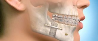
Figure 1. Formation of the fistula tract.
The first clinical sign that the acute stage has not been cured is the appearance of fistulas with pus in the wound area. Sometimes fistulas can open on the skin of the maxillofacial area.
Next, the formation of sequesters is observed, which, depending on their size, either exit through the fistula tracts (small) or are subject to removal by a maxillofacial surgeon (large).
Figure 2. Sequestration formation and rejection.
If the outflow of pus is disrupted and small sequesters are removed through the fistulous tracts, the chronic process worsens, the clinical picture becomes the same as in acute osteomyelitis.
The picture described above is characteristic of destructive or destructive-productive forms of osteomyelitis. The productive form is characterized by the absence of sequesters and an increase in bone tissue in the area of inflammation; it occurs only in osteomyelitis of the lower jaw.
General information
Deep inflammation of bone tissue disrupts trophic processes in the affected area and activates the activity of leukocytes. Decay products begin to decompose the bone with the formation of a purulent cavity, which over time can grow greatly, increasing the fragility of the bone and causing deformities.
If treatment is carried out poorly or incompletely, after the infectious pathogens adapt to the drugs, additional mutant strains appear, which blur the symptom complex of the disease and weaken the body’s response to medications. Over time, this can cause immunodeficiency states and chronicity of the process.
Reference! Anatomically, the jaw bones have the closest possible contact with a potential source of infection - carious teeth and pathogenic microflora of the oral cavity. A small amount of damage is enough for bacteria to gain access to the bone tissue and initiate inflammation. This explains the high percentage of osteomyelitis affecting the maxillofacial region of the skeleton.
Diagnosis of chronic odontogenic osteomyelitis
Diagnosis of chronic odontogenic osteomyelitis consists of collecting a history of the disease, examining the patient and performing radiography.
From the anamnesis we learn that the patient either suffered from acute osteomyelitis and did not seek help, or assistance was provided to him, but the acute form of the process became chronic. In both cases, further examination of the patient is carried out.
The clinical picture is very diverse, so it is difficult to accurately characterize all the signs of the disease.
Externally, the face may be asymmetrical due to soft tissue swelling or bone deformation. In the productive form, asymmetry can be caused by an increase in the volume of bone tissue.
The opening of the mouth is either normal or not fully, which is caused by inflammatory contracture of the masticatory muscles.
Lymph nodes are normal or may be slightly enlarged and painful on palpation.
When examining the oral cavity, an inflammatory infiltrate, hyperemic mucosa, the causative tooth or the socket of an extracted tooth are determined. Fistulas are found on the mucous membrane of the oral cavity or on the skin, through which formed sequestra are probed. Teeth that are mobile in acute osteomyelitis are less mobile in the chronic form of the disease.
Next, an X-ray diagnosis is carried out, preferably an orthopantomogram or radiography in two projections (direct and lateral). In the acute form of odontogenic osteomyelitis, only the source of infection is observed - thinning of the bone tissue in the area of the apex of the root of the causative tooth. If the disease has become chronic, sequestration is visible on the image. But the first manifestations of the disease in the picture appear only at the end of the 2nd, and sometimes 3rd, week. The situation with the destructive form of osteomyelitis is described above.
If we talk about the productive form, then bone sequestration is not observed. But the amount of mineralized tissue increases due to the reaction of the periosteum. The patient's face becomes asymmetrical, the bone increases in volume.
Chronic odontogenic osteomyelitis of the lower jaw
Chronic odontogenic osteomyelitis of the lower jaw often affects only the alveolar part of the bone, less often the body or branch of the jaw. Due to the structural features, the disease is severe with the formation of small and large sequesters. Often, the destruction of bone tissue leads to a pathological fracture (the bone breaks with a weak “blow” to the jaw).
Chronic odontogenic osteomyelitis of the upper jaw
Chronic odontogenic osteomyelitis of the upper jaw develops faster and is easier than in the lower jaw. Sequestra are formed in 3-4 weeks, while in the lower jaw - in 6-8 weeks. With the diffuse nature of the disease, destruction of the anterior wall of the maxillary sinus or even the lower edge of the orbit is possible.
Classification of pathology
Depending on the method of infection, the following types are distinguished:
- Hematogenous - bacteria spread through the circulatory system from other infectious foci. This path is typical for secondary osteomyelitis of the upper jaw, which develops against the background of another disease. This could be scarlet fever, purulent otitis media, tonsillitis, or diseases of the larynx. First, the infection affects the bone tissue of the jaw, and then only the teeth and gums.
- Odontogenic – external route of infection. The cause is an infected pulp or tooth root. Predisposing factors include advanced dental diseases. These are pulpitis, periodontitis, the presence of cystic formations, alveolitis.
- Traumatic – fractures and injuries of the jaw joint with damage to soft tissues and the presence of fragments of the dentition. Often occurs due to late seeking medical help.
According to the course and nature of the manifestation of symptoms, the pathology can be acute, subacute and chronic. Based on the area of distribution, local (limited) and diffuse (diffuse) forms are distinguished.
Treatment of chronic odontogenic osteomyelitis of the jaw
Treatment of chronic odontogenic osteomyelitis of the jaw is complex and includes surgical intervention and drug treatment.
I. During exacerbation of chronic osteomyelitis, the symptoms of acute inflammation are first relieved. If the causative tooth has not been removed previously, then it must be removed this time. Adjacent mobile teeth are trepanned and splinted, if not removed according to indications (after assessing their viability and x-ray examination). Sanitation of the oral cavity is mandatory and all chronic sources of infection are removed to prevent complications during subsequent procedures.
To facilitate the outflow of pus, fistulas or wounds are expanded, and primary surgical treatment of subosseous and perimandibular purulent foci is performed.
An important part of the surgical stage of treatment is sequestrectomy. After evaluating the radiograph, the formed sequesters are removed. Removal is performed through an intraoral or extraoral incision. Large sequestra in the area of the body and ramus of the lower jaw, as well as in the area of the infraorbital margin and zygomatic bone, are removed extraorally. Sometimes large necrotic areas of bone are broken into several parts for ease of removal. Incisions are made along the natural folds of the face for better aesthetics.
After removal of sequesters, attention is paid to granulations and sequestral capsule. Using a curettage spoon or even a milling cutter, pathological tissue is removed until the signs of healthy bone are revealed: alveolar bleeding, white bone, hard bone tissue.
The free space is filled with a biosynthetic osteotropic drug: kolapol, kolapan, etc. The wound is tightly sutured, drainage is left. The stitches are removed after 7-10 days.
II. Next, let's move on to drug treatment. As with other purulent diseases, etiotropic, pathogenetic and symptomatic treatment is carried out.
To eliminate the cause of the disease, the surgeon removes the causative teeth. But the infection remains in the blood, so the patient is prescribed antibacterial drugs: macrolides, cephalosporins. It is also worth prescribing the patient antifungal agents (Deflucan 150 mg once a week).
Since the patient’s immunity is reduced, it is recommended to prescribe immune drugs such as thymalin, T-activin, levomisol, staphylococcal toxoid.
In case of extensive damage to bone tissue, the patient is recommended to follow a gentle diet to prevent a pathological fracture of the jaw.
To reduce the symptoms of inflammation, detoxification and anti-inflammatory therapy is carried out. Physical therapy exercises and physiotherapy are individually selected for the patient to restore function.
Chronic osteomyelitis of the jaw. Outcomes and complications
Outcomes:
- Favorable - if the patient contacts a maxillofacial surgeon in a timely manner and receives adequate treatment, the patient’s full recovery is possible.
- Unfavorable – if treatment is insufficient and the patient contacts a doctor late, the following may occur:
- Exacerbation of the disease
- Jaw deformity,
- Jaw fracture - occurs due to minor physical impact, from which a healthy jaw would not be damaged,
- Complications of osteomyelitis: Abscesses and phlegmon of soft tissues of the face,
- Thrombosis of facial vessels and cavernous sinus,
- Mediastinitis,
- Death.
Prognosis and prevention
With timely diagnosis and compliance with all instructions, osteomyelitis responds well to treatment. Otherwise, there is a risk of the following complications:
- phlegmons, abscesses, adenophlegmons - if the affected area reaches the orbit, there is a risk of partial or complete loss of vision;
- severe forms of sinusitis with damage to the maxillary and even frontal sinuses;
- thrombophlebitis and thrombosis of large facial vessels;
- spread of the infectious process to the brain - meningitis, brain abscesses;
- infectious lesions of the lungs (pneumonia, pleurisy), liver, spleen.
A frequent consequence of osteomyelitis of the jaw is serious cosmetic defects with impaired functionality of the maxillofacial apparatus. For a full recovery, the patient may require the services of an oral and maxillofacial surgeon and a plastic surgeon. To avoid an unfavorable scenario, carefully monitor your health and strictly adhere to preventive measures:
- Treat any infectious and inflammatory processes in a timely manner, regardless of whether it is caries or pyelonephritis.
- Follow the rules of personal hygiene - try to brush your teeth at least 2 times a day, use mouthwash. Don’t forget about general hygiene - wash your hands more often, change your linen, do wet cleaning in the house.
- Strengthen your immune system - eat well, strengthen your body, move more.
- Avoid injuries to the maxillofacial area and be attentive to their treatment if this cannot be avoided.
Remember that osteomyelitis is an extreme stage of inattention to one’s health. This is a disease of a neglected organism with a weak immune system and lack of nutrition.
Symptoms
The clinical picture depends on the stage
of Acute Pain, mobility of the causative tooth and those located nearby. Opening the mouth, difficulty swallowing, breathing with a putrid odor. Swelling and redness of the gums. Feverish state, lack of appetite, lethargy, headache. Inflammation of the lymph nodes. Subacute
The inflammatory process subsides, suppuration decreases, the mouth opens freely.
Teeth mobility is maintained. Improvement in general condition, decrease in temperature (slightly above 37°C). Regional lymph nodes are dense but painless. Chronic
Reduced tissue infiltration, subsidence of pain.
The jaw in the area of the inflamed lesion is thickened; upon incision, fistulas with purulent contents are revealed. CT scan reveals roughness and unevenness of dying bone tissue. Exacerbation
The clinical picture of the acute stage is repeated - general signs of intoxication suddenly appear. Local symptoms increase - swelling of the inflamed area, soreness and mobility of teeth, the formation of purulent fistulas.
Stages of treatment
Treatment at our Center is comprehensive; whenever possible, we strive to combine all activities into one visit.
- Oral preparation It is important to establish a sterile environment before surgery to avoid re-infection. Hygienic cleaning and retreatment of teeth are carried out.
- The operation uses ultrasound to remove teeth that have become a source of infection. According to the selected protocol, access to the sinus is formed, infected areas and elements that support inflammation are eliminated.
- Prosthetics If a tooth was removed during the treatment of an odontogenic form of osteomyelitis, on the same day we manufacture and install temporary orthopedic structures to hide the defect.
After surgery, X-ray monitoring is required to assess the quality of the operation.