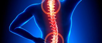
Chest pain ( thoracalgia ) is one of the most serious symptoms a person can experience. Sometimes even a doctor cannot immediately determine the cause of chest pain and find out whether this symptom is a sign of a life-threatening condition.
- Chest pain can be in any part and is caused by diseases of the heart, lungs, esophagus, muscles, bones, and skin.
- Due to the complex innervation of the body, chest pain may come from another part of the body.
- Chest pain may be caused by diseases of the stomach or other abdominal organs.
Symptoms of the disease
The symptoms of this disease are quite simple and it is with this variant of thoracic osteochondrosis that the diagnosis is usually quickly and accurately established. The clinical picture is represented by two main and a number of accompanying symptoms:
- Pain in the thoracic spine. The pain may intensify with movement, after static physical activity.
- Muscle tension in the thoracic spine.
- Crunching in the vertebrae when moving.
- Discomfort, crawling sensation in the interscapular area.
- An indirect symptom is an increase in the above complaints after hypothermia, prolonged exposure to a monotonous position (working at a computer).
How does vertebrogenic thoracalgia differ from thoracalgia due to ischemic heart disease?
*IHD - coronary heart disease.
Osteoarticular pain in the thoracic spine and chest
N.V. PIZOVA
,
Doctor of Medical Sciences, Professor, State Budgetary Educational Institution of Higher Professional Education "Yaroslavl State Medical Academy" of the Ministry of Health of Russia The article discusses in detail the causes and mechanisms of the occurrence of vertebrogenic pain syndromes.
A differentiated approach to therapy is presented, taking into account the pathogenesis and stage of the underlying disease. The advantages of using combination drugs (diclofenac and B vitamins) in the treatment of patients with back pain are described. Back pain is one of the most common sufferings of modern people. It is associated with significant economic losses caused by temporary and even permanent loss of ability to work in young and middle-aged people. Back pain accounts for up to 6% of all direct costs for the treatment of various diseases, 15% of all days of disability and 18% of causes of disability [1]. Acute and chronic back pain can be manifestations of any disease. And a doctor of any specialty faces two main tasks - to identify the cause of pain and to find a medicine that adequately helps with it.
All back pain syndromes can be classified into the following categories:
1. by reason – vertebrogenic and non-vertebrogenic; 2. by mechanism – reflex, compression, against the background of instability of the spinal motion segment (SMS), vascular, inflammatory; 3. by localization - local, reflected and irradiating; 4. by duration – acute and chronic.
During life, back pain occurs in 70–90% of the population, and in 20–25% it is registered annually. In the vast majority of patients, as a result of the therapy, pain is relieved within 4 weeks. However, 73% of patients develop at least one exacerbation during the first year [2, 3, 4]. Thus, according to a Russian study, the most common location of pain (576 patients - 60.6%) was the back. According to medical documentation, back pain was regarded as dorsopathy in 417 (72.4%) patients and as a consequence of intervertebral disc herniation in 104 (18.1%). 34 (5.9%) patients had osteoporotic fractures of the vertebral bodies, and 21 had other causes of back pain [5].
Chronic back pain is observed in 20–25% of cases, and this category of patients accounts for up to 80% of the economic costs associated with pain in this localization [6]. Chronic pain is the result of a complex interaction between biological, psychological, social and cultural factors that make its diagnosis and treatment difficult [7, 8]. Chronic back pain can be classified as nociceptive, neuropathic, inflammatory, dysfunctional, or can be mixed when there are characteristics of several types of pain [9]. Although their causes and clinical manifestations are different, the mechanisms by which these types of pain occur may overlap, and a patient may develop chronic back pain with components of more than one type of pain.
The source of back pain can be pain impulses associated both with the spine itself - vertebral factors (ligaments, muscles, periosteum of processes, fibrous ring, joints, roots), and with other structures - extravertebral factors (muscles, visceral organs, joints). Traditionally, thoracalgia, like other pain syndromes, depending on the cause, is divided into vertebrogenic (pathogenetically caused by changes in the spine) and non-vertebrogenic pain syndromes. Vertebrogenic thoracic syndromes include damage to the thoracic roots due to intervertebral disc herniation, spinal canal stenosis, spondylolisthesis and instability, and arthropathic syndrome due to degenerative lesions of the facet and costotransverse joints. Vertebrogenic causes of pain in the thoracic spine also include relatively rare malignant neoplasms of the spine (primary tumors and metastases), inflammatory (spondyloarthropathy, including ankylosing spondylitis) and infectious lesions (osteomyelitis, epidural abscess, tuberculosis), as well as compression fractures of the vertebral bodies due to osteoporosis. The cause of non-vertebrogenic pain syndromes can be pathology of internal organs and muscle pain syndromes, which can be formed under the influence of both vertebrogenic and non-vertebrogenic changes. Therefore, the division into vertebrogenic and non-vertebrogenic pain syndromes can be considered quite relative. In addition, psychogenic pain syndromes (panic attacks and hyperventilation disorders) are a possible cause of nonvertebrogenic chest pain.
Clinical syndromes in spondylogenic thoracalgia include [10]:
1. local vertebral syndrome, often accompanied by local pain, tension and soreness of adjacent muscles, pain and deformity, limited mobility or instability of one or more adjacent segments of the spine; 2. vertebral syndrome at a distance; 3. reflex (irritative) syndromes: referred pain, muscular-tonic, neurodystrophic syndromes, autonomic (vasomotor, etc.) disorders, etc.; 4. compression (compression-ischemic) radicular syndromes; 5. spinal cord compression syndrome (ischemia).
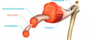
Vertebrogenic pain syndromes are conventionally divided into reflex (occurring in 85–90% of cases) and compression (observed in 10–15% of cases). Reflex pain syndromes arise due to irritation of the receptor apparatus in muscles, tendons and fascia, ligaments, spinal joints, intervertebral discs, etc. due to the formation of nociception sites with a local nonspecific inflammatory reaction. Under conditions of activation of the synthesis and release of pro-inflammatory and algogenic substances (substance P, kinins, prostaglandins, leukotrienes, cytokines, nitric oxide, tumor necrosis factor, etc.), the excitability (sensitization) of nociceptors increases. As a result, a powerful flow of nociceptive afferentation is formed, which enters through the dorsal roots into the neurons of the dorsal horns of the spinal cord, from where it reaches the central parts of the nervous system (reticular formation, thalamus, limbic system and cerebral cortex) along the ascending nociceptive pathways, causing an NMDA-dependent increase in these structures intracellular calcium concentrations and activation of phospholipase A2. The latter stimulates the formation of free arachidonic acid and the synthesis of prostaglandins in neurons, which, in turn, increases their excitability [11, 12]. At the same time, in the posterior horns of the spinal cord, the flow of pain impulses through interneurons activates the neurons of the lateral horn with activation of adrenergic (sympathetic) innervation and motor neurons of the anterior horns of the spinal cord. Activation of the latter leads to spasm of the muscles innervated by this segment of the spinal cord (sensorimotor reflex). Muscle spasm is an additional source of pain due to the activation of muscle nociceptors due to its shortening, the development of neurodystrophic changes and impaired microcirculation in muscle tissue. As a result, a vicious circle closes: “pain, neurogenic inflammation - increased protective muscle tension, pathological changes in the muscles - increased pain,” which contributes to the development of persistent reflex muscle-tonic syndrome [11, 12, 13]. Normally, there is a strictly balanced relationship between the intensity of the stimulus and the response to it. The antinociceptive system exercises descending inhibitory cerebral control over the conduction of pain impulses and inhibits the transmission of pain stimuli from primary afferent fibers to interneurons. The interaction of these structures results in a final pain assessment with an appropriate behavioral response. However, long-term preservation of nociceptive impulses leads to the formation of stable pathological connections, the appearance of pronounced dystrophic changes in surrounding tissues, which, in turn, become a source of pain signals, thereby increasing peripheral pain afferentation, which contributes to the depletion of the antinociceptive system [11, 12, 13] . The chronic course can be facilitated by inadequate treatment of acute pain, excessive limitation of physical activity, a “painful” personality type, low mood, in some cases the patient’s interest in long-term disability, aggravation of existing symptoms or a “rental” attitude towards the disease. With a long course of the disease, pathological changes occur in the roots with the development of axonal and/or demyelinating processes.
The term “osteochondrosis” was proposed in 1933 by the German orthopedist Hildebrandt to designate involutional changes in the musculoskeletal system [14]. Osteochondrosis (Greek osteoon - bone, chondros - cartilage, osis - suffix denoting a pathological condition) is understood as a congenital or acquired degenerative-dystrophic cascade process, which is based on disc degeneration with subsequent secondary involvement of adjacent vertebral bodies, intervertebral joints and ligaments . In turn, the intervertebral disc is a component of the intervertebral symphysis - a complex connection of the vertebrae in the spinal column. In the symphyses of the spine, unlike synovial joints, between the surfaces of the vertebrae covered with hyaline cartilage, there is not synovial fluid, but a specific formation of a cartilaginous nature - an intervertebral disc consisting of a nucleus pulposus and an annulus fibrosus. The first is close in morphological structure to hyaline cartilage due to the high content of proteoglycans, hyaluronic acid, type II collagen and water. While the annulus fibrosus refers to fibrous cartilage with a high collagen content (up to 68%), mainly due to type I collagen in the outer plates of the ring, and rich in sulfated glycosaminoglycans integrated into large proteoglycan molecules, with their characteristic ability to retain water [11, 15]. However, despite the existing differences in the morphological structure of synovial joints and intervertebral symphyses, there is an opinion about the similarity of arthrosis changes occurring in them, expressed in an imbalance of anabolic and catabolic processes in the cartilage matrix [16]. An imbalance of the most important homeostatic processes leads to a decrease in the synthesis of complete collagens and proteoglycans by chondrocytes. Non-sulfated glycosaminoglycan, hyaluronic acid, is no exception, ensuring the formation of matrix proteoglycan aggregates and the hydration of the nucleus pulposus, which plays the role of a protective cushion (due to hydration and changes in its volume). Changes in the quality and quantity of hyaluronic acid in osteochondrosis lead to a decrease in the content of bound water in the nucleus pulposus and destruction of the collagen network, especially in the pericellular zones of chondrocytes [17, 18, 19]. The latter is primarily associated with an increase in the synthesis of metalloproteinases (collagenase, stromelysin), ultimately leading to the complete disappearance of the pericellular collagen network and the loss of shock-absorbing properties of the intervertebral disc as a whole.
Osteochondrosis and spondyloarthrosis are provoked by identical pathogenetic factors, in response to which in the involved structures of the SDS (including two adjacent vertebrae and the intervertebral disc, the own articular, musculo-ligamentous apparatus, the vascular system, as well as the section of the spinal cord corresponding to this level, roots and spinal autonomic ganglia with their connections within this segment) there is a release of biochemically and immunologically active mediators that interact with sensitive receptors, which, in turn, triggers complex and not yet fully understood neurophysiological mechanisms of the formation of pain sensations [11]. It has been established that the source of pain can be an anatomical structure innervated by unmyelinated fibers or containing substance P (or peptides similar to it) [20, 21]. The intervertebral disc has long been considered a formation indifferent to the generation of pain impulses, since no nerve endings were found in it. More detailed anatomical and histochemical studies showed the presence of thin nerve endings in the outer third of the fibrous ring - 1-2 segments above or below its exit [11, 18, 19].
Another source of pain is considered to be the facet (facet) joints, the synovial capsule of which is richly innervated by articular nerves, which are branches of the posterior branches of the spinal nerves, and small accessory nerves from the muscular branches. Due to their vertical orientation, the facet joints offer very little resistance to compression, especially during flexion. Most likely, this slight resistance is due to stretching of the capsular ligaments. Under conditions of extension, the facet joints account for 15 to 25% of compression forces, which can increase with disc degeneration and narrowing of the intervertebral space [11, 21]. With sudden unprepared movements associated with rotation of the body, lifting heavy objects, when working with arms raised above the head, facet syndrome often occurs. The pathogenesis of this syndrome is associated with the convergence of the articular surfaces of the facet (facet) joints and their blocking when the load on the joint and its ligamentous apparatus increases. Pain associated with facet syndrome in the thoracic spine can range from mild discomfort to severe intensity and be severely disabling. It usually increases with extension and decreases with flexion of the spine and can be reflected on the anterior surface of the chest. Below and above the level of joint blocking, a reflex spasm of the erector spinae muscle is often detected [22, 23, 24, 25, 26].
One of the causes of chest pain may be Tietze's syndrome, first described by Tietze in 1921. This syndrome is a relatively rare condition characterized by the presence of nonspecific benign reversible painful edema in the area of the II (in 60% of cases) or III costal cartilage. In 80% of cases there is a unilateral lesion limited to one costal cartilage. The pain is usually well localized, but can radiate along the entire anterior surface of the chest wall, as well as into the shoulder girdle and neck. There are no redness, increased temperature and other changes in the skin over the affected area. The pain usually resolves spontaneously after 2–3 weeks, but often persists for several months, and residual swelling can persist for up to several years. The disease usually develops at a young or childhood age. Its causes are unknown [22, 23, 24, 25, 26].
One of the most common causes of chest pain is costosternal syndrome. This syndrome is much more common than Tietze's syndrome. With costosternal syndrome, palpation in 90% of cases reveals multiple areas of pain: in the left parasternal region, below the left mammary gland, in the projection of the pectoral muscles and sternum. There is no local edema in costosternal syndrome. The cartilages of the II and V ribs are most often affected. When the upper costal cartilages are damaged, pain irradiates to the heart area. The pain usually worsens with chest movement. The disease is more common in women after 40 years of age [22, 23, 24, 25, 26].
Another common cause of chest pain is slipping rib syndrome. The syndrome is characterized by intense pain in the projection of the lower edge of the costal arch and increased mobility of the anterior end of the costal cartilage, usually the X and less often the VIII and IX ribs. This condition is believed to be of traumatic origin and is associated with recurrent subluxation of the costal cartilage during trunk rotation. Unlike the overlying ribs, the cartilaginous parts of which form the sternocostal joints, the cartilaginous parts of the VIII–X ribs form articulations with the cartilaginous parts of the overlying ribs using the external intercostal membrane. This zone is anatomically the weakest area of the chest, prone to injury. Following damage to the cartilage joint, the free cartilaginous part of the rib deviates upward, moving in the vertical or anteroposterior direction when breathing relative to the overlying cartilage, which is accompanied by pain and a characteristic clicking sensation. The pain, as a rule, is sharp or shooting in nature, localized in the upper quadrant of the abdominal wall and provoked by hyperextension of the chest when raising the arms up. In the acute stage of the disease, the patient often takes a forced position with the torso tilted forward and to the painful side to reduce tension in the abdominal wall muscles attached to the costal angles. In some cases, the displaced costal cartilage can injure the perichondrium of the overlying rib and the intercostal nerve. Pathognomonic for this condition is the test described by Holms, which consists of pulling the edge of the rib anteriorly with a bent finger. In this case, a typical pain pattern is reproduced, accompanied by a characteristic click. Carrying out such manipulation on the healthy side is not accompanied by the described phenomenon. The diagnosis can also be confirmed by infiltrating the space between the separated cartilage and the rib with 5 ml of 0.5% lidocaine solution, leading to complete regression of pain 10 minutes after the procedure [22, 23, 24, 25, 26].
Diffuse idiopathic skeletal hyperostosis (Forestier disease) is a disease that is relatively common in middle-aged and elderly people, mainly in men. The main symptoms are usually mild to moderate pain and a feeling of stiffness in the thoracic and lumbar spine. Upon examination, increased thoracic kyphosis, limited range of motion in the thoracic spine and chest excursion are determined. Local pain is often detected on palpation of the thoracic and lumbar spine. To confirm the diagnosis of diffuse idiopathic skeletal hyperostosis, it is necessary to conduct radiography of the spine, which reveals hyperostosis, most pronounced in the thoracic region and manifested by linear ossification along the anterior surface of four adjacent vertebrae or more with preservation of radiological clearing between bone deposits and vertebral bodies, as well as relative preservation of the height of the intervertebral gap. Also characteristic is the formation of osteophytes between the bodies of neighboring vertebrae, interlocking with each other in the form of “bridges” [22, 23, 24, 25, 26].
Diagnosis of vertebrogenic pain syndromes includes establishing the nature of pain and their relationship with static and dynamic loads, identifying trigger points, and symptoms of tension in nerve trunks. Computer and magnetic resonance imaging and radiography are important for determining the nature of the process and assessing the extent of existing changes. To determine the functional state of the roots, determine the location and stage of their damage, electroneuromyography is used [2, 6, 12, 13]. Already during the first examination of the patient, one should exclude danger symptoms (“red flags”) that are generally recognized for dorsalgia, namely, pay attention to the presence of fever, local pain and local increase in temperature in the paravertebral region, which are characteristic of an infectious lesion of the spine. A tumor (primary or metastatic) may be supported by an unreasonable decrease in body weight, a history of malignant neoplasm of any location, persistence of pain at rest and at night, as well as the patient’s age over 50 years. Spinal compression fractures are more common with trauma, corticosteroid use, and in people over 50 years of age. With a tumor lesion of the spinal cord, pain can be constant or recurrent, appear at rest and decrease with movement, often leads to sleep disturbance, forcing you to move or sleep in a forced position, for example, sitting. Against the background of constant pain, lumbago, provoked by coughing or sneezing, is often noted. Motor and sensory disturbances corresponding to the level of damage are identified. With syringomyelia and multiple sclerosis, pain may also be observed, the localization of which depends on the area of damage to the spinal cord.
Other causes of damage to the thoracic roots may be herpes zoster with the development of postherpetic neuralgia, diabetes mellitus, and fractures of the thoracic vertebrae. The pain in these cases, as a rule, is long-lasting, intense, localized in the area of the corresponding segment, has a tightening or burning character, is often accompanied by short shooting pains, and can be lancinating. The pain intensifies at night and with movements in the thoracic spine. Hyperesthesia, hyperalgesia and hyperpathy are often detected in the affected segments. To clarify the diagnosis, radiography, CT, and MRI of the thoracic spine are necessary. In case of rib injuries, the interosseous nerves can be affected, which is accompanied by acute superficial, burning pain in the area of their innervation. The pain intensifies when inhaling or when moving the chest, reminiscent of the pain of pleurisy. As a rule, a small area of segmental hyperalgesia or hyperesthesia is detected, which occurs even when one nerve is affected.
After identifying the cause, mechanism, nature and duration of the pain syndrome, the issue of selecting adequate therapy is decided. Treatment should always be individualized. It depends on the nature of the underlying disease and is divided into undifferentiated and differentiated therapy. The main objectives of undifferentiated therapy are to reduce pain or the patient's reactions to pain and eliminate autonomic reactions. The main direction of differentiated therapy for back pain syndromes is the influence on their pathogenetic mechanisms; treatment also depends on the phase of the underlying disease. The basic principles of conservative treatment include: 1) drug treatment using analgesics, nonsteroidal anti-inflammatory drugs (NSAIDs), dehydration, vascular and phlebotonic agents, angioprotectors, antihypoxants, antioxidants, muscle relaxants, B vitamins, biostimulants, chondroprotectors, desensitizing agents, immunocorrectors, neuroprotectors , metabolites, anticholinesterase, absorbable and vegetotropic drugs; 2) reflex treatment using acupuncture, laser therapy, physiotherapy, massage (segmental), thermal procedures, physical therapy (physical therapy), manual therapy, local irritants; 3) orthopedic treatment involving immobilization, traction therapy, massage, exercise therapy, manual therapy; 4) local anesthetic treatment with the appointment of irrigation with chlorethyl, blockades, dimexide applications, analgesic and anti-inflammatory ointments, gels, and patches.
The first and fundamentally important task facing the doctor is pain relief as quickly and effectively as possible. NSAIDs are the most widely used drugs for symptomatic pain management [27]. They are quite effective, easy to use, inexpensive and generally well tolerated. It should be noted that the effectiveness of NSAIDs as analgesics is determined not only by the peripheral effect associated with a decrease in the synthesis of prostaglandins, as well as other mediators of pain and inflammation in damaged or inflamed tissues.
The most widely used NSAIDs are from the group of non-selective cyclooxygenase (COX) inhibitors. Basic recommendations for the use of NSAIDs (in monotherapy or in combination with other analgesic drugs): 1) their use is advisable for acute or chronic diseases and pathological conditions manifested by pain associated with both inflammatory and degenerative damage to the musculoskeletal system, acute trauma and surgical interventions; 2) the duration of NSAID use depends on the duration and intensity of pain in a particular situation; 3) to relieve acute pain, it is advisable to prescribe parenteral forms of NSAIDs or those that give the most pronounced analgesic effect with a minimal risk of side effects; 4) for a long course of treatment, NSAIDs with a medium or long half-life are recommended, taken orally or in the form of rectal suppositories. These drugs are characterized by good analgesic and anti-inflammatory effects and provide relatively quick relief from pain.
In modern medicine, diclofenac, which is one of the most frequently prescribed painkillers, is recognized as the recognized standard for the treatment of diseases with severe pain. Like other NSAIDs, dilofenac inhibits the activity of the COX enzyme, which is involved in the formation of prostaglandins from arachidonic acid. Diclofenac also inhibits the activity of the enzyme lipoxygenase. According to Russian studies, with the simultaneous administration of diclofenac and B vitamins, a higher Cmax value was observed compared with the use of diclofenac alone [28]. To increase the therapeutic effect of diclofenac, while minimizing its dose as much as possible, using B vitamins, a highly effective combination drug Neurodiclovit was created, one capsule of which contains 50.0 mg of diclofenac, 50.0 mg of vitamin B1, 50.0 mg of vitamin B6 and 250.0 mcg vitamin B12. B vitamins have a wide range of pharmacodynamic properties and participate as coenzyme forms in most metabolic processes. It is known that thiamine (vitamin B1) has a significant effect on the regeneration processes of damaged nerve fibers, provides energy for axoplasmic transport, regulates protein and carbohydrate metabolism in the cell, affects the conduction of nerve impulses, and promotes the development of an analgesic effect. Pyridoxine (vitamin B6) is a cofactor for many enzymes acting in nervous tissue cells, participates in the synthesis of neurotransmitters of the antinociceptive system (serotonin, norepinephrine), supports the synthesis of transport proteins and sphingosine, a structural element of the nerve fiber membrane. ensures the delivery of fatty acids to cell membranes and the myelin sheath. Cyanocobalamin (vitamin B12) ensures the delivery of fatty acids to cell membranes and the myelin sheath. The use of vitamin B12 promotes not only remyelination (due to the activation of the transmethylation reaction, which ensures the synthesis of phosphatidylcholine in nerve cell membranes), but also a decrease in the intensity of pain, which is associated with the intrinsic antinociceptive effect of high doses of cyanocobalamin [29]. Experimental studies have shown that vitamin B1 alone or in combination with vitamins B6 and B12 is able to inhibit the passage of pain impulses at the level of the dorsal horns of the spinal cord and thalamus [30]. B vitamins function as coenzymes in metabolism, in particular in nervous tissue, which enhances the analgesic effect of diclofenac. When using B vitamins in the treatment of pain, one should remember that their analgesic properties decrease accordingly: B12˃B6˃B1 and that the multivitamin complex (B1+B6+B12) has a more pronounced analgesic effect than monotherapy with vitamin B1, B6 or AT 12. In the treatment of acute back pain, a combination of B vitamins with NSAIDs is more effective than NSAID monotherapy [31]. The drug is used for inflammatory and degenerative diseases of the joints and spine (chronic polyarthritis, rheumatic and rheumatoid arthritis, ankylosing spondylitis, osteoarthritis, spondyloarthrosis). Neurodiclovit has a low incidence of unwanted complications and good individual tolerability, which allows it to be recommended for widespread use in the treatment of patients with osteoarticular pain in the thoracic spine and chest.
The main therapeutic measures of individual forms are presented in Table 1.
Table 1. Basic treatment measures
| Nosological form | Therapeutic measures |
| Tietze syndrome | Local warming procedures and the use of NSAIDs. With high pain intensity, infiltration of the affected joints with local anesthetics (0.25–0.5% novocaine solution), sometimes in combination with corticosteroids. |
| costosternal syndrome | Intercostal nerve blocks with local anesthetics along the posterior axillary line |
| slipping rib syndrome | Explain to the patient the benign nature of the condition, NSAIDs, blockade with local anesthetics and corticosteroids. If the above measures are ineffective, sometimes they resort to resection of the rib edge. |
| sternoclavicular hyperostosis | NSAIDs, warming physiotherapy and exercises aimed at strengthening the back muscles. |
| facet syndrome | Infiltration of the affected joints with a solution of local anesthetic, warming the painful area and active physical therapy aimed at strengthening the muscles of the abdominal wall and the erector spinae muscle. |
Literature:
1. Statistisches Bundesamt. Health report for Germany: Federal Health Bulletin. Wiesbaden: Metzler-Poeschel; 1998. 2. Putilina M.V. Features of diagnosis and treatment of dorsopathies in neurological practice. Consilium medicum: a journal of evidence-based medicine for medical practitioners. 2006;8(8):4448. 3. Fedin A.I. Dorsopathies (classification and diagnosis). Atmosphere. Nervous diseases. 2002;2:28. 4. Manek N., MacGregor AJ Epidemiology of low back disorders. Curr. Opin. Rheumatol. 2005;17(2):134140 5. Naumov A.V., Semenov P.A. Pain in Russia: facts and conclusions. Consilium Medicum 2010;12(2):38–41. 6. Buchner M., Neubauer E., ZahltenHinguranage A., Schiltenwolf M. Age as a predicting factor in the therapeutic outcome of multidisciplinary treatment of patients with chronic low back pain a prospective longitudinal clinical study in 406 patients. Clin. Rheumatol. 2007;26:385392. 7. Hainline B. Chronic pain: Physiological, diagnostic, and management considerations. Psychiatr Clin North Am. 2005;28:713-5. 8. Morley S. Psychology of pain. Br J Anaesth. 2008;101:25-31. 9. Costigan M., Scholz J., Woolf C.J. Neuropathic pain: A maladaptive response of the nervous system to damage. Annu Rev Neurosci. 2009;32:1-32. 10. Handbook on the formulation of clinical diagnosis of diseases of the nervous system / Ed. V.P. Shtoka, O.S. Levina. - M.: MIA, 2006. - 520 p. 11. Ivanova M.F., Evtushenko S.K. Dorsalgia caused by degenerative pathology of the spine. News of medicine and pharmacy. 2010;15(335):1617. 12. Pain (a practical guide for doctors) / Ed. N.N. Yakhno, M.L. Kukushkina. - M.: Publishing House of the Russian Academy of Medical Sciences, 2011. - 512 p. 13. Damulin I.V. Back pain: diagnostic and therapeutic aspects. - M.: RKI Severo press, 2008. - 40 p. 14. Mendel O.I., Nikiforov A.S. Degenerative diseases of the spine, their complications and treatment. Russian medical journal. 2006;14(4):34-39. 15. Shostak N.A. Modern approaches to the treatment of pain in the lower back. Consilium medicum. 2003;5(8):457-461. 16. Khodyrev V.N., Golikova L.G. Clinical effectiveness of alflutop for spinal osteochondrosis (12-month study). Scientific and practical rheumatology. 2005;2:33-36. 17. Zborovsky A.B., Mozgovaya E.E. Alflutop: experience of many years of clinical use. Pharmateka. 2006;19:35-40. 18. Mense S. Pathophysiology of low back pain and transition to the chronic state - experimental data and new concepts. Schmerz. Der. 2001;15:413-420. 19. Kamchatov P.R. Acute spondylogenic dorsalgia - conservative therapy. Russian medical journal. 2007;15(10):64-74. 20. Golubev V.L. Neurological syndromes: A guide for doctors / Ed. V.L. Golubeva, A.M. Wayne. — 2nd ed., add. and processed - M: MEDpress-inform, 2007. - 736 p. 21. Podchufarova E.V. Chronic back pain: pathogenesis, diagnosis, treatment. Russian medical journal. 2003;11(25):1395-1401. 22. Danilov A.B. Cardialgia and abdominalgia. Pain syndromes in neurological practice / Ed. Veina A.M. - M.: Medpress-inform, 2001. 23. Khabirov F. A. Clinical neurology of the spine. - Kazan, 2003. - 472 p. 24. Podchufarova E.V., Yakhno N.N. Pain in the back and limbs // Diseases of the nervous system: A guide for doctors / Ed. N.N. Yakhno. - M., 2005. - T. 2. 25. Bonomo L., Fabio F., Larici AR Non-traumatic thoracic emergencies: acute chest pain: diagnostic strategies. Eur. Radiol. 2002;12:1872-85. 26. Ho KY, Kang JY, Yeo B. Non-cardiac, non-oesophageal chest pain: the relevance of psychological factors. Gut. 1998;43:105-10. 27. Use of non-steroidal anti-inflammatory drugs: Clinical recommendations. Nasonov E.L., Lazebnik L.B., Belenkov Yu.N. and others. M.: Almaz, 2006. 28. Zhuravleva M.V., Makhova A.A., Shikh E.V. The place of milgamma in the complex therapy of back pain. Pharmateka. 2013;19:1-4. 29. Zaichenko A.V., Barinov A.N., Makhinov K.A., Bryukhanova T.A. Treatment of back pain refractory to NSAIDs Medical advice. 2013;12:2-8. 30. Pizova N.V. Milgamma and Milgamma compositum in the treatment of neurological diseases. Neurology, neuropsychiatry and psychosomatics. 2009;3-4:75-81. 31. EF. Neurology and psychiatry. 2012;4:1-7.
Treatment
Unfortunately, it is the localization of osteochondrosis in the thoracic region that suggests the least effectiveness and the speed of onset of the effect of therapy. Also, often, the treatment time for thoracic osteochondrosis increases to a month or more. Treatment of thoracalgia should be carried out by a neurologist. As with other localizations of the pathological process, therapy uses standard anti-inflammatory drugs (meloxicam, diclofenac, Celebrex, Airtal and others), muscle relaxants for existing muscle-tonic disorders (mydocalm, baclosan, sirdalud), neuroprotective drugs (B vitamins, thioctic acid etc.).
Chronic thoracalgia – pain syndrome of vertebrogenic origin
Chronic vertebrogenic thoracalgia is characterized by a recurrent long-term course. periods of exacerbation of pain are followed by remission, in which there are no unpleasant sensations and the patient considers himself completely healthy. Despite the feeling of general well-being, even during the period of remission, the syndrome of vertebrogenic thoracalgia is constantly developing. Further destruction of the nerve fiber occurs. The longer this syndrome lasts, the more difficult it is to restore the process of normal innervation of the soft tissues of the chest.
Thoracalgia as a pain syndrome can be present in a number of pathologies. For example, pain in the chest can be observed with pathologies of the pancreas, large intestine, gallbladder and liver. This syndrome may indicate general intoxication of the body and be observed with dysfunction of the endocrine system.
Mild thoracic thoracalgia syndrome may be a consequence of osteoporosis. This fact should be paid attention to by patients whose age has exceeded the threshold of 50 – 5 years. During this age period, secondary calcium deficiency may occur. It is important to monitor the condition of the large intestine, periodically check the condition of the thyroid gland, and take a blood test to determine the level of vitamin D.
In children, thoracalgia of vertebrogenic origin may be associated with rickets, a disruption of the process of formation of physiological curves of the spinal column. In adolescence, in almost 100% of cases, developing thoracalgia indicates poor posture. As a result of curvature of the spinal column, overstrain of the back muscles occurs. This causes sharp pain.
Exercise therapy exercises
Physical therapy exercises are very important, but they are ineffective in the acute stage. After all, the thoracic region has limited mobility and muscle spasms can sometimes only be relieved with medications. However, as a means of preventing the recurrence of thoracalgia, exercise therapy exercises come first. In case of illness, sets of exercises are indicated to strengthen the muscle corset, swimming, and cardio exercises in order to reduce excess body weight. It is undesirable to engage in martial arts (risk of injury), weightlifting (overload), basketball (frequent vertical loads).