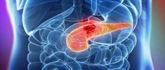
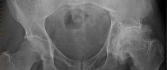
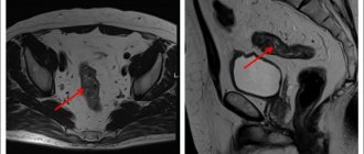
The phrase “oncological disease” hides many pathologies of various kinds. More than a hundred pathologies share only a few common features - abnormal cell structure under the influence of a certain genetic mutation, which is expressed in uncontrolled reproduction and lack of an adequate response to external signals. This is how a malignant tumor settles in the human body. Over time, it increases in size, invading the space of neighboring tissues and organs. Some malignant cells, breaking away from the tumor, migrate to other organs, and secondary foci of the disease appear, called metastases.
The importance of identifying stages of the disease
Without proper treatment, the disease spreads progressively and becomes more difficult to treat. In the initial stages, cancer is quite easily treated by removal, but in the presence of metastases, achieving remission becomes almost impossible.
To correctly determine the stage of disease development, a cancer staging system was developed. Of course, universal staging levels are used for all diseases, but in each specific type of disease they have their own differences. Correctly determining the stage of the disease is the primary task for the oncologist at the examination stage.
Breast implants lead to breast cancer
In fact, women who have and those who do not have implants are equally at risk for breast cancer.
But there is a very rare disease - anaplastic large cell lymphoma of the breast. This is not a disease of the breast tissue, but of the lymph nodes. And it is the presence of breast implants that influences the development of this disease.
The basic risk of developing this disease is three per 100,000,000 cases, and the risk of development in the presence of implants is already 203 per 100,000,000 cases, i.e. the risk increases by 67 times. However, the absolute incidence of this disease is extremely small.
Thus, breast implants are not associated with breast cancer, but do increase the risk of breast lymphoma.
The need to divide cancer diseases into stages
Dividing cancer into stages is necessary to solve many problems: planning treatment, determining the prognosis for the patient, since each stage has certain statistical indicators of survival, remission and the possibility of relapses in the future, monitoring the course of the disease and the effectiveness of the therapy used, ensuring continuity in the provision of care by oncologists . After all, a universal classification erases the boundaries between the different languages of the world spoken by doctors.
TNM classification
TNM is the fundamental classification used throughout the world. It was developed back in the mid-20th century. Last revised in 2009. Subsequent revisions are planned as information about innovations in the diagnosis and treatment of cancer accumulates.
The classification abbreviation is informative; each letter carries information about the characteristics of a malignant formation:
- T (tumor) is a description of the initial tumor;
- N (nodus) – tumor migration to regional lymph nodes;
- M (metastasis) – presence of metastases.
When determining what stage the disease is at, the corresponding number or special designation is assigned to the letter.
Legend:
| Characteristics of the initial tumor | TX – the primary tumor has not been assessed. T0 – the primary tumor was not found. Tis - literally means: “cancer in situ”; a cluster of cancer cells has been detected that do not grow into neighboring tissues, that is, this is the initial stage of cancer. T1-4 – the number indicates the size of the tumor and the degree of its penetration into neighboring tissues. |
| Spread to regional lymph nodes | NX – it is impossible to evaluate regional lymph nodes. N0 – there are no cancer cells in the regional lymph nodes. N1-3 – the number indicates the degree of damage to regional lymph nodes by cancer cells. |
| Metastases | M0 – no metastases. M1 – metastases detected. |
Often, an additional letter is placed before the TNM designation, which is responsible for information about the method of diagnosis:
- p – established by a pathologist when studying the tumor under a microscope after removal;
- c – clinically diagnosed before biopsy.
Sometimes doctors prescribe an additional test called a sentinel biopsy. In essence, this is the study of the sentinel lymph node, that is, the closest lymph node in the direction of outflow of the malignant tumor. This study allows us to determine the condition of regional lymph nodes and the need for their removal and direction of treatment.
When to see a doctor
A reasonable question arises: when should you contact a specialized specialist without wasting precious time? After all, many types of cancer pathology are asymptomatic for a long time, not showing themselves in the first stages.
A reliable way to determine the first symptoms of cancer is regular preventive examinations.
They are especially important for people at risk of developing cancer. Risk factors include:
- genetic predisposition;
- detection of certain genetic mutations during diagnosis;
- frequent contact with carcinogens and other toxic substances (occupational hazards);
- unfavorable environmental conditions;
- age factor.
The medical community has developed special diagnostic programs - screening studies that determine the presence or absence of a malignant neoplasm. Timely detection of cancer symptoms and undergoing a screening examination makes it possible to carry out the most effective treatment, achieve stable remission or complete recovery.
Doctors recommend the following screening tests:
- Mammary cancer. Women at any age are at risk of developing breast cancer. Every representative of the fairer sex should conduct a self-examination of the mammary glands once every couple of months. It is advisable to consult a mammologist at least once a year. After 40 years, you will need to perform mammography (a type of radiography), and, if indicated, an ultrasound examination of the breast.
- Stomach cancer. Stomach problems are determined by fibrogastroduodenoscopy (FGDS). Using the endoscopic method, one can easily diagnose certain changes in the mucous membrane of the esophagus, stomach, and duodenum.
- Colon cancer. People of both sexes after 50 years of age should undergo fibrocolonoscopy. Examination of the mucous membrane with an endoscope reveals precancerous conditions (ulcerative colitis or Crohn's disease) or a cancerous tumor itself.
- Lungs' cancer. Medical experts recommend low-dose CT scans once a year for the following groups of people: ages 55 to 75 years; smokers aged 40 years and older; former smokers (quit within the last 15 years).
- Skin cancer. Alarming changes in moles, warts or birthmarks can be diagnosed by a dermatologist using dermatoscopy. It is advisable to undergo the procedure annually.
General diagnostics of oncopathology is also carried out. The examination involves performing a number of diagnostic measures:
- Lab tests:
- Determination of the level of tumor markers in the blood.
- Molecular genetic analysis. It allows you to identify specific mutations that predetermine the risk of developing an oncological process.
- DNA diagnostics of closest blood relatives. The procedure makes it possible to find out the risk of the disease in all family members.
- Ultrasound. The undoubted advantages of the method: it is carried out quickly, safely, and is affordable.
- MRI, . An examination will be required if previous diagnostic results raise suspicions among the attending physician about the presence of an oncological process.
- Endoscopic diagnostics. With its help, you can see the tumor with your own eyes through the optics of the endoscope.
- Carrying out a biopsy. The purpose of the event is to analyze a tissue sample of the affected organ. This is followed by a conclusion about the presence or absence of atypical structures.
Simplified classification
The simplified classification of cancer stages is based on the global TNM classification, but involves designating 5 stages of the disease from 0 to IV in Roman numerals.
Stage 0 cancer practically means that cancer cells have not spread to other tissues and are small in size, located in the place of their initial appearance. At its core, this is the initial stage of cancer, when the tumor is easily removed and remission occurs with a 100% probability.
Stage 1 cancer is characterized by a small tumor within a single organ. This is an early stage cancer that can be successfully treated.
Stage 2 cancer, compared to stage 1 cancer, has a larger tumor size, and it takes root deep into the organ without affecting neighboring tissues, but in some cases affects the lymph nodes.
Stage 3 cancer involves proliferation beyond one organ and damage to regional lymph nodes.
Stage 4 cancer is characterized by the presence of distant metastases.
Types of malignant neoplasms
The structure of a malignant formation primarily depends on the tissues in which it was formed. Based on this feature, the following types of malignant neoplasms are distinguished:
- muscular;
- epithelial;
- nervous;
- connective tissue;
- bone;
- vascular;
- endothelial.
Based on the principle of division of malignant cells, histological, clinical and morphological types of tumors are distinguished.
According to the complexity of the structure, there are simple (formation from one type of tissue) or complex types of malignant neoplasms. Cancer is also distinguished by the organ or system in which it was formed: bones, intestines, ovaries, skin, stomach, mammary glands, prostate, cervix, lungs, etc.
Types of malignant neoplasms based on the cell structure on which they develop:
- carcinoma - epithelial tissue;
- leukemia - oncology of the hematopoietic system;
- sarcoma - cancer of the musculoskeletal system and connective tissue;
- melanoma - skin cancer of melanocyte cells;
- lymphoma - infection of lymphatic cells;
- choriocarcinoma - affects the placenta;
- glioma - degeneration of glial cells of the brain;
- Teratoma - a cancerous process affecting gonocytes.
Degree of tumor malignancy
The success of the chosen therapy, the likelihood of relapses and the chances of remission depend on how aggressively the affected cells behave. Because of this, experts have developed a system for assessing the degree of malignancy, in other words, aggressiveness:
- GX is assigned when the degree of aggressive impact cannot be assessed;
- G1 means that the tumor tissue is similar to normal tissue, but still behaves aggressively (albeit to a lesser extent). At this stage, metastases do not appear;
- G2 indicates that significant differences between cancer and normal cells are visible under the microscope;
- G3 and G4 indicate that there is no possibility of treatment, since the cells behave extremely aggressively and practically do not respond to therapy.
Causes of malignant neoplasms
Scientists have identified a number of causes of malignant neoplasms that can trigger the development of cancer. For convenience, they are usually divided into two large groups: endogenous and exogenous.
Let's take a closer look at each of them.
Endogenous causes of malignant neoplasms:
- immunological hereditary abnormalities (combined immunodeficiency, X-linked agammaglobulinemia, etc.);
- formation of hereditary neoplasms (diffuse polyposis, multiple endocrine adenomatosis);
- an increased risk of malignant formation, but with an unproven hereditary connection, is present in organs such as the lungs, mammary glands, endometrium, large intestine, stomach;
- disruption of the endocrine system;
- the presence of pathologies predisposing to malignant degeneration of cells (for example, xeroderma pigmentosum, Peutz-Jeghers or Down syndrome, Recklinghausen syndrome, multiple exostoses).
Exogenous causes of malignant neoplasms:
- acquired immunodeficiencies;
- radiation with a low dose of exposure (diagnosis using X-ray devices, ultraviolet irradiation, etc.);
- radiation with a high degree of radiation exposure (nuclear weapons, radiation therapy, nuclear power plant accidents);
- the influence of chemical carcinogens, in particular industrial products (inorganic compounds - nickel, chromium, arsenic, cadmium, etc., organic compounds - asbestosis, gasoline, vinyl chlorides, coal tars, benzidine, etc.);
- negative effects of certain medications (immunosuppressants, alkylating medications, testosterone, estrogen, procarbozines, phenacetin, etc.);
- exposure to bad habits (drinking alcohol and smoking);
- incorrect and irrational diet;
- improper processing of products and poor hygiene;
- infection with infectious elements - fungi (aflotoxin), parasites (schistosomiasis), hepatitis B, African Burkitt's lymphoma, hepatocellular cancer, nasopharyngeal carcinoma.
Tumor assessment after surgery
Postoperative classification allows oncologists to assess the success rate of treatment performed through surgery:
- RX reports that it is not possible to assess the presence of tumor cells in the body after surgery;
- R0 states that there is no tumor in the body after surgery;
- R1 says that microscopic examination revealed residual tumor foci;
- R2 disappointingly reports that even without a microscope, a tumor is detected in the patient’s body.
A few words about other types of malignant tumors
A number of diseases are so non-standard that it is not possible to apply the TNM system to them. Let's talk about them. Tumors in children are very different from malignant tumors in the body of an adult and deserve consideration in a separate article. Blood cancer has different names (leukemia, lymphoma, myeloma, etc.), but invariably does not have a specific density and specific localization, due to which it cannot be classified according to the TNM system. Tumors of the nervous system are localized in the brain and spinal cord. They do not have their own generally accepted classification, so the letter T is used to determine the stage.
Depression contributes to cancer
There is a misconception that negative emotions and severe depression can lead to cancer.
A study was conducted in which people with severe forms of depression took part. As a result, it was proven that the risk of developing a malignant tumor in people with depressive disorders is exactly the same as in people without such disorders.
Thus, a bad mood has absolutely no effect on the occurrence and development of a tumor and is not even a risk factor for its development.
Methods for diagnosing cancer diseases
There are many different diagnostic methods, each of which can determine the diagnosis with varying degrees of accuracy. In this case, when making a diagnosis, a classification is used that allows one to understand the diagnostic methods and the degree of accuracy:
- C1 – standard diagnostic methods (examination, radiography, endoscopy) are less accurate;
- C2 – more accurate diagnostic methods were used (biopsy, CT, MRI, endosonography and others);
- C3 – cytological and histological studies of damaged tissue obtained through surgery.
- C4 – the diagnosis was established only after a full surgical operation to remove the tumor.
Rate of cancer development
The process of the appearance of “wrong” cells in the human body is inevitable and almost everyday, since DNA copying failures quite often occur during their reproduction. There is a special type of immunity for this case. However, it happens that abnormal cells have a high survival rate and begin to actively multiply, forming a tumor. One thing can be said with certainty: before the first manifestation, cancer cells remain in the patient’s body for several years.
What is the period per month for cancer to appear?
Cancer at an early stage is practically undetectable. Because of this, it is impossible to accurately determine at what time the abnormal cells that formed the tumor appeared. Even the tumor itself may not show its presence in the body for years, while rapid proliferation of cancer cells is also quite common.
How long does it take for stage 4 cancer to develop?
In this matter, much depends on the stage at which the patient went to the doctor, how correct the diagnosis turned out to be and the susceptibility of the tumor to treatment.
Mutation theory
This theory is often called one of the most widespread. According to it, due to the accumulation of mutations in specific areas of cellular DNA, a tumor begins to develop. Against this background, the appearance of defective proteins is noted in the body. This theory was first put forward by the German biologist Theodor Boveri . In 1914, he suggested that abnormalities in chromosomes cause the formation of cancer cells. Today, scientists have already identified more than 100 genes that are associated with cancer. This theory was supported by Hermann Müller , Bert Vogelstein , Eric Faron and other scientists who for a long time regularly found evidence that cancer depends on genetic mutations.
Article on the topic Five ailments of our time. The most popular diseases and their symptoms The discovery of proto-oncogenes and suppressor genes is evidence of the mutational nature. It is their change due to mutational events that becomes the cause of cell malignancy. Proteins appear in cells due to certain transformations. Each cell has its own life cycle, in addition, it performs certain functions at different stages of development. The cycle program is written in DNA, which can change under the influence of certain factors.
There is also a theory of random mutations, its author is called Lawrence Loeb from the University of Washington. Geneticists note that during the entire life of a cell, a random mutation occurs in only one gene. But sometimes, under the influence of carcinogens or other provoking factors, the frequency of mutations may increase. There is a version that is both difficult to confirm and refute - that cancer occurs against the background of a huge number of mutations, measured in tens of thousands per cell.
Curability of cancer
In oncology, it is not customary to use the terms “cure” and “recovery.” The thing is that the process of reproduction of the abnormal cells that appear cannot be stopped forever. Therefore, the term “remission” is used, which means that at the time of the examination no traces of the presence of cancer were found in the patient’s body. However, the risk of relapse remains with the patient forever.
The highest probability of remission is observed with stage 1 and zero cancer. For patients with stage 2 cancer, the prognosis is not so good, but there is still a chance of achieving remission. Stage 3 cancer leaves virtually no chance of remission and involves treatment aimed at stopping and slowing down the progressive development of the disease, as well as reducing the impact of symptoms in order to prolong life. Stage 4 cancer does not give a chance for remission, but miracles do happen in life.
The chances of recovery depend on the timeliness of the diagnosis and appropriate adequate treatment. To do this, if the slightest symptoms appear, you need to contact a specialist. You can make an appointment at the oncology department by phone. Be attentive to your health!
People with dark skin do not get skin cancer
Statistically, skin cancer occurs more often in people with fair skin.
The main cause of skin cancer is exposure to ultraviolet rays. Protection against them is provided by melanin, which is less in people with fair skin.
A study by scientists has confirmed that skin cancer occurs in people with dark skin. But the survival rate of such dark-skinned patients is much lower than that of lighter-skinned patients. This is due to the difficulty of detection and the frequency of detection, since it is more difficult to detect a neoplasm on dark skin than on light skin.
One notable example is reggae musician Bob Marley, who died at the age of thirty-six from late-diagnosed acral melanoma, a melanoma that develops on the skin of the nails.
Thus, skin color has no effect on the risk of skin cancer, and people with dark skin can also be diagnosed with it.


