A third of the world's population suffers from inflammatory processes in the kidneys. But not many people know that these problems, for the most part, are associated with pyeloectasis of the right kidney and only sometimes - with the left. With this anomaly, the renal pelvis expands. This happens due to the accumulation of fluid in it, which then travels through the ureters to the bladder.
- Causes and forms of the disease
- Symptoms and complications of pyeelectasis
- Establishing diagnosis
- Therapy (treatment) and prevention
What is pyelectasia?
Pyeloectasia is an expansion of the renal pelvis (pyelos (Greek) - pelvis; ectasia - expansion). In children, as a rule, pyeelectasis is congenital. If the calyxes are dilated along with the pelvis, then they speak of pyelocalicoectasia or hydronephrotic transformation of the kidneys. If the ureter is dilated along with the pelvis, this condition is called ureteropyeloectasia (ureter), megaureter or ureterohydronephrosis. Pyeelectasis is 3-5 times more common in boys than in girls. There is both unilateral and bilateral pathology. Mild forms of pyelectasis often go away on their own, while severe forms often require surgical treatment.
Characteristic symptoms of pyelectasis
Let us remember that pyeelectasis is not a disease, but a concomitant condition resulting from some kind of kidney pathology.
Such diseases include:
- urolithiasis, which manifests itself as renal colic (recurrent intense pain in the lower back);
- kidney tumors, which cause aching pain in the back, radiating to the groin and abdomen; possible appearance of scarlet blood in the urine;
- chronic inflammation of the kidney, which leads to intoxication, lower back pain, cloudy urine with the appearance of sediment and mucus in it.
It happens that pyeelectasis occurs without symptoms and is accidentally discovered on an ultrasound.
If the dilated collecting apparatus is infected, this can lead to the following symptoms:
- fever up to 38.5-41 ° C;
- chills;
- dizziness;
- nausea, vomiting that does not bring relief;
- loss of appetite;
- increased fatigue.
What can be an obstacle to the outflow of urine?
Often, an obstacle to the outflow of urine from the kidney is a narrowing of the ureter at the junction of the pelvis and the ureter, or when the ureter flows into the bladder. Narrowing of the ureter may be a consequence of its underdevelopment or compression from the outside by an additional formation (vessel, adhesions, tumor). Less commonly, the cause of impaired urine outflow from the pelvis is the formation of a valve in the area of the ureteropelvic junction (high ureteric outlet). Increased pressure in the bladder, resulting from a disruption of the nerve supply to the bladder (neurogenic bladder) or from the formation of a urethral valve, can also impede the flow of urine from the renal pelvis.
Symptoms and clinical picture of pyeloectasia
For some time, manifestations of pyelectasis may be absent, and pathological changes are detected only with the help of instrumental studies. Some patients experience decreased appetite and swelling of the face.
As pathological abnormalities develop in the body, these symptoms of pyelectasis are joined by aching pain in the lumbar area and urination problems. In rare cases, a rise in body temperature, vomiting and nausea may occur.
What diagnostic methods are used for pyeelectasis in a newborn?
For mild pyelectasis, it may be sufficient to conduct regular ultrasound examinations (ultrasounds) every three months. If a urinary infection occurs or the degree of pyelectasia increases, a complete urological examination is indicated, including radiological research methods: cystography, excretory (intravenous) urography, radioisotope study of the kidneys. These methods allow you to establish a diagnosis - determine the level, degree and cause of the disturbance in the outflow of urine, as well as prescribe reasonable treatment. The research results themselves do not constitute a verdict that unambiguously determines the fate of the child. The decision to manage the patient is made by an experienced urologist, usually based on observation of the child, analysis of the causes and severity of the disease.
Classification of the syndrome
There are two forms of this condition depending on the degree of damage to the urinary organs:
- unilateral pyeelectasis of either the left or right kidney;
- bilateral pyelectasis of both kidneys. This condition requires immediate measures to solve this problem, since enlargement of the pelvis of both kidneys at once has a detrimental effect on the entire urinary system, and on the general condition and functionality of the body.
This syndrome is classified according to severity - mild, moderate or severe. It is important to take into account the volume of active preserved tissue and the presence of an inflammatory process, as well as pay attention to the presence of signs of renal failure.
If the syndrome progresses very quickly, the doctor diagnoses hydronephrosis, which has a unique set of symptoms that helps identify the presence of the disease. For example, these are nagging pain in the lower back, frequent urination and high blood pressure, which leads to fatigue, headaches, and a complete decrease in performance.
If the pelvis and calyx increase in size at the same time, this can lead to complete transformation of the kidney.
What diagnoses are made based on the examination?
Some examples of common diseases accompanied by pyelectasis:
- Hydronephrosis caused by an obstruction (obstruction) in the area of the ureteropelvic junction. It manifests itself as a sharp dilation of the pelvis without dilatation of the ureter.
- Vesicoureteral reflux is the backflow of urine from the bladder into the kidney. It manifests itself as significant changes in the size of the pelvis during ultrasound examinations and even during one examination.
- Megaureter - a sharp dilation of the ureter can accompany pyelectasis. Causes: severe vesicoureteral reflux, narrowing of the ureter in the lower section, high pressure in the bladder, etc.
- Posterior urethral valves in boys . Ultrasound reveals bilateral pyelectasis and dilatation of the ureters.
- Ectopic ureter - The flow of the ureter not into the bladder, but into the urethra in boys or the vagina in girls. Often occurs with double kidneys and is accompanied by pyeelectasis of the upper segment of the double kidney
- Ureterocele - the ureter, when it enters the bladder, is inflated in the form of a bubble, and its outlet is narrowed. Ultrasound reveals an additional cavity in the lumen of the bladder and often pyelectasis on the same side.
Prevention and treatment of pyelectasis
As a rule, in the case of congenital pyelectasis, surgical intervention is not possible. In this case, the narrowing of the ureter will be expanded using a stent - a frame that expands during installation and maintains the expanded position.
If pyeloectasia is formed as a result of urolithiasis, then treatment is aimed at eliminating the stones. Most often, doctors try to solve the problem without surgery; surgical methods are used in extreme cases.
It is worth noting that self-medication for pyeelectasis is very dangerous. The doctor prescribes treatment only after passing all the examinations and passing all the necessary tests.
Prevention of the anomaly includes timely identification of the problem, as well as complete treatment with impeccable compliance with all the doctor’s instructions, restriction in the use of liquids, and so on.
Pyeelectasia is an enlargement of the renal pelvis, which causes urine to accumulate in the pelvis before being discharged through the ureter. Pyeelectasia is not a disease, but a syndrome resulting from other kidney diseases. Timely detection of the problem and proper treatment can relieve pyeelectasis and protect against the development of complications. Be healthy!
Criteria for the differential diagnosis of pyelectasia and hydronephrosis in children
Department of Pediatric Surgery of the Russian State Medical University (head of the department, doctor of medical sciences, prof. Razumovsky A.Yu.) Morozov Children's City Clinical Hospital of the Children's Health Department, Moscow, Children's City Clinical Hospital No. 13 named after. N.F. Filatova House of Health, Moscow.
Pyeelectasia is often considered a stage of hydronephrosis. At the same time, the existing expansion of the pelvis in infants and young children is often transient in nature, can spontaneously decrease and even disappear completely, which makes it difficult to choose an adequate tactic.
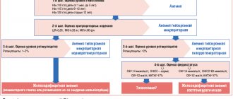
Target. The purpose of our study was to establish differential diagnostic criteria for pyelectasia and hydronephrosis based on determining the parameters of renal blood flow and assessing the condition of the renal parenchyma and CL, and studying the growth factors that determine nephrogenesis.
Materials and methods. A total of 98 patients with isolated dilatation of the pelvis aged from 3 months to 14 years were examined. For ultrasound of ureterovesical discharges and diuretic ultrasound (DUUS), previously developed techniques were used. Epidermal growth factor (EGF) in urine. Changes in the size of the CL were detected in utero - in 36 patients, discovered by chance, during routine ultrasound - in 62 children.
Urinary epidermal growth factor (EGF) was studied preoperatively in children with hydronephrosis, as well as in patients with pyeelectasis.
Research results. With pulsed Doppler (PDM), hemodynamics were not impaired - the resistance index (IR) at all levels fluctuated within minimal limits. A significant increase in IR of ureterovesical emissions was established in all age groups from 0.75 to 0.82-0.86 relative to the indicators on the contralateral side, where no dilatation of the pelvis was observed. An analysis of the studies showed that by the age of 6 months in 10 children (8.5%) the pelvis had shrunk, its size corresponded to the age norm. By 1 year, in 25 children (21.2%), the pelvis did not exceed standard values; by 3 years, in 80 patients (67.8%), the size of the pelvis did not exceed 4-5 mm. EGF levels in urine were 8.7 – 18.5 ng/ml (with the norm being 8 – 19 ng/ml), average – 13.5 ng/ml.
In three patients, an increase in the transverse size of the pelvis was recorded from 8-10 mm to 18 mm and a sharp decrease in the concentration of EGF in the urine (1.4 - 1.4 - 2.2 ng/ml), which was regarded as the presence of an obstructive process. When further surgical correction was performed, it was established that the cause of obstruction in all cases was an aberrant vessel.
Thus, in young children, pyeelectasis in the vast majority of cases is a functional condition and does not require surgical correction. In the absence of changes in the parenchyma, indicators of renal blood flow and urine tests, or an increase in IR of ureterovesical emissions, dynamic observation is possible. A decrease in EGF in urine made it possible to distinguish from the group of children with pyeelectasis patients with a tendency to develop hydronephrosis who required surgical interventions.
Topics and tags
Abstracts: V Anniversary All-Russian School of Pediatric Urology-Andrology - 2016
Comments
To post comments you must log in or register
Establishing diagnosis
During a routine ultrasound examination of a pregnant woman, the doctor may detect pyeelectasis of the fetal kidney. Boys are more susceptible to this disease. In addition, statistics claim that the cause is most often dilation of the pelvis. In some cases, such diseases occur at a time when the child’s internal organs are growing too quickly. For the most part, pyeelectasis in children is congenital. This disease affects an adult as a result of a stone entering the ureter, which then becomes blocked. Accordingly, for any variant of the development and course of urolithiasis in the patient, an ultrasound examination of the affected organs is indicated.
Such an examination allows for accurate diagnosis and allows one to determine how static the size of the pelvis is before and after urination. At the same time, the progress of possible changes in organ size is monitored throughout the year. In certain cases, the doctor prescribes studies such as urography and cystography. They are necessary because the course of pyeloctasia changes greatly all the time. Accordingly, diagnostic methods need to be improved. In the case when pyeloectasia develops in both the right and left kidneys, the disease is severe, frequent relapses are inevitable, and even in the case when a complete cure occurs.