Anatomy of the ankle joint
The ankle connects the bones of the foot to the bones of the lower leg. The ankles are the distal portions of the tibia and fibula.
The structure of the ankle is quite complex: it is formed by the talus and tibia bones, which are connected by cartilage and muscle tissue. Each joint has an extensive network of blood vessels and nerves, which provide tissue trophism and coordinated movement of the limb.
In the event of an injury, all this is disrupted and, if not treated correctly, is never fully restored.
Anatomical features
According to its structure, the ankle joint is a trochlear joint: between a kind of “fork” of two ankles, a block of the articular surface of the talus is inserted. Stability is provided by numerous ligaments: they are arranged in several layers, powerful and durable.
Fig.1. Ankle structure
Freedom of movement is somewhat limited: the joint allows the foot to flex and extend with good amplitude only in one plane.
The ability to move the foot to the right and left is small, but the supporting ability allows it to cope with loads reaching ten times the body weight. True, sometimes even such a safety margin is not enough.
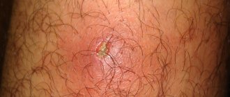
Causes and symptoms of an ankle fracture
The main causes of ankle fractures are direct (impact) and indirect (twisting of the lower limb) injuries.
Indirect injuries are more common. They can occur when falling during icy conditions, sports training, roller skating and snowboarding, slipping on a wet floor, etc.
Causes of direct ankle injuries can include car accidents, something heavy falling on your foot, etc. An ankle fracture is accompanied by:
- Pain syndrome;
- Swelling of soft tissues;
- Hematomas;
- Impaired joint function.
Diagnostic measures
Questioning and inspection cannot provide the necessary information. The key role is played by x-ray methods. As a rule, a radiograph of the injured limb is required in two projections.
Moreover, for better visualization of individual parts of the joint, different techniques are used (displacement, rotation, tilt of the foot). When necessary, a photo of the healthy leg is taken for comparison. There may be a need for CT or MRI.
Fractures of the talus require a more careful choice of diagnostic technique, as they are more complex in terms of treatment.
Fig4. A. CT scan shows a fracture of the talus and tibia, b. MRI revealed a fracture of the talus
First aid for a fracture of the outer ankle
If the ankle is broken, the victim must be taken to the emergency room. It's better to call an ambulance. When this is not possible, you will have to organize a stretcher from available materials and deliver the victim to the nearest medical facility.
Before the doctors arrive, the patient needs to be provided with pre-medical care:
- Free the leg from traumatic factors, of course, if this is possible without additional damage to the limb.
- Elevate the affected leg. To do this, you can make a roller out of clothes.
- If bleeding occurs, apply ice or something cold, if possible - a hemostatic tourniquet, which should be loosened for 20 seconds, every 20 minutes.
- If the integrity of the skin is damaged, under no circumstances should you try to set bone fragments.
- If the pain is severe, give the victim an analgesic.
- When there is no specialized splint for immobilization, and the patient needs to be taken to a medical facility independently, you need to apply a splint from improvised means.
Gently bend the injured limb at the knee joint and position the foot so that the heel is at a right angle to the shin. Apply an improvised splint and secure it with a bandage, belt, belt, etc.
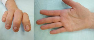
Symptoms
Certain aspects of external manifestations depend on the type and extent of injury. Attention should be paid to the following symptoms:
- Pain in the injured joint.
- Swelling in one or both ankles.
- Atypical foot orientation.
- Joint deformity.
The tricky part is the risk of low-symptomatic variants. You may encounter a break-off of a small fragment of bone, which does not lead to gross external manifestations, which means there is a delay in seeking medical attention after a fracture and, accordingly, treatment.
Treatment of an ankle fracture
An ankle fracture is treated conservatively and surgically.
Conservative treatment methods are used in the following cases:
- Closing fracture without displacement;
- Displaced fracture, but immediate closed reduction is possible;
- Minor damage to ankle ligaments;
- The operation cannot be performed (risk of anesthesia for serious diseases, patient refusal, some pathologies of the cardiovascular system and central nervous system, decompensated diabetes mellitus).
Stages of conservative treatment of an ankle fracture:
- Closed reduction (reposition) of the bone.
- Application of a plaster cast.
- Removing the plaster.
Indications for surgical therapy:
- Open fracture;
- Ineffectiveness of closed reduction (reduction);
- Old injuries;
- Damage to both limbs;
- Rupture of the ligamentous apparatus of the ankle joint;
- Damage to the ankle with simultaneous fracture of the tibia and fibula, rupture of ligaments and outward subluxation of the foot.
Mechanism of injury
Fractures of the ankle joints occur under the influence of multidirectional forces:
- Axial force perpendicular to the sole (causing wedging of the talus between the ankles).
- Excessive bending (flexion) and extension (extension).
- Turning inward (supination) and turning outward (pronation).
- Rotation of the foot inward (inversion) and outward (eversion).
If a sufficiently large amount of energy is applied in any of these directions, the joint structure may fail and break.
Diagnostics
The doctor will discuss with the patient the medical history (how long the pain has been, what happened before the injury) and symptoms. He will also ask how the injury occurred and examine the affected area. If your trauma surgeon thinks you may have broken your ankle, he or she will order a series of tests to more fully understand the injury.
X-rays : X-rays can show whether the ankle bone has been broken and how many pieces of broken bone are there. They can also determine if there is displacement (a gap between broken bones). The doctor may also take X-rays of other parts of the leg or foot to make sure nothing was damaged as a result of the injury.
Stress test : A stress test is performed to determine whether surgical procedures are necessary to heal the injury. The doctor will apply some pressure to the ankle and take a special x-ray to determine the severity of the injury.
CT scan (computed tomography): If the fracture extends beyond the ankle, a CT scan may be needed to further examine the injury. A CT scan allows you to obtain a series of images in different planes, from which the doctor can determine the severity of the injury. Source: Comprehensive diagnosis of ankle joint injuries. Kim L.I., Dyachkova G.V. Genius of Orthopedics, 2013. p.20-24.
MRI (Magnetic Resonance Imaging) Scan: If your doctor suspects ligament damage has occurred, he or she may order an MRI scan to get a better look at the affected area. MRI can look deeper into bones, soft tissues, and ligaments to create higher-resolution images than most other tests.
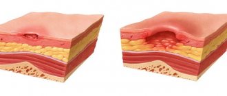
Consequences
If you do not consult a doctor in time, even a simple ankle fracture can complicate your future life.
A common consequence of delayed treatment is ankle pseudarthrosis, fusion in the wrong position, post-traumatic arthrosis.
Insufficiently thought-out treatment leads to impaired ankle stability and deformation. In old cases, eliminating such consequences is not easy.
Gross defects are much more dangerous:
- Persistent impairment of the supporting function of the foot.
- Due to blood supply disorders, aseptic necrosis of bones occurs.
- Infectious complications in the affected area.
The loss of physiological gait affects the entire human skeleton, primarily the spine. In some cases, ankle replacement may be required. .
Dietary recommendations
There is also a “whole science” here - due to injury, not only the limb suffers, but the entire body! It needs to be saturated with a number of nutrients, starting with minerals and ending with amino acids, in order to bring them to the damaged area where the regeneration process is taking place. For such patients, a diet is developed individually, which includes “dos and don’ts” and “don’ts.”
Yes, although an injured person is prescribed vitamins and minerals, one should not neglect the body’s ability to consume them naturally, that is, through food. Here's what you can and should eat:
- poultry meat - chicken and turkey are ideal;
- rabbit meat, it is often recommended to patients not only with fractures;
- fermented milk products;
- various nuts and seeds;
- berries and fruits, both fresh and dried;
- vegetable stews, salads, you can add a little vegetable oil;
- fish, preferably river;
- cereal porridge;
- parsley, dill;
- clear soups - you can use the same chicken, turkey or rabbit.
- slightly dried white bread.
Important! This is an approximate list; for each person, in accordance with his characteristics, his own diet is compiled. By the way, after receiving such an injury, patients need not only to eat, but also to consume a sufficient amount of fluid (mandatory for joints). What drink:
- mineral waters, weakly alkaline ones are needed;
- sweet and sour fruit drinks, juices;
- milk jelly - tasty and healthy (calcium);
- compotes.
On the day the cast is removed, be sure to drink 2.5 liters of fluid! A decoction of rose hips and chamomile is often used as a folk remedy. What not to eat and drink:
- fast food (burgers, shawarma, etc.);
- semi-finished products;
- smoked and fatty foods;
- all kinds of saltiness - no chips, crackers, dried squid;
- fried potatoes and pasta in any form;
- pickled products;
- Under no circumstances should you drink alcohol! The recovery rate will decrease by an order of magnitude;
- Black tea;
- coffee;
- energetic drinks.
The last three drinks also slow down the recovery process. Below we will talk about exercise therapy as one of the stages of rehabilitation after an ankle fracture - there are patients who use “energy drinks” in order to increase their tone. This cannot be done.
results
- 212 of 247 patients completed the study (86%).
- At week 52, the mean OMAS score was 87.6 (±18.3) in the 6-week cast group, 91.7 (±12.9) in the 3-week cast group, and 89.8 (±18.4) in the 3-week cast group. group using an orthosis for 3 weeks. The difference between groups was 3.6 points between 3 and 6 weeks cast immobilization (95% CI −1.9 to 9.1, P=0.20) and 1.7 points when comparing 3 weeks cast immobilization. and 6 weeks of cast immobilization (95% CI −4.0–7.3, P=0.56). Thus, in both comparisons the CI did not reach −8.8 points.
- Statistically significant differences between groups were found in small improvements in plantar flexion and the incidence of deep vein thrombosis (between the 3-week orthosis immobilization group versus the 6-week cast immobilization group).
Possible complications
After treatment, it is important to follow your surgeon's instructions. Failure to do so can lead to infection, deformity, arthritis, and chronic pain. Source: Mistakes and Complications in the Treatment of Compound Ankle Fractures. Salikhov R.Z., Pankov I.O., Plakseychuk Yu.A., Soloviev V.V. Practical medicine, 2014. p. 128-131.
Article sources:
- Errors and complications in the treatment of complex fractures of the ankle joint. Salikhov R.Z., Pankov I.O., Plakseychuk Yu.A., Soloviev V.V. Practical medicine, 2014. p. 128-131
- Analysis of ankle joint injuries. Taylashev M.M., Salatin P.P., Sobolev V.V., Pozikov V.V., Kolesnikov A.S. Acta Biomedica Scientifica, 2008. p. 144-145
- Comprehensive diagnosis of ankle joint injuries. Kim L.I., Dyachkova G.V. Genius of Orthopedics, 2013. p.20-24
- Fractures of the ankle joint and treatment methods. Cherednik A.A., More Gautam Saherbao, Al-Fakih Abdulaziz. Bulletin of Medical Internet Conferences, 2014. p.432
- Therapeutic tactics for intra-articular fractures of the ankle joint (literature review). Zedgenidze I.V., Tishkov N.V. Acta Biomedica Scientifica, 2013. pp. 178-182
| Name of service (price list incomplete) | Price, rub.) | In installments* |
* You can read more about the conditions here - Treatment on credit or in installments.
A set of exercises to develop muscles after a leg fracture
You should regularly perform exercises that will help your muscles quickly gain tone and restore normal balance when walking. It is recommended that you first seek the help of a doctor or exercise therapy instructor, who will monitor the correct execution and explain in detail how to develop an ankle after a fracture.
With a ball
The patient presses a special ball, a physioball, against the wall with his lower back. The feet and torso are pushed forward. You should squat smoothly so that the ball does not fall. The knees should bend until a right angle is formed (90°).
On the platform
Standing on an unstable surface, the healthy leg should be slightly bent at the knee. First you just need to learn how to balance and keep your balance. After mastering it, you can take the ball and, without losing a stable position, throw it at the wall and catch it. Exercise activates stabilizer muscles and restores balance.
Jumping
You need to jump on each leg alternately along the line marked on the floor. With each jump you need to land on different sides. The hip muscles are strengthened, balance and coordination of movements are trained. By how easily this can be done, one can assess the degree of development of the joint after an ankle fracture.
On a roller
Alternately stand with your foot on the bolster, and with the other foot you need to perform moderate swings back and to the side. For stability, the leg standing on the roller must be slightly bent, while keeping it straight. You can use a loop of elastic tape attached to the bottom of the wall. It is put on the leg that swings.
You can simply stand on your leg while maintaining your balance using the elastic loop on your other leg. Legs must be alternated.
If you follow the doctor’s advice and your own desire and perseverance, joint mobility is completely restored in a fairly short time.
Surgical measures
If it is obvious that closed reduction will not be effective, plaster is not used: they resort to open osteosynthesis surgery and installation of metal structures directly on the bone tissue.
Violations of the integrity of the talus are preferable to be treated surgically due to the risk of aseptic necrosis. Reduction and reposition of her fracture-dislocations must be carried out by repeating the mechanism of injury in the opposite direction.
Fig.5 a. osteosynthesis of the talus and calcaneus with screws, b. fracture of both ankles; osteosynthesis with plate and screws.
Medicines
Recovery from an ankle fracture includes taking medications. In this section, we will warn you right away: never “prescribe” medications to yourself or a relative without consulting a specialist! The course of treatment is prescribed on an individual basis - the doctor takes into account all factors, including the presence of other diseases in the patient. Before starting rehabilitation after an ankle fracture (immediately after the injury), medical workers use strong painkillers (injections), such as:
- "Ketanov";
- "Tramala";
- "Tramadol".
They are needed to relieve severe pain, and later the patient should switch to lighter drugs in tablets so that he does not develop drug dependence on potent painkillers in ampoules.
Also, at the first stage, antibiotics are prescribed to prevent the development of various types of infections in the victim. Non-steroidal drugs are:
- "Diclofenac";
- "Ibuprofen";
- "Ortofen";
- the familiar “Paracetomol” to everyone.
These drugs are needed to relieve swelling and inflammation. Plus, the patient will need to take medications that reduce congestion, improve blood flow and normalize metabolic processes, such as Trental and Ascorutin. The fact is that blood carries the function of transporting nutrients that cells need to regenerate damaged tissues!
To restore cartilage tissue, so-called chondroprotectors are needed, for example, “Structum”. The medical professional will also prescribe vitamins and minerals (the same calcium necessary for fusion), including B12 (“Cyanocobalamin”), which has hematopoietic properties.
Again, the correct course of taking a set of medications will only speed up the rehabilitation process, otherwise, with self-medication, the patient’s condition can significantly worsen! The doctor will take into account all contraindications, for example, the possible presence of renal and liver failure, which will require a more careful selection of medications.
How long does it last
To answer the question: “How long does rehabilitation last after an ankle fracture?” three criteria are always taken into account:
- the age of the injured patient and his lifestyle;
- the presence of chronic diseases that can (and will) slow down the process of tissue repair;
- the nature of the fracture, there are two of them, open and closed.
Now, in order. The older a person is, the more worn out the body is, and we emphasize that this includes his lifestyle - yes, this is relevant in our time, because now a huge number of people lead a sedentary lifestyle! Also, do not forget about one more thing - we are talking about the possible presence of bad habits that slow down the recovery process.
It’s worse when a patient, especially an elderly one, has diseases of the musculoskeletal system or diseases that can affect recovery processes - diabetes mellitus is a prime example! It has long been noted that diabetes significantly complicates treatment for other diseases and slows down the regeneration of damaged tissues.
Here is the nature of the fracture, if it is open (plus possible displacement, rupture of ligaments, blood vessels), then the recovery will become longer and more painful - about 6 months. In the best case, without complicating factors, a person will be able to get back on his feet after about 6 weeks. In children, the rehabilitation period is halved.