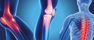
Leg pain is the third most common complaint among our patients. The reason for this is the high prevalence of varicose veins, arthritis and the seeming insignificance of such a problem as pain in the legs. At the same time, if your legs hurt from the knee to the foot, you cannot ignore this symptom as unimportant, since it may be the beginning of a more serious disease.
Pain below the knee in the front tends to occur in a few common situations. These include varicose veins, chronic venous insufficiency, myofascial pain syndrome (in which the pain is accompanied by cramps) and, less commonly, deep vein thrombosis, which is essentially an emergency condition.
At the appointment, we explain to our patients that pain in the legs must be treated and all necessary measures must be taken to eliminate it, since it deprives a person of the main factor of health - his mobility.
Pain below the knee with varicose veins
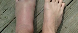
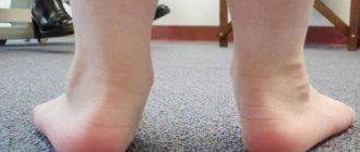
A typical situation in which a person is bothered by heaviness in the calves, swelling, “roaring in the legs,” and sometimes cramps. This picture is due to poor outflow of venous blood from the calf muscles, resulting in swelling, fluid accumulation, heaviness and pain. Superficial veins swell, blue venous nodes appear, and the skin of the lower leg takes on a brownish tint (pigmentation). The causes and treatment of varicose veins are described in detail in our article Varicose veins: causes, treatment, symptoms. Osteopathy helps not only to get rid of the symptoms of this disease, but also to prevent its further development, and often, the need for surgery.
Diagnosis in the early stages
A medical specialist conducts diagnostics to determine the cause of the disease. Collecting anamnesis, analyzing complaints, studying hereditary pathologies become the basis for determining the type of diagnosis. Possible causes identified by studying symptoms:
- Phlebeurysm
. Swelling appears, blueness of the limbs appears, and a pattern of veins appears. - Inflammation of the ankle joint
. Swelling and redness in the affected area. The feet swell and numbness appears. - Degenerative process in joints
. The pain intensifies in the evening and in the morning, the patient limps on one leg. - Diabetes
. Redness of the skin, formation of weeping ulcers.
For diagnosis, a medical specialist prescribes a general blood test to detect hidden inflammation. If a certain disease is suspected, ultrasound examination, MRI of joint pathologies and computed tomography may be prescribed. In rare cases, a cell biopsy is prescribed.
Myofascial pain syndrome
Overexertion of the lower leg muscles is the main cause of myofascial pain in the legs. Even in a healthy person, the deep muscles of the lower leg, as a rule, are in spasm, and if you run your hand over them, an acute characteristic pain appears. It is distinguished from venous pain by its greater severity, localization in one or several points, and irradiation into the foot or deep into the lower leg. Often muscle overstrain turns into spasms and cramps. In this case, neither taking calcium tablets nor increasing the dose of vitamins and microelements helps.
Myofascial pain is eliminated using osteopathic methods in 2–3 sessions, and from the first appointment the patient already feels significantly better. But you will have to be patient a little: trigger points on the legs are quite painful when touched, and their relaxation is associated with pressing on the spasmed area of the muscle. However, the positive effect quickly and completely covers the unpleasant sensations from the manipulations.
Osgood-Schlatter disease
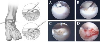
Osgood-Schlatter disease (sometimes also called Schlatter disease or osteochondrosis of the tibial tuberosity) is an inflammation in the upper part of the tibia, where the patellar tendons attach to the tubercle called the tibial tuberosity. This disease is often the cause of knee pain in adolescence (from 10 to 15 years), is associated with the growth of the musculoskeletal system, and as growth completes, the symptoms may disappear.
Osgood-Schlatter disease can cause pain, swelling, and tenderness in the area of the front of the knee, below the kneecap. As a rule, one knee is affected (sometimes it happens on both sides). Symptoms worsen when the tendon attached to the tibial tuberosity is stressed (for example, when jumping). The diagnosis is made on the basis of radiography. The main goal of treatment is aimed at preventing symptoms (reducing certain types of stress, immobilization during exacerbation, physiotherapy). Surgical treatment methods are rarely used.
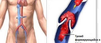
Acute thrombosis of the veins of the leg
An emergency condition that requires immediate hospital treatment. Hospitalization cannot be delayed; a blood clot can completely block the outflow of venous blood and lead to gangrene of the limb. In addition, a detached blood clot can clog the blood vessels of the lungs and cause thromboembolism. If a patient comes to us with symptoms of acute thrombosis, we immediately send him to the hospital. It will be possible to continue osteopathic treatment after discharge - eliminate the causes that led to the development of thrombosis, improve venous outflow from the lower extremities, eliminate blood stagnation at the pelvic level, etc.
Arterial diseases
Pain in the legs is caused by:
- atherosclerosis of the arteries of the lower extremities;
- obliterating endarteritis;
- thromboangitis obliterans;
- embolism and arterial thrombosis.
Atherosclerosis of the arteries of the lower extremities
Among diseases of the arteries of the lower extremities, atherosclerosis is the most common - in almost 90% of cases. It refers to obliterating vascular diseases (obliterating - leading to closure, fusion). Previously, the disease mainly affected older men. Unfortunately, there is now a tendency towards rejuvenation of this disease, and atherosclerosis occurs even in middle age. Cases of obliterating atherosclerosis in women have also become more frequent.
Risk factors are well known - smoking, low physical activity, lifestyle changes, hereditary factors, hypertension, coronary heart disease, diabetes mellitus, liver and biliary tract diseases, obesity, high cholesterol in the blood.
During the disease process, plaques consisting of cholesterol, other fats, calcium and connective tissue form on the inner walls of the arteries. The lumen of the arteries narrows. At rest, arterial circulation in the lower extremities may be sufficient, but during physical activity the increased oxygen demand of the muscles cannot be satisfied. The muscles respond to this with pain, which is how intermittent claudication appears. When walking, especially uphill, pain occurs in the calf muscles, forcing the patient to periodically stop for it to pass. If pain occurs in the entire leg, it means that the arteries at the pelvic level are affected by the pathological process. Unlike radiculitis, pain does not depend on sudden movements or turns of the body.
Instead of pain, you may experience cramps, weakness, or a feeling of heaviness in your legs.
The legs become cold and pale, and no pulse can be felt on them. At the last stage, trophic ulcers and foci of gangrene may form. If gangrene develops, amputation of the affected limb may be necessary to save the patient's life. Often, especially with high occlusions, when necrosis develops on the fingers, amputation is performed at the level of the upper third of the thigh, since with lower amputations it is difficult to achieve healing of the postoperative wound.
Find out more about the promotion >>>
Obliterating endarteritis
The International Classification of Diseases ICD-10 does not provide for such a diagnosis. It is necessary to use other diagnoses specified in this document - “arterial atherosclerosis”, “thromboangiitis obliterans”, “other specified peripheral vascular diseases”, “other unspecified peripheral vascular diseases”.
The disease affects mainly middle-aged and even young men. The exact cause of the disease is still not known. The role of cold and mechanical trauma, as well as autoimmune processes, is suggested.
The small arteries of the legs are affected. Unlike atherosclerosis, which develops gradually, obliterating endarteritis is characterized by an undulating course - exacerbations alternate with remissions. The symptoms and complications and outcomes are almost the same - intermittent claudication, possible amputation due to gangrene, etc.
Thrombangitis obliterans (Berger's disease)
This is a “disease of young men who smoke.” An autoimmune mechanism of development is suggested. Exacerbations alternate with remissions. Every second patient undergoes amputation at the level of the fingers, foot or thigh.
A feature of thromboangiitis obliterans and endarteritis is the symmetry of the damage to the extremities and the possible involvement in the process of not only the lower, but also the upper extremities. At the same time, atherosclerotic lesions are often unilateral and affect almost exclusively the legs.
Embolism and arterial thrombosis
In embolism, an artery is blocked by some substance (in thrombosis, a blood clot).
In the area fed by the blocked artery, diffuse pain occurs. The limb first turns pale, then turns blue. Her pulse can no longer be felt. Numbness appears, and in severe cases paralysis may develop. In such cases, if treatment is not started in a timely manner, everything ends with rapidly developing ischemia, wet gangrene and amputation of the limb.
LUMBOSACROSIS OSTEOCHONDROSIS
Shooting pain spreading along the back of the leg from the buttocks to the heels is characteristic of lumbosacral osteochondrosis. Many diseases come from a problematic back - tension, poor lifestyle, and little physical activity aggravate the poor condition of the spine and lead to pain in different parts of the body, including the legs from the lower leg to the foot.
Neurologists, vertebrologists and osteopaths deal with osteochondrosis: you can always contact our doctors for advice and help, which will not only relieve pain, but also eliminate the original cause of its occurrence.
Osteopathy is one of the most effective methods of treating osteochondrosis, relieving pain literally after the first appointment and helping, in addition, to eliminate the original cause of the painful condition.
Manifestations and symptoms
The diagnosis is made on the basis of research and analysis of the current condition. The medical specialist pays attention to concomitant pathologies. Worthy of attention:
- severe progressive swelling of the calves;
- intense pain in the arms;
- swelling around the affected part;
- change of skin color;
- purulent lesions of the integument.
If small areas of the skin turn purple or acquire bright red or dark burgundy shades, you should immediately consult a doctor. Severe swelling of the legs, visible patterns of capillaries and non-healing ulcers indicate a severe pathological process that occurs in the body.
The presence of temperature indicates an inflammatory process and possible infection.
If pain occurs, the patient should independently analyze the previous days. If intense training or unusual physical activity contributed to the illness, then there is nothing to worry about. Pay attention to various ailments that could provoke complications in the form of aching joints.
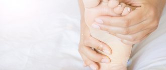
FLAT FOOT
The cause of the painful condition of the legs from the feet to the legs can be “normal” flat feet. This pain is characterized by an increase in the evening and aggravation during long walking and other physical activities; it is aching, tiring, and spreads throughout the lower part of the limbs. The feet may become visually larger, and the bone often begins to protrude.
Flat feet are “treated” by choosing shoes after consultation with a doctor, physical therapy complexes, and osteopathy also helps to cope with it. Osteopaths work on the foot, improving blood circulation, eliminating congestion, and stimulating the production of synovial fluid, which works as a lubricant for the joints. After just a few techniques, even an adult who has suffered from flat feet all his life becomes easier and more comfortable to shift.
Treatment
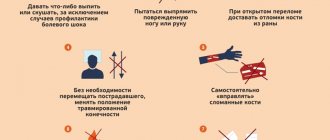
Help before diagnosis
For minor injuries and non-traumatic lesions, it is recommended to ensure rest and an elevated position of the limb. For tibia fractures, temporary immobilization using splints or improvised materials is required. The leg is fixed from the foot to the upper third of the thigh. The victim is given an analgesic. For pain of non-traumatic origin without signs of severe inflammation, it is possible to use local anesthetics. In case of intense pain or a violation of the general condition, an immediate examination by a specialist is necessary.
Conservative therapy
For patients with tibial fractures, a blockade is performed, followed by fixation using skeletal traction. For other diseases and injuries, depending on the severity of the pathology, a plaster splint is applied, rest or a gentle regimen of physical activity, and the use of orthopedic devices are recommended. The following methods are used:
- Drug therapy
. The list of drugs is determined by the etiology and symptoms of the disease. For intense pain, analgesics are prescribed. For purulent lesions, antibiotics are necessary. For vascular pathologies, antiplatelet agents, anticoagulants, and antispasmodics are indicated. - Exercise therapy
. Therapeutic exercise is a mandatory part of rehabilitation measures. Allows you to maintain muscle strength and joint mobility, prevent the development of complications, and improve limb function. - Physiotherapy
. Physiotherapeutic procedures reduce pain and inflammation, activate blood circulation, and stimulate recovery processes. Widely used techniques include medicinal electrophoresis, UHF, and magnetic therapy. Electrical stimulation has been successfully used for some diseases.
Patients are prescribed massage. According to indications, manual therapy is performed. For a number of pathologies, kinesio taping is used. Patients are referred to sanatorium-resort treatment.
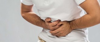
LEGS HURT RIGHT NOW! WHAT TO DO?
1. Relax. Lie down with your ankles on a bolster to elevate your legs above heart level. The blood will drain, the swelling will decrease, and the muscles will relax.
2. Apply a cold compress. For example, a thin towel with ice or some package of semi-finished products taken out of the freezer. Keep the compress for up to 20 minutes, repeat several times a day.
3. Take an over-the-counter pain reliever such as ibuprofen. But take only those medications that you are sure of, and if the pain does not go away, be sure to visit a doctor.
4. Get a massage. This is true if your legs have cramps, or if they “hum” after a long period of exercise. You can stretch the muscles yourself, or you can consult a specialist, including an osteopath.
Juvenile idiopathic arthritis
Juvenile idiopathic arthritis, often called juvenile rheumatoid arthritis or juvenile chronic arthritis, is a childhood disease characterized by inflammation of the joints, which become swollen and painful (and therefore impair joint mobility). It manifests itself in children as pain in the joints, impaired gait, due to decreased mobility in the joints and stiffness in the joints, usually in the first half of the day, with regression within 1-1.5 hours and after normal physical activity. The causes of this disease are not entirely clear. Most researchers believe that the disease is caused by a combination of factors that cause excessive activation of the immune system. There are several types of juvenile arthritis. The division into types is based on the number of joints involved in the process during the first 6 months from the onset of the disease, the area of the body where the disease manifested itself, and the presence of other symptoms. Types of juvenile arthritis: oligoarthritis (affects no more than 4 joints), polyarthritis (affects 5 or more joints), rheumatic-positive and rheumatic-negative (depending on the presence of rheumatic factor in the blood), systemic (accompanied by rashes on the body, fever and lesions). eye).
The benefits of osteopathy in the treatment of foot diseases
- Osteopathy restores not only the function of the damaged area, but also the biomechanics of your entire body.
- We find the reason that led to your dysfunction and eliminate it.
- We do not give you injections into the joint cavity, we do not use hormonal drugs, which means you have no risk of infection or other complications.
- We provide all patients with a full osteopathic examination and examination, regardless of complaints. This allows for correction at a higher level, including the body’s reserves and directing them towards self-healing.
Medicines
Some medications can cause leg problems due to side effects. Combination with smoking or drinking alcohol increases the risk of drug side effects. The main groups of drugs that can cause leg problems:
- Drugs that cause blood clots (for example, contraceptives)
- Neuroleptics (aminazine, haloperidol)
- Beta-2 receptor agonists (terbutaline or albuterol)
- Antihypertensive drugs (nifedipine, amlodipine or nicardipine)
- Statins are drugs that lower blood cholesterol levels (simvastatin or atorvastatin).
- Estrogens
- Lithium preparations
- Diuretics (Lasix and others)
- Opiates
- Steroids
If you are taking medications (especially long-term), you should inform your doctor about the occurrence of pain or cramps in the legs.
Transient ischemic attacks
Transient ischemic attacks are a temporary disruption of the blood supply to the brain. They are harbingers of a stroke. The only difference from a stroke is that the symptoms gradually disappear. The following symptoms should alert you:
- Sudden numbness, tingling and weakness in half the body or limb
- Gait disturbance
- Impaired perception of reality
- Speech Impairment
- Strong headache.
If these symptoms occur, seek emergency medical attention
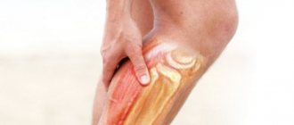
Muscle rupture
Most often, muscle ruptures occur in the lower leg (possibly in the thigh). A rupture occurs as a result of a sudden stretch or contraction of a muscle. Typically, a small section of muscle is torn where it connects to the tendon. But there are also large ruptures, up to separation of the muscle from the tendon or separation of the tendon. Muscle rupture occurs mainly when the limb is sharply flexed in the direction opposite to the acting force. The rupture is accompanied by acute pain immediately after excessive stress. The pain may subside for a while, but as the hematoma grows, the pain will gradually intensify. Swelling occurs in the area of the damaged muscle and range of motion is limited.
Crick
Muscle pain can appear after fast walking, running, playing sports, or walking in poorly fitting shoes. As a rule, discomfort occurs immediately after exercise or after 12-24 hours. Most often, pain occurs in the legs, sometimes in the hips. Possible pain on palpation. Sometimes swelling of the lower leg occurs. If there is a clear connection with physical activity and the pain is bilateral, then the diagnosis is beyond doubt. In cases where the pain is one-sided, it is necessary to exclude possible venous thrombosis using objective diagnostic methods.