In the medical field, gallbladder cancer is considered a rare malignant tumor. Often occurs in older people and is accompanied by the presence of gallstones. Experts identify neoplasms in the wall of an organ of various histological structures. Gallbladder cancer is characterized by active growth into the liver. It quickly affects the lymph nodes and metastasizes to the lungs and bones.
By its nature, in 80% of cases it occurs in the form of adenocarcinoma. This tumor is represented by glandular cells. Rarely develops as a classic carcinoma.
Risk factors for gallbladder cancer
As mentioned earlier, gallbladder cancer most often begins and develops in older people (the average age is 50-60 years). But there are other risk factors:
- heredity. If there have already been similar cases among relatives, then the person’s risk of developing gallbladder cancer increases by 60%;
- sufficiently long contact with carcinogens;
- harmful working conditions for many years. We are talking about work in places for the production of rubber products, metal smelting;
- parasitic infections that people have had. Most often, gallbladder cancer develops against the background of opisthorchiasis;
- all kinds of chronic inflammatory diseases of the gastrointestinal tract. In a special risk group are people diagnosed with Crohn's disease, nonspecific ulcerative colitis;
- alcohol addiction and smoking are also causes of gallbladder cancer;
- weakened immune system.
Poor nutrition can also cause gallbladder cancer. Especially if the diet is dominated by fatty and spicy foods, but there are practically no cereal products. Preservatives, chemical additives, smoked food - all this is the main provocateur of the development of the disease.
Background pathologies also contribute to the development of gallbladder cancer. These include:
- polyps;
- polycystic gallbladder;
- calcification (stones in the biliary tract);
- biliary cirrhosis;
- sclerosing cholangitis (catarrhal process in the liver);
- carriage of salmonella or past salmonellosis
Stages
Like any other cancer, gallbladder cancer has its own stages.
- Tis or stage zero gallbladder cancer. It stands still, does not grow and does not spread to neighboring organs and tissues.
- T1 or first stage. Characterized by an increase in malignant cells. The tumor is beginning to grow, but so far only slightly.
- T2 or second stage gallbladder cancer. Caused by significant changes in size. The tumor grows to the serous layer.
- T3 or third stage. The tumor affects the liver and radiates to the gastrointestinal tract. Metastases form.
- T4 or stage four. The tumor increases in size up to 2 cm. It grows into the stomach, pancreas, and duodenum.
- N0. Metastatic damage begins.
- N1. The lymph nodes are largely affected.
- N2. At this stage, metastases reach the head of the pancreas, as well as the duodenum and the celiac artery.
- M0. Characterized by strong metastases to other organs.
- M1. Distant metastases are detected.
But what are the symptoms of gallbladder cancer?
Cholecystitis
Which doctors should I contact?
After detecting symptoms indicating cholecystitis, you should immediately consult a doctor.
Usually in such cases they turn to a surgeon, therapist, gastroenterologist, and in acute cases call an ambulance. All patients with acute cholecystitis and suspected of having this disease are subject to referral to a surgical hospital with a team of qualified surgeons and anesthesiologist on duty around the clock. In severe cases, the participation of a resuscitator is required. Treatment of cholecystitis
The main goals of treatment of chronic acalculous cholecystitis are relief of pain and inflammation in the gallbladder, correction of digestive and metabolic disorders, treatment of complications and concomitant diseases.
- During the period of exacerbation, patients with chronic cholecystitis are recommended bed rest, fasting for 1-3 days, then diet therapy - limiting fatty foods and their calorie content (diet No. 5, 5A), eating 4-6 times a day.
- Antispasmodics and analgesics are the main drugs for the treatment of exacerbation of cholecystitis, allowing to alleviate biliary colic.
- Analgesics are used to relieve pain.
- To stop the infectious inflammatory process, broad-spectrum antibiotics are prescribed.
- When helminthiasis is confirmed, anthelmintic drugs are used.
- Relief of dyspeptic disorders is carried out with the help of enzymatic, antiemetic drugs that suppress gastric secretion, etc.
- Violation of the outflow of bile is corrected with choleretic agents.
- If the stones are cholesterol, then there is a possibility of dissolving them with the help of drugs based on ursodeoxycholic acid.
Lithotripsy is a shock wave method that is used to crush stones, which are subsequently safely removed from the body or dissolved.
The selection criterion for patients for this procedure is limited by the size of the stones - single stones no more than 2 cm, a few up to 1 cm. The standard solution for the treatment of chronic calculous cholecystitis is surgical intervention, which eliminates new exacerbations and possible complications. Laparoscopic cholecystectomy is the main surgical method for treating chronic calculous cholecystitis. Removal of the gallbladder is performed with special instruments through one or more small incisions in the abdominal wall. Complications
Cholecystitis can lead to a number of serious complications.
- Due to inflammation, the gallbladder can stretch and increase in size (dropsy), which increases the risk of its rupture (perforation) and the development of peritonitis.
- Stagnation of bile in the gallbladder is a risk factor for infection of bile by bacteria, as a result of which the infection can enter the bloodstream with the development of sepsis.
- Violation of blood microcirculation in the wall of the gallbladder with the further development of ischemia and focal or total necrosis of the wall is the cause of gangrene of the gallbladder.
The following complications of cholecystitis require emergency surgical care:
- empyema of the gallbladder - accumulation of pus in the cavity of the gallbladder;
- peri-vesical infiltrate and abscess - an inflammatory process that spreads beyond the gallbladder;
- perforation is a rupture of the wall of the gallbladder with the release of its contents into the abdominal cavity.
Intraoperative complications include bleeding and bile duct injury.
Postoperative complications include pneumonia, myocardial infarction, thromboembolic complications, and pulmonary embolism. Prevention of cholecystitis
Primary prevention consists of eliminating risk factors leading to the development of chronic cholecystitis - timely sanitation of the biliary tract, elimination of dyskinesia, early detection of stones in the gallbladder and timely adequate treatment, including surgery.
Secondary prevention involves reducing the frequency of relapses, preventing the progression of the disease and the development of complications. Active identification of patients with clinically pronounced forms of chronic cholecystitis, frequent exacerbations and their adequate non-drug and drug therapy.
Diet plays a significant preventive role - low-calorie, predominantly plant-based food is useful, the use of vegetable fats containing polyunsaturated fatty acids, phospholipids, vitamin E, which help normalize cholesterol metabolism, participate in the synthesis of prostaglandins that dilute bile, and increase the contractile function of the gallbladder. Vegetable fats are especially important for bile stagnation. Vegetables, fruits, and bran promote the discharge of bile, reduce the cholesterol content in bile, thereby reducing the likelihood of stone formation.
Sources:
- National clinical guidelines “Acute cholecystitis”. Russian Society of Surgeons, 2015.
- Beburishvili A.G., Panin S.I., Zyubina E.N., Bykov A.V. Optimal timing of surgical treatment of acute cholecystitis according to evidence-based studies. Annals of surgical hepatology. 2020;25(3):12-19.
- Vorotyntsev A.S. Modern ideas about the diagnosis and treatment of cholelithiasis and chronic calculous cholecystitis. "Attending doctor". No. 02, 2012.
IMPORTANT!
The information in this section cannot be used for self-diagnosis and self-treatment. In case of pain or other exacerbation of the disease, diagnostic tests should be prescribed only by the attending physician. To make a diagnosis and properly prescribe treatment, you should contact your doctor.
Symptoms and signs of gallbladder cancer
Clinical manifestations include:
- swelling is noticeable in the epihystric zone;
- There is a feeling of heaviness under the ribs (on the right side). Sometimes this can manifest itself as a feeling of “bloating” in the area. This is one of the main symptoms of gallbladder cancer;
- frequent attacks of nausea;
- Dull pain is felt under the ribs (in the right side);
- bowel disorder. Moreover, with gallbladder cancer this can manifest itself in different ways. Some people have diarrhea, while others have constipation;
- frequent fatigue and severe weakness;
- sudden and unexplained weight loss.
All symptoms of gallbladder cancer are the first sign that you should undergo a medical examination. More serious manifestations of the disease are also possible.
It should be understood that as the first symptoms of gallbladder cancer progress, they begin to acquire the greatest clinical significance:
- low-grade body temperature (it can rise to 38 degrees and remain at this level for quite a long time);
- itchy skin;
- adynamia, lethargy (lethargy may appear);
- presence of bitterness in the mouth;
- anorexia (this is already a very serious symptom of gallbladder cancer);
- the appearance of yellowness on the skin;
- darkening of urine and discoloration of stool.
Symptoms and diagnosis of the disease
The bile ducts have a small diameter, so even with a minimal tumor size, signs of blockage begin to appear. They are expressed in aching pain in the right hypochondrium, jaundice, lack of appetite, sudden loss of body weight, and itchy skin. Laboratory diagnostics can detect an increase in direct and total bilirubin, serum alkaline phosphatase, and a slight change in the concentration of serum transaminases. Instrumental studies are also informative - endoscopic retrograde cholangiopancreatography and percutaneous transhepatic cholangiography. Both methods also involve the collection of affected tissue for histology.
When to see a doctor
Already at the first symptoms of gallbladder cancer (even the most minor ones), you should seek help from a specialist. The doctor will conduct all the necessary tests and be able to diagnose the disease. This will allow you to start treatment on time, which will give the most favorable prognosis for gallbladder cancer.
The oncology department is located in the central district of Moscow (Mayakovskaya metro station). Highly qualified doctors with extensive experience and all the necessary knowledge work here.
Classification of the disease
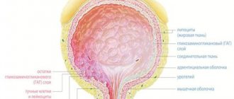
The most widely used international classification of oncological diseases is TNM, derived from the Latin terms tumor, nodus and metastasis. The first letter describes the size of the tumor and the degree of its growth into healthy tissue, the second - damage to the lymph nodes, the third - the presence of metastases. Within each characteristic there is also a separate gradation of the attribute, for example:
- TIS or carcinoma in situ - the tumor does not extend beyond the lesion;
- T1 - the muscle layers and mucous membrane of the gallbladder are affected;
- T2 - the tumor grows into the perimuscular connective tissue;
- T3 - the neoplasm metastasizes to the visceral peritoneum and regional organs, for example, to the liver;
- T4 - the tumor grows into the liver by more than 2 cm, or spreads to at least two nearby organs: omentum, stomach, duodenum, pancreas, colon, extrahepatic bile ducts;
- N1 - metastasis involves the porta hepatis and lymph nodes of the cystic and common bile ducts;
- N2 - metastases spread to the lymph nodes of the head of the pancreas, portal vein, superior mesenteric and celiac arteries, duodenum.
Diagnosis of gallbladder cancer in the oncology center
Symptoms of gallbladder cancer do not always indicate the presence of this particular disease. To confirm the diagnosis, it is necessary to undergo a comprehensive examination.
To diagnose gallbladder cancer, you must:
- conduct a series of laboratory tests;
- do an ultrasound;
- perform cholecystography;
- perform diagnostic laparoscopy.
Also, when gallbladder cancer manifests itself, they resort to the diagnostic method of MRI and CT.
A full examination can be done at the oncology department, which is located in the center of Moscow.
Establishing diagnosis
It is recommended to carefully collect complaints and medical history from patients to determine factors that influence the choice of treatment tactics. A physical examination is required to assess nutritional status to determine functionality.
Diagnosis of gastric cancer includes:
- Laboratory research. They are carried out at primary signs of the disease and regularly during treatment to predict the course and determine risk factors. Laboratory diagnostics include a CBC (complete blood count) and a biochemical test to check liver function.
- Instrumental diagnostics (ultrasound and CT). The structure of the gallbladder, bile ducts, as well as the size and location of the tumor are determined.
Additional studies - endoscopy (choledochoscopy, endoscopy). Direct contrast methods and angiographic examination are recommended. For morphological verification of the diagnosis, a tumor biopsy is indicated.
Treatment of gallbladder cancer
If gallbladder cancer is confirmed by ultrasound or the oncology is detected in another way, then treatment is prescribed immediately. The main way to get rid of cancer is surgery, after which additional measures are prescribed to destroy the tumor:
- chemotherapy;
- radiation therapy;
- radiation therapy with complex administration of sensitizers.
The treatment method is determined individually depending on the manifestation of gallbladder cancer and its stage. Rehabilitation is required, which may take a certain period of time (until the body is completely restored).
TNM classification of the disease
In accordance with the international classification of stages of development of oncological diseases, the following types of cholangiocarcinomas are distinguished:
- TIS or carcinoma in situ - the tumor does not extend beyond the affected organ;
- T1 - malignant tumor spreads in the muscular-connective layer of the organ or in the subepithelial connective tissue;
- T2 - carcinoma grows into the perimuscular connective tissue;
- T3 - carcinoma metastasizes to adjacent structures;
- N1 - the tumor metastasizes to the lymph nodes in the hepatoduodenal ligament;
- N2 - metastasis spreads to the lymph nodes near the celiac and mesenteric arteries, head of the pancreas, duodenum and peripancreatoduodenal lymph nodes.
How to make an appointment with a specialist in oncology
To make an appointment with a specialist for a consultation or to carry out a full diagnosis, you can use a special form that is available on the website of the oncology center. You can see a doctor by calling: +. We are located in the central district of Moscow, at the address: 2nd Tverskoy-Yamskaya lane 10 (Mayakovskaya metro station, Belorusskaya metro station, Novoslobodskaya metro station, Tverskaya metro station, Chekhovskaya metro station). When a disease is detected, doctors immediately prescribe treatment and further monitor the patient.
Causes of the disease
There are certain factors that increase the risk of developing gastric cancer:
- age 65 years and older;
- hereditary factor;
- excessive alcohol consumption, smoking, exposure to harmful chemicals;
- diseases that cause inflammation and scarring (colitis, primary sclerosing cholangitis).
- liver fluke infection.
Almost 2/3 of cases of gastric cancer develop with a long previous course of chronic cholecystitis or cholelithiasis. Carcinogenesis is promoted by injury to the mucous layer by moving stones.
Treatment tactics and survival prognosis for common bile duct cancer
Only surgical removal of the tumor can increase the chance of recovery, and its resectability is no more than 10%. For resection of distal cancer, the so-called Whipple operation is performed - pancreaticoduodenectomy, after which the patency of the biliary tract and gastrointestinal tract is restored. If the tumor is localized in the proximal parts, restoration of the general bile flow is required after removal of the tumor. When tumor removal is not possible, tunneling is performed using drainage. On one side it opens into the common bile duct, on the other - into the intrahepatic ducts. External drainage is sometimes used in percutaneous transhepatic cholangiography. After surgery, life expectancy increases by an average of 23 months. Chemotherapy helps to slightly extend this period.
According to Praderi, drainage is performed with a U-shaped tube. The manipulation is carried out for palliative purposes, with its ends being exposed to the skin. In cases of blockage, drainage can be replaced with tissue debris. The new tube is sutured at one end to the old one, and replacement is done by pulling it up. Drainage makes it possible to prolong the patient’s life by 6-19 months.
The prognosis for cancer of the common bile duct is disappointing. Metastasis occurs quite late. The causes of death in this disease can usually be general exhaustion, sepsis, liver infection, abscesses and biliary cirrhosis.