Sign up
+7
Pericarditis is a heart disease manifested by an inflammatory process localized in the serous membrane of the organ. Acute pericarditis is an inflammatory syndrome with or without pericardial effusion (effusion). The diagnosis of subacute pericarditis is established when its duration is from 4-6 weeks to 3 months without remission. Recurrent pericarditis is diagnosed after a documented first episode of acute pericarditis and a symptom-free period of 4-6 weeks or longer (usually about 18-24 months).
Chronic pericarditis lasts longer than 3 months without remission. It can develop in various forms - asymptomatic, effusion, with lime deposits - the so-called “shell heart”, constrictive.
In whatever form the disease manifests itself - constrictive, effusion, etc. – it requires quick diagnosis and proper effective treatment.
Make an appointment with a specialist without queues, at a convenient time
Sign up
+7
Reasons for the development of the disease
This cardiac disease has a polyetiological nature:
- Infectious causes include rheumatic, tuberculous, bacterial. In addition, pericarditis can have a fungal or viral etiology, for example, with influenza.
- Aseptic reasons include allergies, diffuse connective tissue diseases, hematological diseases, malignant processes, injuries, radiation exposure, hypovitaminosis.
- Idiopathic pericarditis may also develop, the cause of which cannot be determined. It is these pathologies that are most often prone to relapse.
Forecast
The prognosis of this disease depends entirely on the severity of the disease. So, with a mild form of armored heart, treatment may not be required or it will only be medicinal.
Complex forms of pericarditis are not only difficult to treat, but can also threaten the patient’s life.
With timely treatment, the prognosis is quite good. The recovery period takes from 2 weeks to 3 months. Relapse of the disease occurs in 15 - 30% of cases. When complications such as heart failure, hyperthermia and accumulation of large volumes of fluid occur, the prognosis worsens.
The worst prognosis occurs with constrictive pericarditis. Removal of the pericardium with this diagnosis is successful only in 60% of cases.
In order to prevent undesirable consequences and complications of pericarditis, it is necessary to seek help from a cardiologist in a timely manner. If you have pericarditis, it is very important not to self-medicate , which can only worsen the situation.
Symptoms
Pain syndrome in acute pericarditis can be characterized by varying intensity. This can range from a slight tingling sensation when turning or bending sharply, to severe, all-consuming pain, like a heart attack. The pain may become stronger when sneezing, coughing, or deep breathing. There may be a sensation of radiating to the shoulder, neck, or shoulder blade.
In addition to pain, accompanying symptoms are: shortness of breath, mild headache or dizziness, and a slight increase in body temperature. Despite the fact that this symptomatology is quite “common,” it is extremely important to carry out a differential diagnosis and accurately determine the presence of pericarditis. Thus, acute pericarditis can be diagnosed if at least 2 of the following 4 signs are present:
- Pericardial chest pain.
- Pericardial murmur.
- The ECG interpretation shows signs of pericarditis.
- Pericardial effusion (formed or increased compared to the previous volume).
Additional signs: signs of an inflammatory process in the pericardium during imaging methods (computed tomography - CT, magnetic resonance imaging - MRI, ultrasound examination of the heart).
- Available: doctor’s appointment from 1500 rubles
- Convenient: open daily from 8:00 to 21:00
- Quickly: we will carry out all the diagnostics at the first appointment
- Complete: has all the necessary equipment
Diagnostics
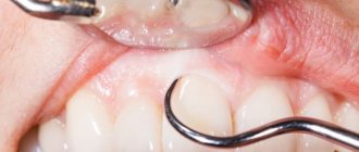
The first signs that may lead a doctor to think about pericarditis are revealed at the examination stage - this is a pericardial friction noise. If necessary, the specialist asks questions to clarify a clearer picture. To confirm the diagnosis, the doctor prescribes instrumental diagnostic methods:
- Echocardiography (ultrasound of the heart) is the most informative method for diagnosing pericarditis. Using ECHO-CG, the doctor can see and describe the contents of the pericardial cavity for the presence of various types of inclusions (blood clots, fibrin).
- Electrocardiogram. The typical sign of acute pericarditis is widespread ST segment elevation on the film.
- Chest X-ray is recommended for all patients. An enlarged heart shadow is detected on an x-ray only if the effusion is more than 300 ml. However, the study reveals signs of involvement of the pleura (the outer lining of the lungs) in the inflammatory process.
Laboratory blood tests are also prescribed:
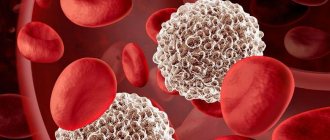
- General blood analysis. An inflammatory reaction of the blood often accompanies acute pericarditis - an increase in the level of leukocytes, the appearance of their young forms (band, juvenile), a high level of ESR (erythrocyte sedimentation rate).
- Biochemical blood test - C-reactive protein (increased), markers of myocardial damage (troponins, MB-fraction of creatine phosphokinase can be increased if the myocardium is involved in the process).
The listed examinations are included in the mandatory list for diagnosing acute pericarditis.
After an examination, clarification of the medical history, and carrying out the listed tests, the doctor usually already has guesses about the cause of the disease. To confirm the diagnosis, the following additional examinations are performed:
- intradermal tuberculin test (Mantoux test, Diaskintest) for the diagnosis of tuberculosis;
- blood culture for suspected infective endocarditis;
- virological studies using ELISA and PCR methods;
- analysis for HIV, hemophilus influenzae infection;
- exclusion of chlamydial and mycoplasma infections using ELISA and PCR methods;
- determination of antinuclear factor, rheumatoid factor, antibodies to cardiolipins (for systemic lupus erythematosus, rheumatoid arthritis and others);
- antistreptolysin-O titer (for rheumatism);
- determination of antibody titers to myolemma and actomyosin in blood serum (if perimyocardial tuberculosis is suspected);
- determination of the level of thyroid hormones (for hypothyroidism).
Sometimes, to make a diagnosis, the doctor requires additional studies with greater resolution to clearly visualize the pericardium and its cavity. Such studies include computed tomography and magnetic resonance imaging.
The countries of the post-Soviet space are characterized by a high incidence of tuberculosis bacillus (the causative agent of tuberculosis) among the population, which often causes inflammation of the outer lining of the heart and associated complications. In this regard, it is recommended to determine the etiology of acute pericarditis in the early stages in the following ways:
- Pericardiocentesis with drainage of the pericardial cavity. Fluid taken from the pericardial cavity is sent for PCR and histochemical analysis to identify the infectious or tumor etiology of the disease. Pericardiocentesis with drainage of the contents is also a mandatory emergency procedure if cardiac tamponade is suspected.
- Pericardioscopy with pericardial biopsy. The specialist inserts an endoscope with a camera into the pericardial cavity, examines it and takes a piece of pericardial tissue for examination for histological and cytological examination.
Types of disease
The clinical picture and course of the disease depends on the underlying disease.
- Nonspecific pericarditis is often accompanied by frequent relapses. The most common cause of its occurrence is complications after viral infections or radiation. Occurs after a viral infection of the upper respiratory tract, insolation. Without timely treatment, it can develop into an exudative or constrictive form.
- With purulent pericarditis, hardware or puncture examination reveals a moderate accumulation of pus in the heart sac.
- Rheumatic pericarditis is a sign of active rheumatism and often occurs against the background of inflammation of the walls of the heart - rheumatic carditis.
- Metastatic pericarditis may be the first sign of lung or breast cancer. A primary pericardial tumor that also causes pericarditis, mesothelioma, is rare.
Classification of pericarditis
There are primary and secondary pericarditis. The disease can be limited, occupying the base of the heart, partial, or general diffuse (the entire serous membrane is involved).
Considering clinical data, pericarditis can be acute or chronic.
Acute pericarditis develops quickly, lasting no more than six months, among them there are:
- Fibrinous pericarditis (dry);
- Exudative;
- Serous-fibrinous pericarditis;
- Hemorrhagic;
- Purulent;
- With and without cardiac tamponade.
Chronic pericarditis develops slowly, lasts more than six months and is divided into:
- Effusion pericarditis;
- Adhesive;
- Exudative-adhesive;
- Asymptomatic;
- Pericarditis with functional disorders of cardiac activity;
- Pericarditis with extracardial adhesions;
- Constrictive;
- Pericarditis with dissemination of inflammatory granulomas.
Non-inflammatory pericarditis includes:
- Hydropericardium;
- Hemopericardium;
- Chylopericardium;
- Pneumopericardium;
- Effusion due to myxedema, uremia, gout.
Pericardial neoplasms include:
- Primary tumors (benign and malignant);
- Secondary;
- Paraneoplastic syndrome.
Among the rare diseases of the pericardium, pericardial and coelomic cysts are distinguished separately. Such cysts can be congenital or acquired.
Treatment
Most patients with acute pericarditis have a good long-term prognosis.
- Anti-inflammatory drugs.
- Immunosuppressive and biological therapy.
- Glucocorticoids.
- Diuretics - for large amounts of effusion.
Treatment will be different for pericarditis of different etiologies. Bacterial pericarditis is treated with antibiotics. If the pathology has a tumor cause, then cytostatics are used; uremic pericarditis requires hemodialysis. In difficult cases, surgery may be required to remove part of the pericardium.
Prices for services
| Name | Time | Price | |
| Appointment with a cardiologist, treatment and diagnostic, primary, outpatient | 1500 | ||
| Treatment and diagnostic appointment with a cardiologist, repeated, outpatient | 1000 | ||
| Preventive, outpatient appointment with a cardiologist | 550 | ||
| Holter ECG recorder with Holter data decoding | 3250 | ||
| Comprehensive consultation with a cardiologist ECG with interpretation + Echo-kg | 3650 | ||
| Call a doctor to your home (Khimki, Starbeevo, Ivakino, Naberezhnye) | 5350 | ||
| Calling a doctor to your home (Skhodnya, Podrezkovo, Novopodrezkovo, Planernaya station, Firsanovka) | 6450 | ||
| Call a doctor to your home (Kurkino, Novogorsk) | 5900 | ||
| Call a doctor to your home (Left Bank, Moscow, within the Moscow Ring Road) | 7500 | ||
| MRI of the thoracic spine. | 4500 | ||
| Medical consultation | 3300 | ||
| Extract from outpatient card | 660 | ||
| Consultation (interpretation) with analyzes from third parties | 1300 | ||
| Consultation on treatment adjustments | 550 | ||
| Repeated reception of CMN | 1600 | ||
| Initial reception of CMN | 2000 | ||
| Express test for determining the level of Troponin in the blood. | 1900 | ||
| Expand all prices | |||
Publications in the media
Exudative pericarditis is an advanced form of inflammation of the pericardium with accumulation of effusion in the cavity of the pericardial sac. If fluid accumulates rapidly, then even with 200 ml of effusion, symptoms of cardiac tamponade may occur. With the slow accumulation of exudate, even a significantly larger volume does not cause clinical symptoms. Exudative pericarditis with cardiac tamponade can be acute and subacute. Etiology. The most common causes of exudative pericarditis: • Acute pericarditis - viral (including as a probable cause in idiopathic pericarditis) or idiopathic • Malignant tumors • Exposure to radiation • Trauma • Diffuse connective tissue diseases (SLE, rheumatoid arthritis) • Postpericardotomy syndrome • Dressler's syndrome • The accumulation of fluid in the pericardial cavity can cause any disease that affects the pericardium • In most patients, the etiology of pericardial effusion cannot be determined even during surgery.
Pathogenesis. The effect of pericardial effusion on hemodynamics largely depends on the rate of its accumulation and the distensibility of the outer layer of the pericardium. Rapid accumulation of fluid in the pericardial sac can lead to severe hemodynamic disturbances, while a gradual increase in its amount can remain virtually asymptomatic for a long time. Pericardial effusion makes it difficult for the heart to fill with blood, with a decrease in its inflow and stagnation, primarily in the systemic circulation. Clinical manifestations • Pericardial effusion is often detected during an x-ray (fluorographic) examination or during echocardiography. Its presence should be assumed in patients with tumors of the lungs or chest, in patients with uremia, with unexplained cardiomegaly, or an unexplained increase in central venous pressure. • Pericardial friction rub is not typical. • The gradual accumulation of fluid in the pericardial cavity is not accompanied by any complaints. An objective examination is usually uninformative. • With the accumulation of a significant amount of fluid •• Pain in the chest, worsens with breathing, coughing, sometimes radiating to the left shoulder, neck, less often to the epigastric region; often begins suddenly; decreases with changes in body position - bending forward and squatting •• Swelling of the face and neck during examination •• Symptoms associated with compression of the heart •• Expansion of the boundaries of relative cardiac dullness in all directions, reduction and disappearance of the apical impulse •• Kussmaul's symptom - increased swelling of the cervical veins during inspiration •• Increased central venous pressure, arterial hypotension, tachycardia (sometimes cardiac arrhythmias, often transient); Paradoxical pulse is characteristic. Additional studies • ECG - decreased voltage of QRS complexes with significant accumulation of fluid in the pericardial cavity. ST segment elevation and signs of complete electrical alternans are also possible: fluctuations in the amplitude of the QRS complex, P waves and T waves (the result of a change in the position of the heart in the chest with a large amount of fluid). • EchoCG is the most specific and sensitive method for diagnosing pericardial effusion: in two-dimensional mode, fluid is detected in the pericardial cavity •• With a small accumulation of fluid, a “free” space appears behind the posterior wall of the left ventricle •• With a moderate accumulation of fluid in the pericardial cavity, a “free” space is determined behind the posterior wall of the left ventricle with a thickness of more than 1 cm and its appearance in the area of the anterior wall, especially during systole •• A significant amount of fluid in the pericardial cavity is characterized by the detection of “free” spaces around the heart in all projections in both phases of the cardiac cycle. • X-ray examination : with a small and moderate accumulation of fluid in the pericardial cavity, the contours of the heart do not change. Cardiomegaly occurs when there is a significant accumulation of fluid in the pericardial cavity. The left circuit of the heart may straighten. Sometimes the heart takes on a triangular shape and its pulsation decreases. • Study of pericardial fluid. To clarify the cause of the hydropericardium, a puncture of its cavity is carried out and the resulting fluid is analyzed (tumor nature of the disease, bacteria, fungi) •• The cytological composition of the fluid is studied •• Bacteriological studies are carried out •• The protein content and activity of LDH are determined •• After centrifugation, an analysis is carried out for atypical cells • • For differential diagnosis with rheumatic diseases, the resulting fluid is examined for ANAT and LE cells •• The presence of hemorrhagic exudate (characteristic of tumors and tuberculosis) may be the result of an accidental puncture of the ventricular wall with a needle (blood from the ventricle coagulates, but not from the exudate). • A biopsy with morphological examination of pericardial tissue is possible.
TREATMENT is carried out in a hospital, if possible, taking into account its etiology. Management tactics depend on the volume of fluid in the pericardial cavity. If the amount of fluid is small, no therapy is required. Drug therapy • NSAIDs are used in average therapeutic doses • It is possible to prescribe GCs, for example, prednisolone at a dose of up to 60 mg/day for 5-7 days, followed by a gradual decrease. The use of prednisolone ensures fairly rapid resorption of effusion. Pericardiocentesis with the introduction of GC into the cavity of the heart sac is indicated if GCs do not have an effect within 2 weeks and a large effusion persists. Complications and prognosis depend on the etiology of the disease. Viral and tuberculous pericarditis are often complicated by cardiac tamponade or result in the development of constrictive pericarditis. Effusion associated with uremia, tumor, myxedema, diffuse connective tissue diseases usually requires specific treatment, much less often - pericardiectomy. Synonym. Pericarditis is effusion. See also Pericarditis, Cardiac tamponade, Constrictive pericarditis.
ICD-10. I30 Acute pericarditis
Make an appointment with a cardiologist
If you need to get advice from an experienced cardiologist, call by phone in Moscow or fill out the form on our clinic’s website. You can undergo diagnostics directly at the clinic so that the doctor has the necessary data to make a diagnosis and develop an effective treatment regimen.
Remember that the lack of timely treatment of pericarditis can lead to serious complications - the formation of calcium-salt deposits in the heart, the development of heart failure, compression of the heart cavities and the inability of normal contractions.
Complications
Pericarditis often leads to serious complications if treatment is delayed.
With the exudative form of this disease, acute tamponade may occur.
With constructive circulatory failure quite often occurs, fluid compresses the vena cava and hepatic veins, the atrium, which causes insufficiency of ventricular diastole, and false cirrhosis of the liver can develop.
Fusion of the myocardium with nearby organs and the chest may also occur due to the formation of scar tissue.
Armored heart: symptoms and treatment
The causes of the development of armored heart can be rheumatoid arthritis, rheumatism, infections (rickettsia, protozoa, fungi, mycobacterium tuberculosis, bacteria, viruses), myocardial infarction, systemic lupus erythematosus, trauma, uremia, vitamin deficiencies B1 and C, tumors.
The mechanism of development of the armored heart is most often autoimmune or allergic.
Causes of pericarditis in adults
The cause of most pericarditis is unknown, but 80 to 90% of cases are thought to be associated with infections (viruses, bacteria, fungi, and parasites).
In addition, pericarditis can be caused by:
- cardiovascular problems (previous heart attack or surgery);
- injuries;
- radiation therapy;
- autoimmune diseases such as lupus;
- some medications, which is rare;
- metabolic disorders such as gout;
- renal failure;
- some genetic diseases, such as familial Mediterranean fever;
- cancer;
- some medications.
How to recognize pathology
Early detection allows you to avoid the development of life-threatening complications. Diagnosis in the early stages of the exudative type with acute tamponade, as well as compressive tumor and purulent varieties, is especially important. It is necessary to determine the cause of inflammation and differentiate it from acute conditions (myocarditis, heart attack). Diagnostics consists of:
- general examination and medical history;
- blood test (general, biochemistry, immunology);
- phonocardiography;
- ECG;
- MRI CT, MSCT;
- fluorography, echocardiography.
VitaPortal - website about health
We provide information under the following main sections.
- News of health, nutrition, diets and healthy lifestyles
- Proper nutrition, weight loss, diets
- Allergies and new treatments
- Bad habits and ways to give them up
- Human diseases, diagnostic and treatment methods
- Having and raising children
- Sports and fitness
- Healthy food recipes
- Free consultations with doctors
- Blogs of doctors, nutrition and fitness experts, interest groups
- Online appointment service with a doctor EMIAS