Neurologist
Chudinskaya
Galina Nikolaevna
Experience 29 years
Neurologist, member of the Association of Interdisciplinary Medicine
Make an appointment
A condition in which the size of the spinal canal or intervertebral foramina decreases is intervertebral canal stenosis. As a result of this process, the roots of the canal and the spinal cord are severely compressed. This disease is most often diagnosed in the lower lumbar vertebrae. But cases when stenosis of the intervertebral canal makes itself felt in the thoracic and cervical spine are no exception.
The spinal canal also has a second name – the spinal canal. It represents a certain space that is located inside the spinal column. It forms:
- in front – vertebral bodies;
- intervertebral discs;
- behind and on both sides - the vertebral arches (they are connected to each other by a special biological yellow ligament).
Looking at it in cross section, you can see that it can be either round or oval.
The spinal canal includes:
- the spinal cord with paired nerve roots that depart from it and extend beyond this canal through their specific openings (they are surrounded by the dura mater);
- loose and fatty connective tissue, which consists of nerves, arteries and veins.
The spinal cord, in turn, performs two main functions:
- conductive (sends and transmits a nerve impulse from the center of its location to the periphery, and then returns this signal back);
- reflex (forms a response to all kinds of irritations from the nervous system).
Spinal stenosis of the lumbar, as well as thoracic and cervical, can be either congenital or acquired.
Symptoms and signs of spinal stenosis
Spinal stenosis at the level of the lumbar, thoracic or cervical spine develops quite slowly. This process can take several years of a person's life. The main symptoms are gradually increasing pain in a certain location. Moreover, discomfort and unpleasant sensations make themselves felt not only in the back, but also in the legs. At first, the disease manifests itself only when walking, and then the pain is present at rest.
Painful sensations do not have a clear localization. When walking, weakness in the legs increases. The person wants to sit down or even lie down. Bend your legs slightly or bend your torso slightly forward to relieve symptoms. Typical sensory disorders with lumbar spinal canal stenosis are numbness, decreased sensitivity in the legs, and a sensation of “pins and needles.”
The function of the pelvic organs can often be impaired. This manifests itself in a decrease in potency in men, defecation, retention, or vice versa - a sudden urge to urinate. With prolonged compression of the nerve roots of the spinal cord, you can notice that the lower limbs begin to gradually lose weight. Symptoms of cervical or thoracic spinal canal stenosis also manifest themselves in increasing spasticity in the legs.
Often this disease goes unnoticed. It is usually diagnosed at later stages of development. Especially when it comes to damage to the cervical spine. Pain may gradually appear in the neck. It can be either one-sided or two-sided. Unpleasant and painful sensations appear in the shoulder blades, shoulders, back of the head and arms. With certain movements of the neck, these pains begin to intensify. There is a feeling of “cottonness” in the legs. Cervical spinal canal stenosis is characterized by constipation and urinary retention.
If compression occurs at the level of 3-4 vertebrae, then respiratory dysfunction is noticeable. Spastic phenomena appear in both the legs and arms.
These are the main symptoms of spinal canal stenosis.
Are you experiencing symptoms of spinal stenosis?
Only a doctor can accurately diagnose the disease. Don't delay your consultation - call
Pathogenesis
The mechanism of initiation and further development of the disease is observed in a certain location. It should be understood that the free (reserve) space around the spinal cord and nerve roots should remain normal, since the vessels are located here. If it decreases or completely disappears, then circulatory disturbances occur both in the spinal cord and in the nerve roots. The circulation of cerebrospinal fluid is also impaired. Pathological narrowing of the reserve space is caused by the introduction of soft tissue, cartilage and bone structures.
The vascular bed begins to experience chronic stagnation due to compression of blood vessels and nerve elements. The spinal cord and roots experience “starvation,” that is, oxygen deficiency and lack of blood supply. As a result of this process, the function of the nerve elements is seriously impaired.
If there is a long-term disruption of the nutrition of the spinal cord and nerve roots, then scar tissue begins to form and grow, and the formation of adhesions is noticeable. Spinal stenosis of the cervical, thoracic, and lumbar regions often causes severe pain. A person develops vegetative, motor, sensory and trophic disorders.
Why does carotid artery stenosis develop?
The disease does not have a single cause. Most often, it is a combination of risk factors. With their prolonged action, sufficient intensity and propensity of the body, the disease develops. This complicates the situation because there is no single factor that needs to be eliminated to correct the situation. There are often situations when the body is affected by several reasons why pathology develops. Let's look at the possible reasons.
First of all, you should pay attention to atherosclerosis. This is the most common condition that leads to arterial stenosis and can cause a complete interruption of blood flow. Atherosclerosis is a disorder of fat metabolism, in which the amount of cholesterol and saturated fatty acids in the blood increases, which leads to the formation of a plaque on the vessel wall, gradually, it becomes larger and interferes with the normal rheological properties of the blood - its flow slows down. The larger the fat accumulation becomes, the more blood elements accumulate on it, increasing in size. A thrombus is formed from platelets and red blood cells that adhere to the site of wall damage, which can lead to embolism. Sometimes atherosclerotic formations can become covered with small ulcers, disrupt the integrity of the vessel, and sometimes block its lumen, stopping the movement of blood.
A blood clot attached to the wall leads to thrombosis. It is worth distinguishing this condition from thromboembolism, in which a blood clot displaces from its place, moves further along the vascular bed and, at the point of narrowing of the diameter, clogs it. The plaque itself can act as an embolus.
Causes of occurrence and development
Before starting treatment for lumbar or cervical spinal stenosis, you should find out what causes the onset and development of the disease.
A congenital or primary type of stenosis is formed during the intrauterine development of the human embryo. This happens in the period from 3 to 6 weeks of development. The genetic factor plays a role here. But absolute spinal canal stenosis can also develop due to infectious, toxic factors that affect the fetus.
Also the cause of congenital stenosis is:
- chondrodystrophy or achondroplasia (chronic). This manifests itself in intrauterine bone growth disorder. In this case, the spinal canal narrows due to shortening or thickening of the vertebral arches, fusion of the vertebrae themselves;
- Diastematomyelia. It is characterized by bifurcation of the spinal cord, separation of the spinal canal by a septum (it consists of either bone or cartilage tissue).
Secondary spinal stenosis appears and develops in childhood or in adulthood.
The following reasons may contribute to this:
- traumatic displacement of the vertebrae or intracanal hematomas;
- changes in the intervertebral joints, which manifest themselves in the growth of bone tissue inside the spinal canal. These changes are degenerative-dystrophic in nature;
- anatomical defect of the spinal arch;
- intervertebral hernia;
- inflammation in ankylosing spondylitis (in this case, the capsules of the intervertebral joints thicken);
- Forestier's disease;
- cysts and tumors that appear inside the spinal canal.
Regardless of the cause, treatment for spinal stenosis should begin as soon as the disease is detected.
Prevention
To slow down the progression of the disease, you should choose the right special exercises.
Every day you should do exercises, dedicating at least 30 minutes to it. Aerobic training (in the form of swimming or walking) is recommended. Proper control of your body and good posture should become a habit.
Examples of treatment of stenosis at the A.N. Center for Pathology and Neurosurgery Baklanova
Risk factors
There is a category of people who are most susceptible to the disease absolute spinal canal stenosis at the level of the lumbar, chest and neck. This disease predominantly affects older people. This is caused by age-related changes and degenerative spinal diseases. In the age group (50 years and older), this figure is 1.8-8%.
Most often, acquired spinal canal stenosis occurs at the level of l5 s1 and l4 l5 of the last stage of spinal osteochondrosis, that is, when the bone tissue of the vertebral bodies and osteophytes actively grows.
Complications
If timely treatment of lumbar, cervical or thoracic spinal canal stenosis is not started, pathological manifestations of this disease are possible. Complications are also observed if there have been other spinal injuries (sports injury, fall from a height, etc.). In this case, increased compression of the spinal cord occurs. The resulting hematomas, scars, fragments of the spine, and displaced vertebrae can put pressure on it.
The most severe complications are:
- paresis;
- paralysis of limbs;
- pelvic disorders that manifest themselves due to damage to the nerve roots of the spinal cord with a surgical instrument;
- a slowly and gradually progressing adhesive process, which additionally compresses the roots and the spinal cord itself.
The most rare inflammatory processes occur in nerve elements, membranes and vertebrae. This is due to the fact that after surgery for lumbar spinal stenosis, strong antibiotics are used. But it is still not uncommon that the consequences after surgery of spinal stenosis of the lumbar, cervical and thoracic region give more severe consequences than the disease itself.
What is laryngeal stenosis?
Laryngeal stenosis is not a separate disease, but a condition in which respiratory function is impaired. This happens due to various diseases affecting the respiratory tract, allergic reactions, damage or congenital pathology of the organ structure. The cause of respiratory dysfunction is partial or complete closure of the lumen (glottis).
There are two main types of laryngeal stenosis depending on the course - acute and chronic form
. In addition, there are four degrees of this dangerous condition.
In modern medicine, laryngeal stenosis in adults and children remains a pressing problem. The rapid acute form of stenosis poses a real threat to human life, especially to a child. The chronic form requires constant medical supervision2.
Diagnosis of spinal canal stenosis
Any signs of spinal stenosis require urgent diagnosis of the disease. This allows you to either identify the disease or refute its presence. But if detected, it is possible to begin treatment to prevent the active development and progressive process of spreading relative spinal stenosis at any level.
In JSC "Medicine" (clinic of academician Roitberg), which is located in the central district of Moscow (not far from the Mayakovskaya, Belorusskaya, Novoslobodskaya, Tverskaya, Chekhovskaya metro stations), you can carry out a complete diagnosis of the spine. Doctors will prescribe the necessary treatment and, if necessary, give recommendations on how to prevent the disease. Our multidisciplinary medical center is located at 2nd Tverskoy-Yamskaya lane 10.
Stenosis of the spinal canal c5 c6 and other levels of various locations can be detected using lateral radiographs by calculating and assessing the M.N. index. Tchaikovsky (this is a certain ratio of the sagittal size of the canal to the sagittal indicator of the vertebral body).
If there are certain symptoms of lumbar spinal canal stenosis, additional examination methods are carried out to measure the size of the spinal canal and identify the main causes that cause compression of all nerve elements included in the system. This allows us to understand how to treat spinal stenosis.
Diagnosis can be carried out using:
- radiography;
- computed tomography (CT);
- magnetic resonance imaging (MRI).
To assess the condition of nerve conduction and the spinal cord, methods are used:
- electroneuromyography;
- myelography;
- scintigraphy.
Stenosis of the spinal canal l5 s1 and other levels is diagnosed based on the totality of identified signs of pathological narrowing. Experts can identify both relative spinal canal stenosis and degenerative spinal canal stenosis.
Classification and stages of development
Lateral and central spinal stenosis can be diagnosed. It depends on the localization. The first type is a reduction in the size of the opening between the vertebrae to 4 millimeters or less. But central spondyloarthrosis, spinal canal stenosis can be either relative (less than 12 mm) or absolute (less than 10 mm).
When all dimensions of the spinal canal change, combined stenosis is diagnosed, that is, spinal canal stenosis at level L4, as well as at other levels of development. Sagittal spinal canal stenosis is also distinguished.
How to treat spinal stenosis
Therapeutic actions for spinal stenosis are determined by the localization of the pathology and the severity of symptoms. The doctor, based on the clinical data obtained, decides which treatment tactics to choose.
Most often, conservative therapy is quite effective in reducing symptoms and maintaining the functionality of the musculoskeletal system. When such methods do not bring results, the doctor may recommend surgery.
Conservative therapy
Conservative treatment for spinal stenosis involves the use of the following groups of medications:
1. Painkillers - analgesics based on ibuprofen, naproxen, acetaminophen, which can temporarily relieve pain caused by discomfort due to spinal stenosis. As a rule, such drugs are prescribed for a short term, since they tend to provoke negative reactions, especially during prolonged use.
2. Antidepressants. Tricyclic antidepressants taken at bedtime (eg, amitriptyline) help relieve chronic pain.
3. Anticonvulsants. Thanks to some anticonvulsants - Gabalentin and Pregabalin - pain caused by disorders in the nerve endings is reduced.
4. Opioids – medications containing codeine (Oxycodone, Hydrocodone) help relieve pain for a while. Such drugs are not recommended for long-term use, as they lead to serious adverse reactions, including addiction.
In addition, the doctor may prescribe physiotherapeutic procedures: electrophoresis, laser therapy, hivamat, cryotherapy, etc. this will help relieve discomfort due to spinal stenosis.
Acupuncture is also used to treat stenosis. The technique is effective for impaired sensitivity. Moreover, when sensitivity stabilizes, pain usually decreases.
In such cases, manual therapy and osteopathy are often prescribed. Such methods help to slightly improve mobility in the elements of the spinal column.
Physiotherapy
Many patients diagnosed with spinal stenosis try to put as little stress on the spine as possible to reduce the risk of increasing pain. Although this can cause the development of weakness in the muscles, which causes pain to increase.
The actions of the physical therapy instructor are aimed at selecting exercises that contribute to:
1. good strong muscle corset and increased endurance;
2. maintaining flexibility and stability in the spinal column;
3. improving balance in the body.
Steroid injections
Nerve receptors in the area of pressure are prone to irritation and swelling. When a corticosteroid is injected into the cavity in the problem area, spinal stenosis cannot be eliminated, but the injection will help reduce the inflammatory process and partially reduce the pain.
Steroid injections do not help every person. Moreover, repeated administration of the steroid helps to weaken neighboring bones and connective tissues, so such drugs can only be injected a couple of times during the year.
Decompression therapy
This treatment involves using needle-shaped instruments to remove some of the thickened ligamentous tissue in the back of the spinal column, this helps in enlarging the cavity in the spinal canal and relieving pressure on the nerve endings. This procedure is prescribed for lumbar spinal stenosis and thickening of the ligament.
The technique is called controlled lumbar decompression.
Since the procedure is performed without general anesthesia, this method may be an alternative for some patients with high surgical risk, which is associated with concomitant pathology.
Surgery
Surgery for spinal stenosis may be prescribed when conservative treatment has failed, or when neurological signs increase. The goal of surgical treatment is decompression of the nervous structure by increasing the cavity in the spinal canal. Decompression in the area of narrowing of the spinal canal is the most radical method of relieving symptoms of spinal stenosis.
Research has proven that surgical intervention in the spinal column has fewer consequences if the treatment is carried out by a doctor. So, if you have any doubts about the surgeon's experience, it is recommended to consult with another doctor.
Treatment of stenosis involves the use of the following methods:
1. Laminectomy. During the operation, the posterior plate of the damaged vertebral element is removed. This procedure is also called decompression surgery, as it reduces pressure on the nerve endings by increasing the cavity near them. Sometimes it is necessary to fix this vertebra to nearby vertebrae using metal implants and bone grafts (spinal fusion). This helps to stabilize the spinal column.
2. Laminotomy. During this operation, only part of the plate is removed, often cutting a hole large enough to relieve pressure on the nerve structure in a particular area.
3. Laminoplasty. This procedure is performed only in the cervical region of the spinal column. The treatment expands the space in the spinal canal and creates a hinge on the plate.
A minimally invasive procedure that removes excess tissue so that damage to nearby healthy tissue is reduced. This reduces the need for spinal fusion. In addition, this technique allows you to reduce the duration of the recovery period.
Most often, surgical intervention to increase the space in the spinal canal helps to alleviate symptoms of spinal stenosis. Although in some cases, after surgical treatment, the symptoms may not only not decrease, but also intensify.
Other surgical risks include infection, rupture of the meninges, blood clots in the lower extremities, and worsening neurological status.
When to see a doctor
At the first symptoms of spinal stenosis in the lumbar, cervical or thoracic region, you should consult a specialist. JSC "Medicine" (clinic of academician Roitberg), located in the center of Moscow (near Mayakovskaya metro station, Belorusskaya metro station, Novoslobodskaya metro station, Tverskaya metro station, Chekhovskaya metro station), offers the opportunity undergo professional diagnostics and effective treatment. The staff consists of highly qualified doctors. Treatment of spinal canal stenosis l4 l5 and other levels is carried out by:
- vertebrologist
- neurologist.
Specialists prescribe the optimal treatment method, taking into account the degree of development of the disease and the individual characteristics of the patient.
Diagnosis of pathology
During an attack in an acute form of pathology, there is simply no time for research and analysis. Doctors who arrive on call diagnose the condition based on the description of the situation by the parents (or other persons present), an external examination of the child and palpation in the throat area.
After providing the patient with medical care and eliminating the threat to life, the child will have to undergo a detailed examination, possibly in a hospital. The purpose of the examination is to identify the cause of the attack and eliminate it by prescribing the most effective treatment.
The main diagnostic measures are as follows.
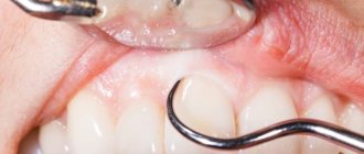
- Examination of the larynx using visual methods - laryngoscopy. Allows you to determine the degree of narrowing of the larynx, the absence or presence of a neoplasm.
- Fibrolaryngoscopy - the study is carried out using a flexible endoscope, at the end of which a video camera is attached. The image is displayed on the computer screen, and the doctor is able to accurately determine the condition and size of the lumen of the larynx.
- A chest x-ray is performed to rule out other concomitant diseases with similar symptoms. For example, with heart disease, shortness of breath is a characteristic symptom.
- Various radiological research methods - computed tomography, MRI. They can be prescribed if difficulties arise with an accurate diagnosis.
- Ultrasound of the thyroid gland.
- Laboratory studies of throat smears are necessary to accurately determine the nature of the infectious disease that provoked laryngeal stenosis.
Treatment
Some cases allow you to resort to treatment without surgery for spinal stenosis. In the early stages of the disease, conservative methods are available. But this is only possible when there are no pronounced neurological disorders, and the patient is only bothered by pain in the lower back and legs.
Drug treatment of spinal canal stenosis l4 s1 and other levels is used:
- non-steroidal anti-inflammatory drugs. They help relieve inflammation and relieve pain;
- muscle relaxants. Relieves muscle tension;
- B vitamins;
- vascular and diuretics;
- drug blockades with local anesthetics and hormones. Relieves pain and swelling.
Also, for spinal canal stenosis at the l4 l5 level, physiotherapeutic procedures are prescribed. This includes:
- electrophoresis;
- amplipulse;
- physical therapy (physical therapy);
- magnetic therapy;
- light massage;
- water and mud therapy.
Exercises that do not put a lot of stress on the back, but at the same time perfectly develop the vertebrae, help with spinal stenosis.
Preventive measures
Some simple steps will help prevent the development of lumbar spinal stenosis or quickly get rid of the problem if it has already arisen:
- It is necessary to visit a specialist’s office if you have any alarming symptoms, pain or discomfort.
- Weight should remain within normal limits so as not to cause excessive stress on the spine and other body systems.
- It is important to ensure that healthy physical activity is maintained - frequent walks, morning exercises, warm-up during the day during sedentary work.
Stenosis reduces the patient’s quality of life and inevitably brings discomfort. If you consult a doctor on time, when the pathology has not developed into a serious stage, it can be eliminated using conservative methods, without surgery. Surgical intervention is required when the situation is advanced, the stenosis progresses and disables the entire body.
Patients who have undergone surgery note that timely seeking qualified help could significantly simplify the situation. For a complete recovery, it is important to maintain a healthy lifestyle and consult a doctor on time.
How to make an appointment with a vertebrologist, neurologist
At the first symptoms of the disease, you need to go to the appropriate specialist: a vertebrologist, a neurologist. In the central district of Moscow, you can make an appointment with a doctor at JSC “Medicine” (clinic of Academician Roitberg), which is located near the Belorusskaya and Mayakovskaya metro stations. You can get to the medical center from the Novoslobodskaya metro station, and the Tverskaya metro station and Chekhovskaya metro station are also within walking distance.
If you doubt your diagnosis, then first you can visit a therapist, who, after an examination, will give a referral to the appropriate specialist at the clinic in the Central Administrative District: a vertebrologist, a neurologist.
You can make an appointment through a special application form, which is available on the clinic’s website. You can also do this by calling: +7 (495) 775-73-60. We are located at the address: Moscow, Central Administrative District, 2nd Tverskoy-Yamskaya lane 10. There is also the possibility of a consultation appointment, where you can receive all the recommendations for preventing the development of the disease.