To the reference book The inner ear, also called the labyrinth, is an auditory and vestibular analyzer.
Author:
- Sokolova Alla Vasilievna
otorhinolaryngologist, otosurgeon, doctor of the highest category
(Voted by: )
- Outer ear
- Middle ear
- Inner ear
, the inner ear is otherwise called a labyrinth.
Located in the thickness of the pyramid of the temporal bone, it consists of 3 parts:
- vestibule;
- snails;
- semicircular canals.
The vestibule is considered the center of the labyrinth and has two pockets. The first, spherical in shape, is located closer to the cochlea. The second, elliptical, is adjacent to the semicircular canals, which are located in three mutually perpendicular planes. Blood supply is carried out through the labyrinthine artery.
The main functions are auditory and vestibular. The auditory analyzer allows you to perceive sound vibrations and ensures the transmission of nerve impulses to the auditory nerve centers, where recognition of the received information occurs. The vestibular analyzer implements sensory, somatic and other reactions.
Causes and course of the disease
Currently, not much is known about the causes of Meniere's disease.
All currently known causes of Meniere's disease can be divided into local, which relate directly to the middle and inner ear, and general, which relate to various pathologies of the whole organism as a whole. Local reasons include:
- dysfunction of the endolymphatic duct - a channel that extends from the labyrinth towards the temporal bone and is a blindly ending duct;
- formation of new vessels inside the endolymphatic sac;
- reduction in size and closure of the vestibular aqueduct (canal in the inner ear);
- reduction in the number of air cavities (cells) in the temporal bone.
Common reasons include:
- circulatory disorders;
- violation of water-salt balance in the body;
- allergic reactions to various compounds;
- hormonal disorders.
Meniere's disease is based on vascular spasm or dilatation, as well as their weak and permeable walls. As a result, the inner ear swells and begins to put pressure on the walls of the labyrinth. In turn, disturbances occur in the labyrinthine fluid at the biochemical level.
As a result, all components of the inner ear expand and increase in size. Due to the increase in fluid volume, the labyrinth stretches, the internal elements of the ear are displaced or damaged. This problem also includes a violation of vascular autonomics, controlled by the central nervous system.
Inner ear
[Home] [To volume contents]
The inner ear lies deep in the rocky part of the temporal bone and consists of the bony and membranous labyrinths. The bony labyrinth is like a capsule for the membranous and is formed from very strong compact bone.
In the inner ear there are: the middle part - the vestibule; in front of it is the cochlea, and behind it is the system of semicircular canals (Fig. 15).
The bony and membranous labyrinths must be described separately, since although the bony labyrinth generally repeats the shape of the membranous labyrinth, the structure of the latter presents great features.
Rice. 15. Cast of the bone labyrinth. 1 - posterior bone ampulla; 2 - oval window; 3 - round window; 4 - place cristae vestibuli; 5 - main curl of the cochlea; 6 - upper curl of the cochlea; 7 - middle curl of the cochlea; 8 - cupula cochleae; 9 - recessus sphaericus vestibuli; 10 - recessus ellipticus vestibuli; 11 - upper bone ampulla; 12 - superior semicircular canal; 13 - common leg; 14 - external bone ampulla; 15 - ampullar pedicle of the external semicircular canal; 16 - smooth leg of the external semicircular canal; 17 - posterior semicircular canal. |
Bone labyrinth - Labyrinthus osseus
Vestibule - vestibulum (Fig. 15)
- a cavity of irregular pear shape, 5-6 mm long, 4-5 mm high and 3-4 mm wide. Its anterior part, which is narrower, communicates with the cochlea, and the posterior part, which is wider, communicates with the semicircular canals through five openings, of which three are more widened for the ampullar legs of the canals, and two are somewhat smaller in size for simple legs. Five, and not six, holes are obtained because the posterior and superior semicircular canals, merging with their simple legs, give one common hole.
The outer wall of the vestibule faces the tympanic cavity and is mostly occupied by the oval window. The medial one faces the posterior cranial fossa and the bottom of the internal auditory canal. It contains two shallow pits, depressions, separated by a noticeable ridge (crista vestibuli), the upper jagged edge of which is called pyramis vestibuli. The anterior of these depressions, the smaller one, closer to the cochlea, spherical in shape, is called recessus sphaericus; it houses the sacculus of the membranous labyrinth; posterior, larger, closer to the semicircular canals, elliptical in shape (recessus ellipticus) for the utriculus.
In the area of the recessus ellipticus there is an internal opening of the vestibular aqueduct (apertura interna aquaeductus vestibuli), through which the ductus endolymphaticus and the vein of the same name pass. In the area of both impressions there is a number of small openings: macula cribrosa superior, media and inferior, through which the corresponding nerve branches pass to the membranous labyrinth.
On the superolateral wall of the vestibule there is an ampullary opening of the superior p.c.; on the lateral, between this last and the oval window, there is an ampullary opening of the posterior s.c. horizontal p.c.
Rice. 16. Axial section through the bony cochlea. 1 - area n. facialis; 2-crista transversa; 3 - area vestibularis inf; 4 - basis modioli; 5 - foramen singulare; 6 - modiolus c. can. centralis; 7-semic. m. ten. tymp.; 8 - septum c. musculotuhatre; 9 - apical curl of the cochlea: 10 - canalis spiralia modioli; 11 - tympanic cavity; 12 - main curl of the cochlea; 13 - lamina spiralis ossea, 14 - area cochleae; 15- meatus acusticus internus. |
Semicircular canals - Canales semicirculares osseae.
The bony semicircular canals, numbering three, have the appearance of semicircularly curved bone tubes with a very narrow lumen, both ends of which open into the vestibule. One of them is flask-shaped swollen in the form of an ampulla, and the other is simple without an ampulla; therefore, there are three ampullary legs, but only two simple ones. Two because the posterior and upper p.c. merge, as said, into one common leg. The channels are located in three mutually perpendicular planes: frontal - vertical, sagittal - vertical and horizontal.
There are upper, posterior and horizontal p.c.
Upper - frontal or anterior p.c. located almost perpendicular to the upper edge of the resp. to the longitudinal axis of the pyramid of the temporal bone. On the upper surface of the pyramid, the convex part of this p.c. protrudes in the form of a noticeable elevation (eminentia canalis semicircularis sup.). It makes an angle of 45° with the frontal plane.
Posterior - sagittal or lower s.c. located parallel to the posterior surface of the pyramid. It makes an angle of 45° with the sagittal plane.
External - horizontal p.c. As already mentioned, its arch projects into the antrum or aditus with a white shiny bone. It makes an angle of 30° with the horizontal plane.
Rice. 17. Right bony cochlea at the apex. 1 - helicotrema; 2 - hamulus lam. spiralis; 3 - lamina spiralis ossea; 4 - scala tympani; 5 - scala vestibuli; 6 - lamina modioli. |
All these p.c. are not located strictly perpendicular to each other; Thus, the upper and horizontal p.c. form angles between themselves of 65-90°, the posterior and upper ones - at 85-115°, only the horizontal and posterior ones are located almost at right angles to each other.
Snail-Cochlea ossea.
The cochlea (Fig. 15) is a bony spirally convoluted canal, making 21/2-23/4 turns, or curls, around the modiolus or columella. Its first or main curl begins from the anterior section of the vestibule, from a special fossa (recessus cochleae), which is located in front of the ampullary opening of the posterior socket. The canal of the cochlea, gradually narrowing, ends blindly at its apex (cupula). The length of the snail is 28-30 mm. Modiolus or columella has the appearance of a short spindle or column located almost horizontally and transversely in the pyramid of the temporal bone. Its base, facing the internal auditory canal, has a depression (fossula cochleae). The modiolus has a canal (canalis centralis) at 3/4 of its length.
Around the modiolus, a thin, narrow bone plate 1 mm wide (lamina spiralis ossea) twists spirally in the form of a spiral staircase and perpendicular to its axis. It begins at the base of the modiolus, from the bottom of the recessus cochlearis, delimiting (anteriorly) the gap, the entrance to the scala vestibuli and, also making 2.5 turns, stretches to the very top of the cochlea, where it ends with the free edge (hamulus) (Fig. 17) . Lamina spiralis ossea is not compact, but consists of two thin plates, upper and lower, the gap between which is cut by radial canals up to its free edge, and the spiral canal of the plate (canalis laminae spiralis osseae), which twists around the modiolus in 2.5 turns. This plate does not reach the opposite wall of the helix by approximately 1/3 of the width of the helix; the remaining space is filled with a special plate (membrana basilaris), which already belongs to the membranous labyrinth.
Rice. 18. Internal auditory canal. 1 - canalis facialis; 2 - area vestibuli sup.; 3 - foramen singulare; 4 - area cochleae; 5 - tractus spir. foraminosus; 6 - crista transversa. |
Lamina spiralis ossea, together with membrana basilaris, divides each curl of the cochlea into two floors: 1) the lower half, to the base of the cochlea (scala tympani), which communicates with the tympanic cavity through the round window, and 2) the upper half, to the apex of the cochlea (scala vestibuli ), which communicates with the tympanic cavity through the oval window. At the apex of the cochlea, both scalae communicate with each other through a special opening - helicotrema (Fig. 17), but at the base they do not communicate with each other. At the very beginning of the scalae tympani, near the round window, there is a small opening for the snail's aqueduct (aquaeductus cochleae).
Internal auditory canal - Meatus acusticus internus.
The internal auditory canal (Fig. 18) is a short bony canal on the posterior surface of the pyramid, generally round in shape, 5 mm in diameter and 8 mm in length. Located almost horizontally and transversely in relation to the entire skull. The rear wall of the pyramid is cut obliquely from back to front, as a result of which its opening (poms acusticus int.) is not round, but oval, and the length of its walls is different: the front is 12-14 mm - almost twice as long as the back, equal to 6 - 7 mm, Bottom the auditory canal, or its inner wall (fundus meatus acustici interni), is at the same time the medial wall of the vestibule and cochlea, and is somewhat depressed. The surface of the wall is divided transversely by a horizontal ridge (crista transversa) into two almost equal parts (Fig. 18). The upper - smaller (fossula superior) has one larger - internal, opening (canalis Fallopii) and behind it several small ones (area cribrosa superior) for the nerves to the utriculus and to the ampullae of the upper and horizontal SC. The lower - large (fossula inferior) with a small roller descending downwards (crista transversa), is also divided into two parts: the front half (fossulla cochlearis) has in the center one larger hole (foramen centrale) for the artery and vein of the cochlea, and around it are located spirally , a beautiful path, many small ones (tractus spiralis foraminulentus) for n. cochlearis; the posterior half in its medial part has a separately located opening (foramen singulare) for the nerve branches to the ampulla of the posterior sac and in front of it several small ones - for the nerve branches to the sacculus.
Membranous labyrinth - Labyrinthus membranaceus
In the membranous labyrinth (Fig. 19) there are two membranous sacs of the vestibule - utriculus and sacculus, three membranous semicircular canals, the membranous part of the cochlea and aqueducts - the vestibule and cochlea. The membranous labyrinth, as already mentioned, generally repeats the shape of its bone capsule, but does not completely fill the gaps of its canals and cavities. This perilymphatic free space is filled with perilymph, with which the membranous labyrinth is washed, being, as it were, suspended in it from the bone wall. The membranous labyrinth is filled with the same light, protein-poor fluid - endolymph.
Rice. 19. Scheme of the structure of the membranous labyrinth. 1 - oval window; 2 - coecum vestibulare; 3 - tympanic cavity; 4 - round window; 5 - ductus perilymphaticus; 6 - cisterna perilymphatica vestibuli; 7 - ductus reuniens Henseni; 8 - round pouch; 9 - scala tympani; 10 - staircase vestibule; 11 - ducus cochlearis; 12 - helicotrema; 13 - coecum cupulare; 14 - bone; 15 - dura mater; 16 - saccus endolymphaticus; 17- ductus endolymphaticus; 18 - superior membranous ampulla; 19 - ductus utriculo-saccularis; 20 - superior semicircular canal; 21 - perilymphatic spaces of the superior semicircular canal; 22 - elliptical sac; 23 - posterior semicircular canal; 34 - perilymphatic spaces of the posterior semicircular canal; 35 - posterior membranous ampulla. |
The vestibule sacs are utriculus and sacculus.
Utriculus is a generally irregular, oval-shaped membranous sac, about 6 mm long; suspended from its medial wall in an oblique position from behind from top to bottom - anteriorly in the recessus ellipticus. Its lateral wall remains free and protrudes into the perilymphatic space (cisterna perilymphatica vestibuli). In the utriculus they open at the previously indicated places. kk. five openings: three ampullary and two simple legs. The ductus utriculo-saccularis opens on the anterior wall. Here, on the front wall from above - from the inside - there is a white elevation - a spot noticeable even with a simple eye - macula acustica utriculi.
Sacculus - spherical, flattened in. in the medial-lateral direction, a membranous sac measuring 3 mm in the long and 2 mm in the transverse axis. On the side of the medial wall, like the utriculus, it is strengthened in the recessus sphaericus, and the outer and anterior walls are freely facing the perilymphatic space (cisterna perilymphatica sacculi). On its posterior wall, the ductus endolymphaticus departs from above and, before entering the apeftura interna aquae-ductus vestibuli, receives the ductus utriculo-saccularis. The ductus reuniens Henseni extends from the lower, narrowed part, connecting the Sacculus with the ductus cochlearis near its blind end (coecum vestibulare). On the medial wall there is a white elevation, a spot visible to the naked eye - macula acustica sacculi.
Both sacs of the vestibule are lined from the inside with simple bridge-like epithelium (Fig. in the next chapter), which in the area of the macula acustica first turns into cylindrical, and this latter into sensory neuroepithelium (on the macula itself). The sensory neuroepithelium consists of two types of cells: supporting cells, which look like an inverted glass - their lower half is expanded and narrowed upward in the form of a process (phalangeal processes); they sit on legs - from one to four. Hair and neuroepithelial cells are located in the spaces between the supporting ones. Their upper half is expanded in the form of a cylinder, and the lower half - the leg - is narrowed. They have one large kernel at the bottom of the expanded part and many small grains. At the upper end of these cells there is a cuticular border, through which short columns and hairs (from 20 to 35) pass, and above them on the hairs, at some distance from the surface of the cells, lies the so-called. otolithic membrane (cupula terminalis) in the form of a gelatinous mass or cloud; hairs penetrate the cupula terminalis. The latter contains, in suspension, crystals of otoliths from carbon dioxide and phosphate of lime. The nerve branches of the corresponding nerves approach the base of the neuroepithelial cells and, passing through the light basal membrane, lose the myelin sheath and, in the form of bare axial cylinders, approach the ends of the hairy auditory cells that close the nucleus.
Membranous semicircular canals - Canales semicirculares membranaceae.
Membranous p.c. exactly repeat the shape of their bone capsule, but are almost three times thinner than their lumens and are suspended in the perilymph; they are attached not in the center of the bone canal, but to the periosteum of the convex side of the canal, i.e. eccentrically. The free surface of the membranous canals, facing the perilymphatic space, is connected to the opposite wall of the bone canal by connective tissue cords in which blood vessels pass (Fig. 20). The membranous ampoules almost completely fill the bone ampoules. There are almost no perilymphatic spaces here.
Rice. 20. Transverse section of the semicircular canal. 1 - membranous canal; 2 - bone canal; 3 - blood vessels; 4 - perilymphatic connective tissue. |
On the outer surface of each ampoule, i.e. corresponding to the convex wall of the p.c., a white, crescent-shaped ridge rises across the longitudinal axis of the ampulla crista acustlca ampullaris, occupying almost half of the lumen of the ampulla.
The histological structure of cristae acusticae ampullaris is similar to the structure of maculae acusticae utriculi and sacculi, with the only difference being that, firstly, the auditory hairs of the hairy cells in them are much longer, reaching almost to the opposite wall of the ampulla and forming the so-called. cupula terminalis, and secondly, they lack an otolithic membrane and otoliths.
Webbed snail - Ductus cochlearis.
As already mentioned, the bony canal of the cochlea is divided into two parts by means of the lamina spiralis ossea and membrana basilaris: the lower - scala tympani - to the base of the cochlea and the upper - scala vestibull - to its apex. In the scala vestibuli from the lamina spiralis ossea, near its free end, a thin membranous plate (membrana Reissneri) extends at an angle of 45° and is attached at the other end to the outer wall of the cochlear canal. The resulting triangular (in cross-section) space (ductus cochlearis) is limited: from above by the membrana Reissneri, from below by the membrana basilaris with the organ of Corti lying on it and partly by the lamina spiralis ossea, on the outside by the outer bony wall of the cochlea with the lig. lying on it. spirale Ductus cochlearis follows the course of the cochlea and its 2.5 whorls and is an endolymphatic sac, blind at both ends: coecum cupulae - at the apex and coecum vestibuli - at the base of the cochlea. Through the ductus reuniens Henseni, the sac is in endolymphatic communication with other parts of the membranous labyrinth, with the sacs of the vestibule resp. with p.c. and with saccus endolymphaticus. Scala vestibuli and scala tympani are perilymphatic spaces and are in connection with other perilymphatic spaces of the labyrinth, and through the aquaeductus cochleae they are openly connected with the subarachnoid spaces of the brain.
Membrana Reissneri is a very thin plate about 3 microns. thickness, almost homogeneous in its base, covered with an epithelial layer in one row on the side of the ductus cochlearis (flat epithelium) and on the side of the scala vestibuli (endothelium).
Rice. 21. Section of the cochlea. 1 - cochlear branch; 2 - staircase vestibule; 3 - crista spiralis 4 - membrane of Corti; 5 - membrana Reissneri; 6 - ductus cochlearis; 7 - stria vascularis; 8 - ligamentum spirale; 9 — drum ladder; 10 - membrana basilaris with the organ of Corti; 11 - lamina spir. ossea; 12 - ganglion cell of the spiral canal. |
Membrana basilaris seu lamina spiralis membranacea consists of three layers:
- The middle one is built from the finest elastic fibers stretched between the free edge of the laminae spiralis osseae and ligam. spirale: the closer to the top, the longer and thicker they are, the closer to the base, the shorter and thinner; therefore, at the apex of the cochlea they are the longest and thickest, and at the base they are the shortest and thinnest (Fig. 21). There are about 20,000 such fibers, or “auditory strings,” in the human ear. A similar device, membranae basilaris, gave Helmholtz the basis to build his theory of sound perception.
- The lower layer - its tympanal surface - is formed by a special layer rich in cells, called stratum tympanale tectorium.
- The upper, vestibular layer, consists of a light, structureless substance on which the organ of Corti rests.
Ligamentum spirale cochleae, to which the membrana basilaris is attached with its outer edge, is a very strong ligament, thickened in section, crescent-shaped or crescent-shaped. On the side of the ductus cochlearis, it is lined with a special stria vasculosa, consisting of numerous vascular plexuses and special vascular cells. Such vascular epithelium is not observed anywhere else in the human body.
Organ of Corti - Organon Corti sive papilla spiralis.
The organ of Corti in its structure is a very complex and original formation, consisting of neuroepithelial cells of various shapes.
It is based (see the figure in the next chapter) on pillar cells in two rows. The inner and outer rows of pillars are inclined towards each other with their upper edges until they touch, thus forming the Arc of Corti. The triangular space or tunnel formed by this arch is already the third canal of the cochlea [1) scala vestibuli, 2) ductus cochlearis and 3) tunnel of the organ of Corti].
Columnar cells are thickened below and above, the nuclei are located at the base of the cells. There are almost twice as many internal pillar cells as external ones.
Inward and outward from the Arc of Corti, resp. from the pillar cells are located the hairy or auditory cells.
Inner hairy cells are arranged in a single row; there are barely times fewer of them than the internal pillar cells, so that each hairy cell lies along two pillar cells. Behind the hairy cells, in three to four rows, lie the internal supporting cells, or Deiters cells. Outside the Arc of Corti, there are four rows of external hairy cells and four rows of external supporting cells, or Deiters cells.
The outer hairy and outer supporting cells lie interspersed: the first on the arc lies a row of hairy ones, followed by a row of supporting cells, then again a row of hairy ones, etc. Behind them lie the outer supporting Hensen'a cells and even further - the Claudius'a cells.
Hairy, or auditory, cells are cylindrical or goblet-shaped; their upper half: more thickened; the cell nucleus is located at the bottom of this thickening; the lower one is elongated into a thin stalk attached to the membrana basilaris; the upper end of the expanded half has from 20 to 25 short columns, or auditory hairs, along its surface.
Supporting or Deiters cells are similar to cells in the macula acustica, with the only difference that they do not have legs, but sit tightly on the membrana basilaris and, unlike hairy ones, have a lower half of a thickened cylindrical shape, and the upper half is elongated into a process (phalangeal process ), reaching the level of the surface of hairy cells. Their core is located at the top of the thickened lower half.
The free surface of Deiters cells is diamond-shaped, with the ends of hairy cells protruding between these diamonds. Between the individual rows of Deiters cells there are gaps - Newel spaces, communicating with the tunnel.
Hensen's cells, or external supporting cells, are arranged in a single row. They begin with a thin stalk from the membrana basilaris and, rising upward, bend outward in an arc, gradually thicken and reach the level of hairy and supporting cells. The level of all these cells, starting from the internal supporting cells, gradually increases outward - to the Nensen cells, so that these latter constitute the highest place in the Organ of Corti.
The free surface of the organ of Corti is covered with a special cuticular film (lamina reticularis). The auditory hairs of the hairy cells pass through the holes in this film.
Claudius cells are located outward from Hensen cells to the ligamentum spirale. They are not of the same size in all curls, but gradually move from high cylindrical in the basal part of the cochlea to cubic at the apex.
Overhanging the organ of Corti is a special striped plate of a cuticular nature, membrana tectoria Corti. It starts from the crista spiralis and covers the entire organ of Corti in the form of a canopy. Its function is still unknown.
Blood vessels of the labyrinth.
The entire labyrinth is supplied with arterial blood from a. auditiva interna. In the internal auditory canal, the artery splits into three branches:
- a. vestibularis - nourishes the upper anterior part of the vestibule and the posterior part of the utriculi and sacculi;
- a. cochlearls - nourishes the entire cochlea with the exception of part of the main helix;
- a. vestibule-cochlearis - nourishes the posterior-inferior part of the vestibule and part of the main curl of the cochlea.
Venous blood is carried out in three ways: 1) into the plexus auditivus internus; 2) in sin. transversus - through v. aquaeductus vestibuli; 3) in sin. petrosus inferior through v. aquaeductus cochleae.
Nerves of the labyrinth.
N. acusticus enters the internal auditory canal together with n. facialis and here it splits into two main branches: ramus cochlearis and ramus vestibularis. The latter, in turn, splits into two branches: the upper one, entering the labyrinth through the area cribrosa superior and supplying nerve endings to the macula acustica utriculi and cristae acusticae ampullares of the upper and horizontal p.c., and the lower one, entering the labyrinth through the area cribrosa media and supplying the macula acustica sacculi and crista acustica ampullaris of the lower p.c.
Rice. 22. Plumbing of the labyrinth. 1 — saccus endolymphaticus (opened); 2 - apertura ext. aquaeductus vestibuli; 3 - thin hair inserted into it; 4 — opened sin. petrosus sup.; 5-6 - sections of the transverse sinus, visible along its entire length between 5 and 6 through the layer of the dura mater; 7 - porus acus. int. with n. acusticus; 8 - apertura ext. aquaeductus cochleae. |
Ramus cochlearis supplies the cochlea and is, in fact, the auditory nerve of the ear. Part of it enters the cochlea through the tractus spiralis foraminulentus (for the first curl of the cochlea), the other through the foramen centrale into the modiolus and further into the canalis laminae spiralis, where it comes into close contact with the nerve ganglia of this canal. A mass of small radial branches is directed between the leaves of laminae spiralis osseae to the cells of the organ of Corti from the side of the scala tympani. These nerve endings approach hairy cells after losing their myelin sheath. Maculae acusticae utriculi and sacculi and crista acustica ampullaris are supplied with the same non-pulp fibers.
Aqueducts of the vestibule and cochlea - Aquaeductus vestibuli et cochleae.
They have already been mentioned more than once when describing parts of the labyrinth (Fig. 22). Physiologically, they are regulators of pressure in the endolymphatic and perilymphatic spaces of the labyrinth.
The vestibular aqueduct (aquaeductus vestibuli) has an external opening in the form of a slit on the posterior surface of the pyramid, behind the internal auditory canal. Ductus endolymphaticus, passing through this canal from the utriculus and sacculus, reaches the dura mater and ends here in the blind sac (saccus endolymphaticus).
The aqueduct of the cochlea (aquaeductus cochleae) opens with a very narrow opening of the aqueduct on the posterior surface and on the lower edge of the pyramid - below the internal auditory canal. As we said, through this hole there is an open, direct connection between the perilymphatic space of the labyrinth and the subarachnoid (subarachnoid) space of the brain.
[to contents]
Clinical picture
Meniere's disease occurs in several stages.
Initial stage. Periodically, the patient experiences tinnitus and a feeling of ear fullness. Then there comes a feeling of swelling inside the ear, and, as a result, the patient feels constant pressure on it. If treatment is not undertaken, then a picture of sensorineural hearing loss develops, a disease in which the auditory nerve is affected and hearing loss occurs. In addition, due to an increase in intralabyrinthine pressure, dizziness and vomiting begin to bother, which can last for several hours, with intense hearing loss. But these are the most initial and weakest manifestations of the disease.
Second stage. Dizziness and vomiting become frequent and occur in the form of attacks, during which significant hearing loss constantly occurs. But in the second stage, the hearing loss is persistent and after the end of the attack, hearing is not restored to the original level. The same goes for noise and ringing in the ears. In the second stage, tinnitus and a feeling of fullness do not occur periodically, but are constantly present.
Stage three: For those who do not take any steps to treat, Meniere's disease becomes irreversible. Dizziness and vomiting will no longer be observed, and upon examination, it will be impossible to detect fluid inside the labyrinth. However, hearing will be progressively lost, and it will no longer be possible to restore it. Hearing loss occurs as a type of sensorineural hearing loss. As a result of irreversible changes in the labyrinth, stability and firmness of gait are lost, which can no longer be restored.
Diseases
Regardless of the nature of the disease, the above analyzers are necessarily involved, therefore the patient’s condition is characterized by vestibular or cochlear symptoms. With such disorders, there is usually a violation of two functions of the ear labyrinth at once.
Diseases can also be divided into inflammatory and non-inflammatory:
- labyrinthitis - inflammation of the inner ear, in which cochleovestibular dysfunction is observed (often occurs as a complication of chronic otitis media; the prevalence is limited and diffuse; occurs in acute and chronic forms; taking into account the clinical manifestations, serous, purulent and necrotic types of lesions are distinguished);
- sensorineural hearing loss (taking into account the degree of damage to the auditory analyzer, there are cochlear, retrocochlear, central and mixed; according to the speed of development, sudden, acute and chronic forms are distinguished);
- Meniere's disease (characterized by periodic attacks of systemic dizziness, deterioration of hearing, first of the conductive type, then of the mixed type, the appearance of tinnitus; the etiology of the disease remains unclear);
- benign paroxysmal positional vertigo (BPPV);
- otosclerosis (a limited process of dystrophic damage to the bone tissue of the labyrinth, develops against the background of a genetic predisposition, with metabolic disorders and hormonal disorders).
Symptoms of inner ear diseases
General symptoms are damage to the facial nerve, dizziness, dyspeptic disorders (nausea, vomiting), imbalance, tinnitus, hearing loss.
Diagnostics
Meniere's disease consists of several stages.
- The patient's complaints of systematic dizziness and poor health are carefully studied, and when examining the labyrinth (additional research), the otolaryngologist discovers swelling of the walls of the inner ear and accumulation of fluid inside the labyrinth (endolymphatic hydrops).
- A caloric test is performed - this is a study of the vestibular apparatus. The doctor evaluates involuntary eye movements (nystagmus) by pouring water into the ear, or by exposing the eardrum to cold and hot air, thereby irritating it.
- Also, the ENT doctor should send the patient an additional test - MRI (magnetic resonance imaging) of the brain and temporal bones, which is a very informative study for this disease;
- Among additional methods, it is necessary to examine hormonal levels, levels and possible metabolic disorders;
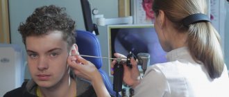
- Instrumental research methods include otoscopy and videotomicroscopy - examination of the external auditory canal and eardrum using an otoscope and a diagnostic ear microscope.
Thus, the otolaryngologist conducts not only an examination of the ear, but also other organs and body functions in order to exclude other diseases. Among them there may be many diseases of the nervous system, which are also characterized by dizziness and other disorders of the vestibular system. These are diseases such as acoustic neuroma, leptomeningitis of the cerebellopontine triangle, specific labyrinthitis and other forms of labyrinthitis.
Friends! Timely and correct treatment will ensure you a speedy recovery!
In addition, when diagnosing Meniere's disease, the otolaryngologist determines the stage of the disease and the degree of hearing impairment.
What is the organ of hearing?
Air and bone sound conduction
Sound energy travels to the structures of the inner ear through air conduction and bone conduction.
Airborne sound conduction is the usual way for sound vibrations to enter the ear - through the auricle and external auditory canal, sound comes to the eardrum. Further, vibrations of the eardrum through the chain of auditory ossicles are transmitted to the fluids of the auditory cochlea - peri- and endolymph, causing the main membrane and structures of the organ of Corti to vibrate.
Bone conduction is the conduction of sound vibration from the surface of the head directly into the cochlea of the inner ear, bypassing the middle ear. When sounds enter the ear through bone conduction, sound vibrations travel through the bones and tissues of the head. Under the influence of bone-conducted sounds, the walls of the cochlea of the inner ear vibrate, which is transmitted to the fluids that fill it. This, in turn, causes oscillatory movements of the basilar membrane and the organ of Corti. Then everything happens in the same way as with airborne sound transmission.
We hear our own voice precisely through bone conduction: the sounds of the voice pass to the cochlea of the inner ear through the tissues of the head. This is why we hear our voice differently than in the recording. This is caused by the fact that the bones of the skull conduct low frequencies better than high frequencies. Therefore, during sound pronunciation, people perceive their own voice as lower and deeper than others perceive it.
Since bone sound conduction practically excludes the middle ear from the process of sound transmission, the study of the auditory perception of air- and bone-conducted sounds during audiometry is very important in diagnosing hearing.
In addition, in cases where it is impossible to use air conduction hearing aids, in particular, in case of certain diseases and after certain operations on the middle ear, the doctor considers the possibility of bone conduction hearing aids.
Intermediate (conducting) section of the hearing organ
The intermediate (conducting) section of the hearing organ begins with the auditory nerve and ends in the cerebral cortex. The neuron bodies of the auditory nerve are located spirally along the axis of the cochlea and form the so-called spiral ganglion. And their long processes - axons - form the auditory nerve, which transmits nerve impulses “up” to the brain. The right and left auditory nerves are called the eighth (VIII) pair of cranial nerves.
The axons of the auditory nerve, like other neurons, are covered with a layer of special tissue - the myelin sheath, which contains “intercepts” - bare sections of the axon. This sheath and its “intercepts” play a key role in the transmission of nerve impulses by the neuron.
The neurons of the auditory nerve switch to the neurons of the medulla oblongata - the cochlear nuclei. Moreover, the cochlear nuclei are the last formations of the auditory analyzer, receiving nerve impulses from only one ear.
The pathways and subcortical centers of the auditory analyzer are part of the central nervous system (CNS) and include the ascending (afferent) and descending (efferent) systems. Anatomically, it is located in the brain stem, subcortical structures of the brain. A simplified diagram of the ascending auditory system is shown in the diagram.
As can be seen from the diagram, the number of nerve cells (neurons) increases many times as one rises from the auditory nerve to the cerebral cortex. There are approximately 35 thousand of them in the auditory nerve, and more than 12 million in the auditory cortex. In addition, as one rises to the auditory cortex, the connection of auditory neurons increases both between both sides of the brain and with neurons of other sensory systems, memory areas, speech and many others.
It is noteworthy that above the right auditory nerve and the cochlear nuclei, in which its neurons switch to the next level, the bulk of the ascending auditory neurons move from this ear to the left side of the brain. And vice versa. Thus, a “crossing” of the conductive pathways of the auditory analyzer occurs, which is clearly visible from the diagram of the brain stem.
Central part of the hearing organ
The central (cortical) section of the auditory analyzer is located in the temporal lobes of the cerebral cortex. Nerve impulses from the right ear go mainly to the left hemisphere of the brain, and vice versa, from the left ear to the right. This is of great importance in hearing care, and here's why. The auditory zones of both hemispheres perform, although similar, but different work.
Research in the 1960s and 70s showed that in most right-handed people, the left hemisphere is better at processing high-frequency, rapidly changing sounds, and is better at perceiving individual sounds, syllables and words of speech. That is why the left hemisphere and, accordingly, the right ear were called dominant in speech perception. And that is why, for the majority of right-handed people, if binaural hearing aid is not possible, hearing aid for the right ear is preferable. For left-handed people, it’s usually the other way around. But since there are many individual differences, an audiometric examination must determine which ear responds better to verbal tests. Assessing the perception of whole speech is a rather long and difficult psychoacoustic study and is not used in audiological practice.
Later studies in the 1970-80s showed that not everything is so simple with the speech dominance of the hemispheres.
Experiments by many scientists have shown that the hemisphere opposite to the dominant one when perceiving individual words (in most right-handed people - the right) perceives intonation and speech rhythm much better, which are necessary for understanding whether the speaker is stating something or asking, whether he is speaking seriously or joking . That is, it better understands sentences as a whole. Moreover, it is the opposite dominant hemisphere that connects all sentences into the general meaning of everything said, for example, the entire story, the entire conversation as a whole. Thus, the hemisphere that was considered “dominant” (the left in right-handed people) carries out a sequential analysis of individual sounds, and the hemisphere that was considered “non-dominant” carries out a holistic perception of speech messages.
Treatment
Treatment for Meniere's disease is as follows:
- Intravenous drips with 5-7% sodium bicarbonate, 100-150 ml. The speed should be quite high - about 120 drops per minute (the norm is 1 drop per 1 second.) The number of such droppers can vary from 1 to 15, depending on the severity of the condition and the stage of the disease.
- Taking B vitamins, as well as Aevita (vitamins A and E), Belloid (ergot alkaloids) and Bellataminal, which is a sedative.
- A 20 ml Glucose solution can be injected intravenously in a slow stream. The number of intravenous infusions will depend on the severity of the patient’s condition and the stage of the disease at which he presented.
- Since attacks of Meniere's disease are associated with dizziness and an unstable, shaky gait, during them it is necessary to maintain complete rest and inject a subcutaneous solution of atropine sulfate in the dosage prescribed by the ENT doctor.
- Reflexotherapy is also used to treat Meniere's disease: acupuncture massage, electro-apuncture, magnetic apuncture and other types of acupressure on certain areas of the human body.
- If drug treatment and reflexology do not bring the desired results (which happens only in rare cases), then treatment of Meniere's disease is carried out by surgical intervention in the labyrinth located in the inner ear of a person. It must be carried out in an ENT hospital, followed by a mandatory rehabilitation period.
Treatment and therapy
Treatment for such manifestations depends on the cause. First of all, in the treatment of all ear infections, decongestant ear drops are always used to better drain the discharge.
For pain, certain analgesics can be used. These drugs are also used as an antipyretic. If a bacterial infection is present, antibiotics are used for treatment. In some cases, local rinsing with both solutions of anti-inflammatory drugs and ordinary boiled water is recommended. This may speed up the healing process.
If a large volume of exudate accumulates behind the eardrum, a puncture may be performed. To improve the outflow of pus and administer medications. Additionally, physiotherapeutic treatment may be used.
Prevention
Due to the unclear etiology of Meniere's disease, its prevention is almost impossible.
However, in some cases, measures to prevent the disease can still be taken. This:
- quitting smoking, which leads to poor circulation and hypoxia of the whole body;
- the use of personal protective equipment when riding a motorcycle or bicycle (helmet, motorcycle and bicycle helmet) in order to avoid traumatic brain injuries, which in some cases can cause the development of Meniere's disease.
- to reduce the number of attacks of dizziness, nausea and vomiting, it is necessary to adhere to a special diet that is low in salt and fluid.
Of course, one of the main measures to prevent the development of Meniere’s disease and its entry into the stage of irreversible changes is a timely visit to an ENT doctor at the first symptoms of hearing loss and signs of dizziness.
Make an appointment right now!
Call us by phone or use the feedback form
Sign up
Complications
Discharge from the ear is not only an unpleasant symptom, but can also cause a serious illness. This is usually otitis externa, but the doctor must rule out more severe pathologies.
The discharge may be bloody or purulent, in which case it indicates inflammation of the eardrum or middle ear. This discharge is accompanied by severe pain, and patients often suffer from dizziness or fever. Discharge is quite an uncomfortable symptom for the patient, even if there are no accompanying symptoms. Because it causes tingling or itching in the ear, and often the fluid has a foul odor.

