Tooth pulpitis, what is it?
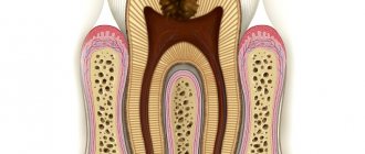
Pulpitis is an inflammatory process involving the pulp, which is the soft tissue inside the root canal of a tooth, consisting of blood and lymphatic vessels, as well as a large number of nerve endings. As a result of inflammation of the nerve fibers, acute toothaches of high intensity occur, which are almost impossible to endure. This pain syndrome is poorly controlled even with potent analgesic drugs.
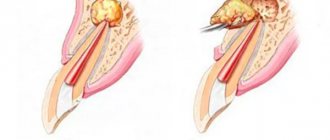
As pulpitis progresses, an infectious process develops in the pulp chamber and harmful bacteria multiply. As a result, nerve endings and connective tissue begin to die, causing pus to form in the root canal. Untimely or improper treatment of pulpitis threatens the death of the nerve and a number of serious complications, as a result of which the nerve and tooth have to be removed. But if you consult a dentist in the early stages of the development of pulpitis, you can cope with the disease with therapeutic treatment, preserving the dental unit.
Causes of pulpitis
The main reason for the development of pulpitis lies in the penetration of pathogenic pathogens into the pulp chamber, which causes an infectious process to develop, under the influence of which the pulp becomes inflamed. There are many factors that contribute to the occurrence of the disease, the main ones being:
- Carious lesion - caries not only spoils the appearance of a smile, with this disease the hard tissues of the tooth are gradually damaged and destroyed. As a result, the protective potential of the enamel is reduced, and channels are formed in the bone tissue through which pathogenic microorganisms can penetrate into the pulp, provoking an infectious and inflammatory process.
- Mechanical injuries to the tooth - as a result of blows and bruises, chips of parts of the tooth, the formation of cracks, there is a possibility of damage to the pulp chamber, which causes an inflammatory process.
- Damage to the pulp during surgery - some dental procedures, such as dental surgery or prosthetics, are accompanied by a risk of damage to the pulp, which leads to the development of pulpitis. A striking example is a burn to the upper edge of the pulp chamber during the process of grinding a tooth before installing a crown.
- Concomitant pathologies - pulpitis can develop even if the integrity of the hard tissues of the dental structures is not compromised. In such cases, the infection enters the pulp chamber through the bloodstream or the root, for example, in the presence of a cyst or advanced forms of periodontal disease.
Biological method of treating acute pulpitis
The treatment plan for this disease depends mainly on its form. The use of a biological treatment method, preserving pulp tissue, is possible in a limited number of cases. In particular, these include:
- reversible inflammatory changes in the pulp;
- accidental opening of the dental cavity during the treatment of caries;
- traumatic nature of pulpitis.
The biological method requires at least two visits to the doctor. First, the dentist expands, shapes and treats the carious cavity with medications. Then a medicinal paste is applied and a temporary filling is installed. After a few days, the final filling is performed if there are no complaints.
Tooth pulpitis - symptoms
Dental pulpitis has a clear clinical picture, which makes it impossible not to notice this disease, and it is extremely difficult to confuse it with another pathological process. The main symptoms of dental pulpitis in dentists include:
- Pain syndrome - in the early stages of the development of the disease, the pain is insignificant, but as it progresses, they become strong and unbearable. The pain intensifies at night, when the tooth comes into contact with cold or bitter food, and can be paroxysmal in nature.
- In the later stages of the development of the pathological process, pain can cover the entire jaw, which makes it difficult to determine its source. However, X-ray examination makes it possible to accurately determine the localization of the inflammatory focus.
- Headaches – damage to the nerve fibers of the pulp in the later stages is accompanied by headaches, which indicates the progression of the disease.
- Increased body temperature – as the inflammatory process develops and purulent deposits accumulate in the area of the pulpal core, the patient’s body temperature may increase to febrile levels.
- An acute reaction to temperature changes - a tooth in which inflammation has occurred in the pulp core reacts with acute attacks of pain when it comes into contact with cold or hot food, drinks, even gusts of cool wind bring noticeable discomfort.
- Deterioration of appetite and quality of sleep are indirect clinical signs that develop against the background of severe pain.
Sometimes chronic forms of pulpitis are encountered in dental practice. They are often accompanied by similar symptoms, however, all clinical signs in such cases are less pronounced. The pain is periodic, not so intense, the reaction to cold or hot is present, but not so acute, etc. The main distinguishing feature of chronic pulpitis from acute pulpitis is the constant presence of bad breath, which occurs due to the accumulation of pus in the pulp chamber or root canal.
Classification of dental pulpitis
Depending on the characteristics of the course, clinical signs and causes of the development of the pathological process, several forms or types of pulpitis are distinguished. In addition, some types of the disease have subtypes, which somewhat complicates the classification. But it is precisely thanks to an accurate understanding of the characteristics of the disease, as well as modern diagnostic methods, that the doctors at the Academy Dent clinic are able to select the most effective and correct treatment.
Acute pulpitis
The most common form of the pathological process is acute. This type of pulpitis develops quickly, has a clear clinical picture and occurs mainly in adults. In dental practice, there are 2 subtypes of acute pulpitis:
- Focal – considered the initial stage of the disease, which lasts from 1 to 2 days. The pathological process is characterized by the maximum proximity of the source of inflammation in that part of the pulp that is close to the carious formation on the tooth. Pain in this case is episodic, lasting no more than 30 minutes, after which it subsides for 1-2 hours and reappears.
- Diffuse is the stage subsequent to chaga, in which the inflammatory process covers the entire pulp core. The pain intensifies and can radiate to various parts of the jaw, as well as the temples, occipital region, and cheekbones. The duration of pain attacks increases and they become more frequent. The duration of this phase is measured for up to 2 weeks. If the disease is not cured during this time, pulpitis progresses to a chronic form.
Chronic pulpitis
Diagnosed after 2 weeks of the acute form of the disease. After 14-21 days, pain and other symptoms of the disease gradually weaken, but do not disappear completely. The tooth reacts sharply to external stimuli, and the pain intensifies. Periodic bleeding from the area of the pulp nucleus is added to the clinical picture, and the process of gradual destruction of the bone tissue of the affected unit of the dentition begins. The classification of chronic pulpitis is more extensive; it includes the following subtypes:
- Fibrous is the initial stage of the chronic form of the pathological process, in which it can occur covertly without causing discomfort to the patient. When examining the oral cavity, the doctor identifies an extensive carious cavity, although externally it may not be connected to the pulp chamber, which may result in incorrect treatment.
- Gangrenous is the next stage after fibrous, which is characterized by atrophy of nerve fibers, as well as a change in the color of the pulp nucleus to dark gray. The disease in this form is accompanied by increased pain in the tooth, and there is a pronounced foul odor from the oral cavity. Upon examination, the dentist reveals not only an extensive, but also a deep carious cavity.
- Hypertrophic is an even more severe form of chronic pulpitis, which is characterized by the formation and spread of granulation tissues with the subsequent formation of a polyp. The latter fills the space of the carious cavity and partially the root canal; when pressed, it responds with acute pain and the appearance of blood. If there is no impact on the tooth, the pain subsides.
- Acute – the last and most severe stage of the chronic form of pathology, in which the symptoms of acute and chronic pulpitis are combined. This stage is especially dangerous due to large-scale destruction of bone tissue and a high probability of periodontal infection, with the subsequent development of periodontitis.
Progression of the pathology to a chronic form (in any manifestation) in most cases threatens the impossibility of therapeutic treatment. The doctor has to remove the pulp core, and in case of large-scale destruction of bone tissue, the tooth itself.
Retrograde pulpitis
Retrograde pulpitis differs from the previously described forms in the cause of its occurrence. Infectious lesions of the pulp occur not from outside, but from within the body (for example, with the bloodstream with concomitant diseases). The main causes of retrograde pulpitis include:
- Periodontal diseases - in cases of progression of periodontitis, periodontal pockets form in the tissues of the oral cavity. In these cavities, which can reach half the length of the root part of the tooth in depth, pathogenic bacteria accumulate. These bacteria are able to penetrate into the cavity of the pulp nucleus through the holes at the apexes of the roots, provoking the development of an infectious process.
- Other pathologies - an infection from the bloodstream can penetrate into the pulp in diseases such as influenza, chickenpox, dental cyst, sinusitis, sepsis, etc.
In addition to the causes of development, the retrograde form of the pathology differs in its clinical picture. First of all, this is expressed in pain; pain is characterized by patients as pulsating, from acute to minor and periodic.
How to treat pulpitis?
Knowing what it is, tooth pulpitis, you need to figure out how to treat this pathological process. The maximum effectiveness of pulpitis treatment at the Academy Dent clinic is achieved thanks to the highest classification of dentists, as well as the use of the latest equipment. Before starting treatment, the doctor makes an accurate diagnosis based on the descriptions of the patient’s complaints, the results of an examination of the oral cavity, as well as diagnostic data.
Based on the information received, he draws up a treatment plan, in total there are 2 possible ways to solve the problem:
- Conservative treatment – used for acute pulpitis, especially effective in the early stages of the disease. The doctor drills and cleans out the carious cavity, after which he places medication over the pulp. In this case, antibiotics, antiseptics and proteolytic enzymes are used. After the use of drugs, a temporary filling is installed to block access to the pulp core and allow the drugs to act. In the future, when the infectious and inflammatory processes are stopped, the temporary filling is replaced with a permanent one. This method preserves the vitality of nerves and other pulp structures, as well as the tooth itself.
- Surgical treatment is a radical solution used in cases where conservative methods of therapy do not bring results. Surgical treatment involves removal of the pulp core and thorough cleaning of the cavity. Depending on the course of the disease, the pulp can be removed only after killing the nerve. If there is large-scale destruction of bone tissue, the tooth must also be removed.
Regardless of the tactics and method of treatment, at the Academy Dent clinic, treatment of pulpitis is as comfortable and painless as possible. Dentists use the latest generation of anesthetic drugs and use modern expert equipment.
Treatment
In dentistry, there are several methods for treating pulpitis:
- complete tooth extraction;
- partial tooth extraction (removal of one of the roots of a multi-rooted tooth);
- endodontic treatment with complete removal of the nerve using the method of vital extirpation or devitalization (necrotization of the neurovascular bundle with special pastes);
- endodontic treatment with partial removal of the nerve (vital amputation method) - the doctor keeps the tooth alive, but removes the coronal part of the neurovascular bundle;
- biological treatment – the source of infection is eliminated using conservative methods, and the tooth is completely kept alive.
On a note! The best option for the patient is to preserve the dental innervation. In this case, the tooth remains alive and, with proper treatment, will not only last for many years, but will also maintain its aesthetics. The tissues of pulpless teeth die, lose their strength and natural shade. They quickly become fragile and are not able to fully withstand chewing loads. You can avoid the depulpation procedure only if you consult a doctor in a timely manner and have modern equipment that provides gentle treatment of hard tissues (without overheating).
Therapy involves a whole range of medications, endodontic treatment, physiotherapy and surgery (if necessary). Standard set of actions in the treatment of pulpitis with nerve removal:
- the procedure for anesthetizing a tooth during treatment - mainly using injection anesthesia methods;
- drilling with a bur the areas of enamel and dentin affected by caries;
- nerve removal;
- mechanical expansion and cleaning of channels;
- canal filling;
- filling the crown part of the tooth.
After filling the root canals, a temporary filling must be placed and an x-ray check of the quality of the work performed must be done. This allows you to correct errors in a timely manner and avoid the consequences of poor-quality treatment.
Attention! The sooner the patient contacts the dental clinic, the higher the chances of avoiding extraction and keeping the tooth alive. If the disease is started and not treated in time, there is a risk of developing complications - periostitis, periodontitis, abscesses, sepsis, infection of internal organs.
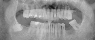
What does pulpitis look like in the photo?
In this section, clinic patients can see what tooth pulpitis looks like in the photo:
It is important to remember that dental pulpitis is a serious and dangerous disease that can cause a lot of discomfort and harm to health. However, with timely treatment of the disease, it is possible not only to save the tooth, but also to leave the pulp completely viable. Every patient can count on high-quality and effective treatment by making an appointment with a dentist. "Dent Academy", where the best specialists work, there is no place for negligence and mistakes.
Prevention
To avoid the appearance of pulpitis, it is necessary to take preventive measures to preserve healthy teeth:
- Carefully follow the rules of hygiene - brush your teeth regularly (at least 2 times a day), rinse your mouth after each meal, use dental floss if possible, remove bacterial plaque in a timely manner.
- Periodically undergo examinations at the dentist's office, have a professional cleaning and remove tartar.
- Eat right - the body must receive a full range of proteins, fats, carbohydrates, vitamins and minerals. In addition, the food should be hard enough to create the necessary load on the teeth without destroying them (you should not chew nuts in the shell and crackers).
- Give up bad habits - smoking, alcohol, and a sweet tooth negatively affect the condition of hard tissues and the blood supply to soft structures.
And, of course, eliminate dental problems in a timely manner, without leading the situation to an inflammatory process. Enduring until the last minute not only makes no sense, but is also dangerous for your health. And taking into account modern methods of anesthesia, this is also irrational, since the anesthetics available to doctors make the treatment painless and as comfortable as possible for the patient.