A tumor is a new growth whose cells have lost the ability to grow under controlled conditions. This group of diseases is very extensive, so it is divided into a separate branch of medicine - oncology. Oncology studies the mechanisms of development, diagnosis, treatment, and prevention of not only malignant, but also benign neoplasms.
Tumors are presented in a very wide variety and can develop in any organ or tissue. For each type of neoplasm, a number of signs are identified, according to which classification is carried out and a diagnosis is established.
- Tumor atypia
- Features of tumor growth
- Types of tumors
- Why do tumors occur?
- Classification of tumors
- Diagnostic methods
- Treatment of tumors
Tumor atypia
Any tumor consists of stroma and parenchyma. The stroma consists of the extracellular matrix, blood vessels, and nerve endings. Parenchyma is the tumor cells themselves. Their structure, as a rule, differs from normal cells - this is atypia. The difference may lie not only in the structure of the cells themselves, but also in their functioning, metabolism, etc. Therefore, there are several types of atypia:
- Morphological atypia. Can be cellular or tissue. In the first case, tumor cells lose the ability to mature and differentiate, as a result of which they acquire a large nucleus, irregular shape, and other characteristics that distinguish them from normal cells. With tissue atypia, the ratio of various tissue elements changes, for example, the ratio of the thickness of the epidermis and dermis.
- Biochemical atypia. Characterized by changes in tumor metabolism. It is this feature that plays an important role in the uncontrolled growth of newly formed tissue. In tumor cells, almost all types of metabolism change, but the most significant is the change in carbohydrate metabolism, which increases several tens of times. Other features include the predominance of protein synthesis over its breakdown, increased absorption of amino acids and water, accumulation of potassium ions and loss of calcium ions.
- Immunological atypia. Tumors are characterized by changes in antigenic structure. For this reason, the immune system cannot effectively “attack” the changed cells, as a result of which the latter can multiply and grow uncontrollably.
- Functional atypia. This is a consequence of the changes described above. Changes in the structure and metabolism of tumor cells inevitably lead to changes in their functions. This may be increased secretion of hormones, loss of the ability to phagocytose, production of substances that are not normally formed, etc.
Some types of atypia have made it possible to develop specific antitumor drugs. In particular, some cytotoxic drugs used during chemotherapy block the uptake of glutamine and glucose. These substances are used by certain tumors in large quantities and are necessary for the growth and division of pathological cells. Immunological atypia formed the basis for cancer immunotherapy. With the help of special drugs, it is possible to eliminate the “defense mechanisms” of the tumor, which make it “invisible” to the immune system.
Types of cancer
Affected cells can occur in any human organ. Therefore, cancer can be called a group of diseases. The specific definition of the disease depends on which organs are affected:
- if the hematopoietic system is affected, it is leukemia;
- if the lymphatic system is affected, it is lymphoma;
- if nervous, connective or supporting tissue is affected, it is sarcoma;
- if the epithelial tissue of an organ is affected, it is carcinoma;
- if the skin is affected, it is melanoma.
There are also other types of cancer such as teratoma, glioma, choriocarcinoma, blastoma. Some types of cancer are less common, some more common. But this is the danger of rare species - they have not been studied so well, so they often lead to death.
Features of tumor growth
The second important feature that is characteristic of all neoplasms is uncontrolled and autonomous growth. That is, the tumor can grow as long as the body exists. At the same time, the regulatory mechanisms that apply to normal cells do not work. The following types of growth are distinguished:
- Expansive growth. Characteristic of benign tumors. The primary focus has clear boundaries due to the capsule. As the tumor increases in size, pushing and compression of surrounding tissues is noted.
- Infiltrating growth. It is characterized by tumor growth into surrounding tissues, which is why it is also called invasive growth. In this case, the boundaries of the primary focus are erased, due to which difficulties arise in determining the exact dimensions. The tumor invades not only healthy tissues of the organ, but also lymphatic ducts and blood vessels, which is a prerequisite for metastasis. This type of growth is observed in malignant neoplasms.
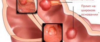
- Exophytic and endophytic growth. It is noted in hollow organs. In the first case, the tumor grows into the lumen of the organ, and in the second - into its wall.
- Unitetric growth - a neoplasm develops from a single focus located in one organ.
- Multicentric growth - the neoplasm originates from several foci located in one organ.
Each type of tumor is characterized by certain growth characteristics. Benign neoplasms in most cases grow slowly, may not increase in size for a long time and even regress. With malignant neoplasms, on the contrary, rapid growth is often observed with infiltration and destruction of healthy tissue.
Book a consultation 24 hours a day
+7+7+78
Angiosarcoma
A malignant neoplasm of blood vessel cells, characterized by rapid proliferation and active spread. The term angiosarcoma applies to a wide range of vascular endothelial cancers that affect various areas of the body.
There are currently 4 known variants of cutaneous angiosarcoma:
- Angiosarcoma of the scalp and face (senile angiosarcoma)
- Lymphangiosarcoma (Stewart-Treves syndrome)
- Angiosarcoma caused by ionizing radiation
- Epithelioid angiosarcoma
The most common variant is angiosarcoma of the scalp and face, which usually occurs in older people. The tumor appears as a large blue area, a blue-black nodule, or a non-healing ulceration ( Figure 6 ). Initially, these lesions can be confused with simple bruising, swelling or infection, making diagnosis difficult.
The death rate from angiosarcoma is high, with a 5-year patient survival rate of only 20–35%. Early diagnosis is critical to the prognosis of the disease.
Rice. 6. Large angiosarcoma on the scalp (Holloway L., et al. Cutaneous angiosarcoma of the scalp: A case report of sustained complete response following liposomal doxorubicin and radiation therapy. Sarcoma 2005; 9: 29–31)
Types of tumors
Tumors are classified according to various parameters. One of the most important is the degree of differentiation. Based on this feature, benign and malignant neoplasms are distinguished. Benign tumors are composed of differentiated cells. Their cells are as similar as possible to the cells of normal tissues. For this reason, such diseases do not pose a serious health threat in most cases. The following signs are characteristic of benign tumors:
- Slow growth.
- No metastases.
- No infiltration of surrounding tissues.
- No tendency to relapse.
Benign tumors respond very well to treatment, which is carried out surgically. Examples of such neoplasms are fibroids, lipoma, adenoma, papilloma, and atheroma.
Malignant tumors consist of poorly differentiated cells that have lost their function and acquired the ability to divide uncontrollably. They have a negative effect on the body, grow into surrounding tissues, quickly spread throughout the body and lead to severe disruption of the functioning of internal organs.
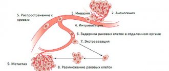
The key difference between malignant and benign tumors is their tendency to metastasize. Metastasis is a secondary tumor site that occurs in a distant organ or tissue as a result of the movement of cancer cells from the primary site. Each type of cancer is characterized by certain localizations of metastases. The liver, lungs, bones, and brain are most often affected. The spread of tumor cells can occur in the following ways:
- Through the blood (hematogenous metastasis).
- Through lymphatic ducts (lymphogenous metastasis).
- Along nerve fibers (perineural metastasis).
- Upon contact with other organs and tissues (contact metastasis).
In some cases, a combination of several options may occur.
Among other features of malignant tumors, there is a tendency to relapse, difficulty in treatment and a significant impact on body functions due to metabolic disorders. In particular, cancer cells actively absorb glucose, vitamins and other nutrients, disrupt natural biochemical reactions and release toxins that are harmful to healthy cells. As a result, such general symptoms of cancer as cachexia (exhaustion), hypoxia, anemia, and intoxication syndrome develop.
Table of contents
- Dermatofibrosarcoma protuberans
- Angiosarcoma
- Merkel cell carcinoma
- Sebaceous gland carcinoma
- Diagnostic imaging of non-melanocytic skin cancer
Non-melanocytic skin cancer (non-melanoma skin cancer) are all types of skin cancers that do not develop from melanocytes. Today, dermatology identifies about 200 such neoplasms, the most common of which are:
- Basal cell carcinoma
- Squamous cell carcinoma
- Cutaneous B-cell lymphoma
- Cutaneous T-cell lymphoma
- Dermatofibrosarcoma protuberans
- Angiosarcoma
- Merkel cell carcinoma
- Sebaceous gland carcinoma
In our company you can purchase the following equipment for diagnosing non-melanocytic skin cancer:
- FotoFinder dermoscope Vexia (FotoFinder)
- FotoFinder ATBM bodystudio (FotoFinder)
- FotoFinder (FotoFinder)
- Handyscope (FotoFinder)
Why do tumors occur?
Until now, scientists have not been able to establish the exact causes and mechanisms of tumor formation, however, thanks to modern molecular diagnostic methods, important details of the process of oncogenesis are known. Disruption of the process of cell division and differentiation develops as a result of changes in DNA molecules. Such a failure can be provoked by various factors called carcinogens. They are divided into three main groups:
- Chemical carcinogens. They are represented by harmful substances that are formed during the combustion of tobacco, are used in production, are present in food, and are released into the external environment. This group includes a very large number of substances, the number of which is approaching 2000. From this list, there are several dozen chemical carcinogens that have the most significant effect on the body and most likely lead to the development of tumors.
- Physical carcinogens. These include radioactive, x-ray and ultraviolet radiation in doses that exceed permissible values. Physical carcinogens, like chemical ones, do not directly affect the formation of tumor cells. They provoke various changes, including disruption of the DNA structure, which cause the development of various neoplasms.
- Biological carcinogens. This group is represented by specific viruses that promote oncogenesis. This group includes some types of human papillomavirus, retroviruses, adenoviruses, hepatitis B and C viruses, etc. Biological carcinogens, unlike the two previous groups, directly affect the genetic apparatus of the cell and change it.
Understanding the mechanisms of tumor development and the discovery of various carcinogens allowed doctors to develop preventive measures. In particular, in order to reduce the risk of developing benign and malignant neoplasms, it is necessary to stop smoking, monitor your diet, avoid working in hazardous working conditions, strengthen your immune system, etc.
Book a consultation 24 hours a day
+7+7+78
How does cancer occur?
As mentioned above, cancer is nothing more than a malfunction of the cellular structure. Each cell of the body has a Heflick limit - this is a certain number of divisions that a cell can make, after which it dies. Due to a number of reasons, a cell may lose this limitation. Therefore, it begins to divide randomly and does not die.
As for fission, a certain amount of energy is spent on it. And if division occurs constantly, then more energy is expended. Because of this, the cell ceases to perform the functions that were originally assigned to it and becomes cancerous.
Cancers are also called malignant tumors. Their difference from benign ones is that the former, although they lose the Heflick limit, do not cease to perform their functions and do not change their structure. And in malignant tumors, cells change in structure and degree of development. Because of this, they can grow into other organs.
Classification of tumors
The simplest and most understandable classification of benign neoplasms. In this case, it is only necessary to know the tissue from which the tumor developed. Taking this feature into account, a diagnosis will be formed. If a benign neoplasm developed from cartilaginous tissue, then the diagnosis will be “chondroma”, from glandular tissue - adenoma, etc.
The division of malignant tumors is much more complex. New growths from epithelial tissue are called cancer or carcinoma (follicular cancer, adenocarcinoma). If the tumor developed from connective tissue, then it is classified as sarcoma (chondrosarcoma, myosarcoma), from nervous tissue - glioma, etc.
The next indicator that is taken into account when making a diagnosis is the degree of cell differentiation. It is graded on a Grade scale and includes the following options:
- GX—cell differentiation cannot be determined.
- G1 - cells are highly differentiated.
- G2 - cells are moderately differentiated.
- G3 - cells are poorly differentiated.
- G4 - cells are undifferentiated.
As the score increases, the aggressiveness of the tumor increases, as does the degree of its malignancy.
Depending on the characteristics of the primary lesion, the presence of metastases in regional lymph nodes and distant organs, malignant tumors are classified according to the international TNM system. The letter T describes the size and growth characteristics of the primary tumor, N indicates the presence or absence of metastases in nearby lymph nodes, and the letter M indicates metastases in distant organs. For each letter a certain index is assigned, as a result the diagnosis may look like this - T3N1M0 or T1N0M0, etc.
Based on the TNM classification, malignant tumors are divided into stages. A localized primary lesion, which does not extend beyond the organ and is small in size, corresponds to the first stage. If the tumor grows into adjacent organs or has distant metastases, then the fourth stage is set.
Cutaneous T-cell lymphoma
A rare type of non-melanoma skin cancer in which the skin is attacked by T cells. Of all primary cutaneous lymphomas, 65% are T-cell. Clinical signs of the disease vary depending on the type—the two most common are mycosis fungoides and Sézary syndrome.
Signs of mycosis fungoides ( Fig. 4 ) :
- Dermatitis, spots on the lower torso and buttocks
- Intensely itchy plaques, lymphadenopathy
- Possible ulceration of lesions
Signs of Sezary syndrome:
- Skin swelling
- Lymphadenopathy
- Palmar and/or plantar hyperkeratosis
- Alopecia
- Dystrophic changes in nails
- Ectropion (eversion of the eyelid)
- Hepatosplenomegaly
The prognosis is directly related to the stage of T-cell lymphoma at which it was diagnosed. In the early stages, the 10-year survival rate of patients reaches 97–98%, while in later stages it drops to 20%.
Rice. 4. Mycosis fungoides (Danish national service on dermato-venereology)
https://www.danderm-pdv.is.kkh.dk/atlas/7-187.html
Diagnostic methods
For modern oncology, the problem of timely detection of tumors, primarily malignant ones, continues to be relevant. In the initial stages, neoplasms do not manifest themselves in any way. The first signs are noted as the tumor grows and the functions of certain organs are impaired. In such cases, patients complain of pain, discomfort, weakness and other nonspecific symptoms, which depend on the exact location of the tumor.
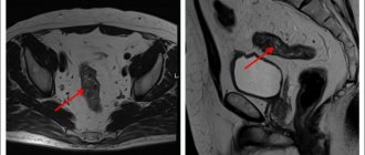
If the tumor is located superficially, for example, in the breast tissue, then it can be palpated. This feature makes diagnosis a little easier, but in order to understand the true nature of the tumor, it is necessary to undergo a comprehensive examination. It includes various imaging methods that help detect the primary lesion: CT, MRI, ultrasound, endoscopy, x-ray.
At the next stage, the doctor is faced with the task of determining the type of tumor, the degree of cell differentiation and other important features. A biopsy, the gold standard for diagnosing tumors, can answer these questions. As a rule, without a biopsy in oncology it is impossible to make an accurate diagnosis and select treatment.
Additionally, to assess the general condition of the patient or to determine sensitivity to a particular drug, laboratory and molecular genetic research methods are prescribed. The exact diagnostic program is always selected individually.
Squamous cell carcinoma
Squamous cell carcinoma (SCC) is the second most common non-melanocytic skin cancer.
Risk factors for squamous cell carcinoma:
- Long stays in the open sun, frequent visits to solariums.
- A special phenotype is light skin of phototypes I–II, the presence of freckles, red hair, blue eyes, and a tendency to sunburn.
- A history of sunburn is included in a separate item because it is of great importance for this tumor. Having one or more burns during childhood or adolescence significantly increases the risk of developing SBS in adulthood. Sunburn in adulthood is also a risk factor.
- The presence of precancerous skin lesions—actinic keratosis or Bowen's dermatosis—increases the likelihood of developing SCC.
- Personal or family history of skin cancer - If the patient or immediate family has had squamous cell carcinoma, the risk of developing it increases significantly.
- Weakened immune system – People with leukemia or taking immunosuppressive medications are at risk for IBS.
- Xeroderma pigmentosum - the skin of such patients has increased sensitivity to sunlight, which increases the likelihood of the appearance of SCC, BCC, melanomas and other malignant oncologies.
About 70% of all SCCs are located on the head and neck, including the forehead, lower lip, pinna, and periauricular area (behind the ear). The classic manifestation of the tumor is a small ulcer with peripheral ridges, often covered with plaque (Fig. 2) . However, the clinical manifestations of SCM largely depend on the location and stage of the disease - these may include the following options:
- Hard red nodule.
- Flat wound area with a scaly crust.
- A freshly inflamed or raised area on an old scar or ulcer.
- A rough, scaly patch on the lip that can develop into an open sore.
- Bright red sore area in the mouth.
- A red, raised spot or sore on the anus or genitals.
The prognosis for patients with SCC is good - 5-year survival rate in the early stages of the tumor is more than 90%, subject to timely diagnosis and adequate therapy. Outcomes for patients with advanced stages of SCC are significantly worse - in the presence of lymph node metastases, 5-year survival is only 25-45%.
Rice. 2. Squamous cell carcinoma in the periauricular region (Danish national service on dermato-venereology)
https://www.danderm-pdv.is.kkh.dk/atlas/7-119.html
Treatment of tumors
The second pressing problem in oncology is the effective treatment of tumors. If a benign tumor can be removed surgically relatively easily, then with a malignant process this is not always possible. Due to the absence of pronounced symptoms, tumor invasion into neighboring organs and vital vessels, and a tendency to metastasize, many patients remain inoperable. In such situations, conservative treatment is used, which includes methods such as chemotherapy, radiation therapy, targeted therapy, immunotherapy, etc. However, in this case, the patient’s chances of a full recovery are reduced. If the tumor has spread beyond the organ, then conservative therapy in most cases will help delay relapse and increase the patient’s life expectancy.
The most effective treatment is possible when the tumor is detected at the earliest stage, after surgical removal of the primary lesion, after which, for some diseases, conservative treatment methods can be used.
Currently, all the efforts of doctors and scientists are focused on solving two pressing problems. On the one hand, methods for early detection of cancer are being developed, and on the other, new and effective treatment methods are being developed.
Book a consultation 24 hours a day
+7+7+78
Diagnostic imaging of non-melanocytic skin cancer
Success in treating most (if not all) types of non-melanocytic skin cancer lies in diagnosing them as early as possible. For the initial detection of malignant skin tumors, dermatoscopy is most often used. The essence of this method is to identify specific signs of a tumor when studying it with a dermatoscope.
To successfully identify these signs, you need not only high-quality equipment, but also experienced personnel. Although a doctor can determine most criteria for skin cancer after basic training in dermatoscopy courses, it is believed that high diagnostic accuracy is achieved provided that the dermatologist has more than two years of experience in treating patients with skin tumors.
Modern technologies can help the doctor determine the degree of malignancy of the detected tumor. These technologies are based on digital dermatoscopy , which allows you to photograph a tumor in high resolution and send it with a detailed description for review by a more experienced colleague. This approach significantly speeds up the diagnostic process - if previously the patient had to wait his turn to see an experienced specialist, now, thanks to the capabilities of digital dermatoscopy and telemedicine, the suspicion of a malignant neoplasm is confirmed in a matter of hours.
The newest direction in identifying skin cancer is the use of artificial intelligence - systems for analyzing images of tumors that use convolutional neural networks (CNN) to determine the likelihood of a malignant process. The leader in digital dermatoscopy today is the FotoFinder company, whose software and hardware systems allow you to examine the surface of the skin, take high-resolution images with maximum transfer of all details, use a second opinion service (when the results of dermatoscopy are instantly sent to an experienced specialist), and also use artificial intelligence, which instantly examines the image of the tumor and gives it an assessment.
Modern technologies open up wide opportunities for doctors who do not specialize in identifying malignant skin tumors - first of all, for cosmetologists. It is especially important to use digital dermatoscopy for those specialists who remove tumors using laser. A doctor inexperienced in the field of dermatoscopy may mistake a malignant tumor for a benign one and remove it, which is fraught with serious consequences for the patient, which can cost him health and even life.
Questions from our users:
- treatment of non-melanocytic cancer
- skin cancer treatment price
- laser skin cancer treatment
- skin cancer treatment with liquid nitrogen


