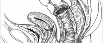
Adenomatous polyps
Precancerous diseases of the colon can become malignant, i.e. transform into a malignant tumor. The most common precancers are adenomatous polyps and hereditary polyposis.
Adenomatous polyps have malignant potential. Typically, about 10 years pass from the moment a polyp appears until cancer develops in it. The likelihood of developing an adenomatous polyp increases with age. Half of people over 80 years old have adenomas, most often they are located in the sigmoid and rectum.
There are three types of adenomas:
— Tubular adenoma is usually pedunculated, so the tumor can be removed during colonoscopy. Of all adenomas, it has the lowest risk of malignant transformation, especially if its size is less than 2 cm.
— Villous adenoma does not have a stalk and is located on a broad base directly on the intestinal mucosa, which is why in some cases it is necessary to remove the polyp along with part of the intestine. Often malignant.
— Tubulovillous adenoma has features of both tubular and villous adenoma.
Symptoms of adenomatous polyps are bleeding from the rectum, anemia, blood in the stool, diarrhea or constipation, abdominal pain, prolapse of the polyp along with the mucous membrane from the anus, intestinal obstruction (for large polyps).
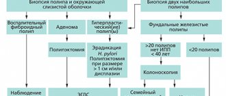
Diagnostic measures for adenomatous polyps and intestinal polyps include digital examination of the rectum, X-ray contrast study with barium, fecal occult blood test, sigmoidoscopy, colonoscopy.
During colonoscopy, detected polyps are removed and sent for histological examination. If cancer is detected in a polyp, a second operation is performed in which part of the intestine is removed. In addition, such an operation is performed for flat polyps (usually villous adenomas), the removal of which, due to their connection with the intestinal wall, by other means can be difficult.
Hereditary syndromes of colon polyposis
With some hereditary syndromes, a very large number of polyps can develop in the intestine. Such syndromes (polyposis) significantly increase the risk of developing colon cancer in humans, so these syndromes are classified as precancerous conditions.
Hereditary polyposis syndromes include:
· Familial adenomatous polyposis, in which hundreds and thousands of polyps are present in the mucous membrane from childhood and adolescence, which without treatment necessarily transform into a malignant tumor.
· Hereditary nonpolyposis colorectal cancer is characterized by fewer polyps. A person with this syndrome has a 70-80% lifetime risk of developing colorectal cancer.
Diagnostic measures, in addition to those listed above, for hereditary polyposis include genetic counseling.
Treatment for familial adenomatous polyposis (which, if left untreated, leads to cancer in 100% of cases by age 40) involves early removal of the colon. This usually occurs between the ages of 15-20 years.
With partial removal of the colon or with complete removal of the colon, there is still a risk of malignancy of polyps in the remaining rectum, so regular examination (colonoscopy, sigmoidoscopy) with the removal of all newly formed polyps is very important. When the colon and rectum are completely removed, an ileostomy is formed (an artificial exit of the small intestine onto the surface of the abdominal wall), but the risk of developing cancer in this case is significantly lower.
In case of hereditary non-polyposis colorectal cancer, prophylactic removal of part (as a rule, with this hereditary syndrome, tumors arise in the right parts of the intestine) or the entire colon is also carried out. In addition, patients with this syndrome have an increased risk of developing cancer of the stomach, small intestine, liver, pancreas, kidney, brain tumors, and in women - cancer of the uterus and ovaries.
Colon
The large intestine is the next section of the gastrointestinal tract after the small intestine and is divided into the cecum, ascending colon, transverse colon, descending colon, sigmoid colon and rectum. The length of the colon is 1.5-2 m, the diameter is 4-6 cm. The wall of the colon is represented by longitudinal muscle fibers, which are concentrated in the form of three ribbons parallel to each other. The width of each of them is about 1 cm. They stretch from the origin of the appendix in the cecum to the initial part of the rectum. The intestine seems to be corrugated, forming protrusions - haustra. The inner (circular) muscle layer is continuous. The mucous membrane of the colon, unlike the small intestine, does not have villi. The submucosa is represented by loose connective tissue containing the bulk of the vessels. The ascending colon in the right hypochondrium forms a hepatic flexure and passes into the transverse colon, the length of which is 50-60 cm. In the left hypochondrium, the intestine forms a left (splenic) flexure and passes into the descending colon. The descending colon becomes the sigmoid colon and then the rectum.
Functions of the colon
The colon produces:
In the colon, water, electrolytes (calcium, potassium, sodium, magnesium salts, etc.), glucose, fatty acids, amino acids, fat-soluble vitamins, etc. are absorbed. Enzymes, cholesterol, heavy metal salts are released into the intestinal lumen, and fiber fermentation occurs. . Intestinal microflora plays an important role in fermentation. The gut contains more than 400 different aerobic and anaerobic microorganisms. Anaerobes predominate, in a ratio of 1:1000. Normal intestinal flora has enzymatic, vitamin-synthesizing (vitamins B, C, K) and protective properties. Anomalies and malformations
|
Polyps and polyposis
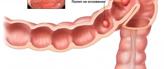
Polyps are classified as benign neoplasms arising from epithelial cells of the colon; some of them are prone to malignancy (malignant degeneration). Polyps are found in 10-12% of proctological patients. When performing preventive colonoscopy, it is 2-4%. In men, polyps are found 2-3 times more often than in women. Polyps can be single or multiple. The diameter ranges from 0.5 to 2 cm, sometimes they reach 3-5 cm. Polyps with a “leg” hang into the intestinal lumen; polyps with a broad base are less common. There are juvenile, hyperplastic, adenomatous (glandular), villous polyps, villous tumors, as well as multiple colon polyposis (true and secondary).
Juvenile polyps
They are observed mainly in children. The mucous membrane of the rectum is most often affected. Macroscopically, polyps have the appearance of a bunch of grapes, their surface is smooth, the color is more intense compared to the surrounding unchanged mucous membrane. These are typical glandular-cystic formations in which the stroma predominates over the glandular elements. As a rule, juvenile polyps do not become malignant.
Hyperplastic (metaplastic) polyps
Small (2-4 mm) formations, most often have a cone shape. They retain the normal structure of the intestinal mucosa with a significant increase in the number of glands, due to which the impression is created of a thickening of the mucous membrane in the form of a polyp. They become malignant very rarely.
Adenomatous (glandular) polyps
Observed in 90%. An adenomatous polyp has the appearance of a tumor with a smooth surface, round in shape, located on a stalk with a wide base, and is an area of hyperplasia of the mucous membrane. With a size of more than 2 cm, malignancy of the polyp occurs in 50% of cases.
Villous polyp (adenopapilloma)
Occurs in 5% of cases of detection of polyps - it has a lobed structure, a velvety surface, and is covered with thin delicate villi. The villous tumor protrudes into the intestinal lumen and is located on a broad base (nodular form). One of the varieties of villous tumors is the creeping, carpet form, in which there is no tumor node. The process spreads over the surface of the mucous membrane, occupying a fairly large area around the entire circumference of the intestine, and manifests itself as villous or finely lobulated growths. The size of villous tumors is 1.5-5 cm. The tendency to malignancy is up to 90%. Most often, villous tumors are localized in the rectum and sigmoid colon. The clinical picture is characterized by the release of mucus during bowel movements, the amount of which can reach 1 - 1.5 liters per day, which leads to water and electrolyte disorders. Slight vulnerability of the tumor villi leads to bleeding. Abdominal pain, constipation, diarrhea, and intestinal discomfort are noted.
Colon polyposis (multiple polyps)
It can be congenital, familial or secondary (as a result of other lesions of the colon, such as colitis). The incidence of malignancy reaches 70% and higher. Polyposis can be considered an obligate (obligatory) precancer. Congenital familial polyposis is inherited, affecting several family members. The disease is usually detected in children and young people. Polyps can be localized throughout the gastrointestinal tract. The combination of multiple colon polyposis with benign tumors of soft tissues and bones is called Gardner's syndrome. The combination of polyposis of the digestive tract with pigment spots on the mucous membrane of the cheeks, around the mouth and on the skin of the palms is called Peutz-Touraine syndrome. Multiple colon polyposis is characterized by abdominal pain without clear localization, diarrhea, discharge of blood and mucus in feces, weight loss, and anemia. Diagnosis of the disease is carried out on the basis of clinical symptoms, anamnestic data and instrumental examination data. The main treatment for colon polyps is surgical removal of the polyp or part of the colon. The volume is determined on the basis of anamnesis, instrumental examination data and the results of histological examination.
Diagnostic measures at PATERO CLINIC:
- Consultation with a surgeon-proctologist.
- Video endoscopic examination.
- in the absence of indications, the first examination is at 45 years of age,
- in the absence of pathology - colonoscopy every three years,
- if pathology is present, treatment is carried out, then colonoscopy every year.
- If you refuse a colonoscopy, perform a virtual colonoscopy (CT colonoscopy)
- Histological examination of the biopsy specimen in the laboratory of PATERO CLINIC.
- If necessary, ultrasound, MSCT, MRI, laboratory diagnostics.