Hyperlordosis is a deformation of the spinal column in the sagittal plane (median, passing in the anteroposterior direction and dividing the human body into right and left parts) in which the concave side of the arch is directed posteriorly.
Natural deflection - lordosis, is present in the spine in the cervical and lumbar regions. These physiological curves distribute the load on the spine and provide support for the head in the correct position.
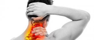
Hyperlordosis of the cervical spine, caused by a deviation of the spinal axis by an angle of more than 40 degrees, is characterized by painful sensations in the cervical spine, fatigue and headache. Pathology that is difficult to treat.
Hyperlordosis of the lumbar spine is a disease in which the natural deflection of the back significantly increases. The greatest load falls on the lumbar spine. The normal angle of curvature of the lumbar spine can range from 20 degrees to 40 degrees. When the curvature exceeds an angle of 45 degrees, we can talk about hyperlordosis - excessive lordosis of the lumbar region. There are two main forms of this pathological curvature - congenital and acquired.
Hyperlordosis is a serious disease that, if not treated in a timely manner, can lead over time to local or complete limitation of movements, even to the point of disability.
What it is
Lordosis is a curve of the spine whose convexity is directed forward. Its name is due to its location. As a rule, the apex of lumbar lordosis is located at the level of 3-4 lumbar vertebrae. A bend of 150 to 170 degrees is considered normal . Any change in the angle up or down is a reason for diagnosing pathological lordosis.
Clinical picture
The formation of pathological curvature of the spine, regardless of the reasons for its occurrence, is accompanied by pain, posture disorders, as well as the functioning of internal organs.
At the same time, the load on the vertebrae increases significantly and becomes uneven. This affects the joints, reduces spinal mobility, and increases the risk of a hernia. In the absence of timely therapeutic measures, the signs of pathology and consequences become increasingly serious.
Below is a detailed classification of lordosis
Classification
Lordosis can be classified according to various criteria.
| From the point of view of the cause: | By degree of manifestation: | By the nature of changes in bending: |
|
|
|
Prevalence and significance
Pathological lordosis of the lumbar region is the most common type of this disease.
It poses a danger of developing irreversible negative consequences, such as:
- spondyloarthrosis;
- spondylosis;
- spinal cord compression;
- malfunctions in the internal organs of the abdominal cavity;
- signs of chronic fatigue.
Lordosis in children
Lordosis often appears in childhood without any obvious cause. This condition is called benign juvenile lordosis. Its development is associated with the fact that the muscles around the child’s hips are weak or tight. Benign juvenile lordosis usually resolves on its own once the child's growth is complete.
Lordosis can also be a sign of hip dislocation, especially if the child has had an accident or fallen somewhere.
Other conditions that can cause lordosis in children are usually related to problems with the nervous system and muscles. These conditions are quite rare and include:
- Cerebral paralysis
- Myelomeningocele, a congenital abnormality in which the spinal cord protrudes through unfused vertebrae
- Muscular dystrophy, a group of inherited diseases that cause muscle weakness
- Spinal muscular atrophy, an inherited disorder that causes involuntary movements
- Arthrogrypposis is a congenital pathology in which the joints cannot move fully.
Risk factors, causes
The main risk factors for the development of this pathology include:
- heredity;
- injuries received during childbirth;
Pay attention to the list of factors that provoke the development of lordosis in the lower back; physical inactivity;- weakness of muscles and ligaments;
- pathological or congenital dislocation of the hip;
- immobility of the hip joint;
- osteochondrosis;
- intervertebral hernia and other chronic pathologies of the spine;
- spinal injury;
- malignant neoplasms in the spine or nearby organs;
- tuberculosis;
- pregnancy.
The risk of hyperlordosis increases significantly if a person is overweight, has bad habits, including smoking and drinking alcohol, or endocrine pathologies.
The reasons for the development of lordosis in the lumbar region may be:
- excessive load on the spine;
- pathologies of the musculoskeletal system;
- cerebral palsy;
- tuberculosis;
- brucellosis;
- rheumatism;
- lupus erythematosus;
- diabetes;
- vertebral fractures.
Video: “What does straightening lordosis mean?”
Lordosis in pregnant women
Many pregnant women experience back pain and show signs of lordosis, a protruding belly and buttocks. But numerous studies have shown that lordosis during pregnancy is actually a compensatory reaction of the spine, regulating the alignment of the center of gravity.
Lordosis in pregnancy is characterized by lower back pain - it can be caused by changes in blood flow in the body and stress on the lumbar spine, and the pain usually disappears after birth.
Consequences
The lack of timely treatment for the diagnosis of lordosis leads to a significant weakening of the spinal ligaments and growing muscle tension. All this leads to severe pain and constant spasms.
At the same time, the condition of the spine itself deteriorates, which manifests itself in the form:
- erasure of intervertebral discs;
- increased mobility of the vertebrae;
- inflammation of the iliopsoas muscle;
- development of deforming arthrosis in the joints of the spine;
- increasing the risk of developing intervertebral hernias.
Due to negative changes occurring in the body, cardiovascular pathologies, breathing problems, and diseases of the gastrointestinal tract may appear.
Myofascial pain syndromes localized in the back
O. V. Vorobyova, Doctor of Medical Sciences, Professor of the State Budgetary Educational Institution of Higher Professional Education First Moscow State Medical University named after. I. M. Sechenova Ministry of Health of the Russian Federation, Moscow
At an outpatient clinic, patients with back pain make up from 30% to 50% of patients, depending on the doctor’s specialization. The etiology and mechanisms of formation of back pain are extremely variable.
There are at least four most significant factors leading to pain:
structural changes in cartilage tissue (pathology of intervertebral discs, degenerative arthritis);
chronic muscle dysfunction (tension, spasm);
damage to the nerve fiber primarily due to compression (disc herniation, osteophyte, spinal canal stenosis);
psychological factors contributing to the complex components of psychosocial dysfunction.
In most patients, pain is caused by a combination of several factors that are the source of primary pain and/or support the persistence of pain. Musculo-ligamentous disorders almost obligately accompany back pain, and sometimes are the root cause of pain. They often remain unrecognized, which is due to the low awareness of medical specialists. The pathology of the musculo-ligamentous apparatus of the back is most clearly reflected by myofascial pain syndrome (MPS), characterized by muscle dysfunction and the formation of local painful compactions in the affected muscles. Approximately a quarter of all unilateral back pain syndromes are caused exclusively by MFS. How to diagnose myofascial pain?
Diagnosis of myofascial pain is based on anamnestic characteristics of pain and clinical examination. It is important to determine the type, intensity, duration and location of pain, as well as the factors influencing the intensity of pain. What facts should be clarified in the anamnestic? Particular attention should be paid to the facts of possible muscle injury. For acute pain, it is important to determine what movement caused the pain and test the muscles involved in that movement. With the gradual development of pain, it is important to examine chronically overworked muscles that are subject to microtrauma.
What examination should the clinician perform? Clinical examination necessarily includes assessment of passive and active movements and muscle tone. MFS is characterized by an asymmetric restriction of the motor pattern. An integral part of the diagnosis of MFS is muscle palpation and identification of trigger points (TP). When examining the effector muscle, extremely sensitive “nodules” called trigger points are palpated within the spasmodic muscle cords. Most researchers recognize palpation as the main method for diagnosing MFS; with sufficient knowledge of this technique, it is possible to identify 85–90% of TT. TTs localized superficially or in the area of localized spasm are most easily detected. To more accurately identify the localization and activity of TP, it is advisable to first relax the spasmodic, painful muscles. For this purpose, the post-isometric relaxation technique or, in the absence of special skills, passive mechanical relaxation can be used.
Depending on the location and volume of the muscle, various palpation techniques can be used (direct pressure on the TT with fingers, superficial palpation, pinch palpation). For superficially located small muscles, gentle palpation is performed with the fingertips. Easily accessible muscles (eg, sternocleidomastoid, upper trapezius, hip adductor, and others) are grasped between the thumb and fingers and the muscle fibers are passed between the fingers (pinch palpation). Finally, deep palpation is used for deep-lying muscles (gluteal, piriformis and others). It is necessary to wait 2–5 seconds after finger pressure on the TT and evaluate the reproducibility of the referred pain. The effectiveness of the method increases when using topographic maps of the favorite location of TP in muscles. Associated dermatomal sensitization and trophic swelling can be assessed using skin plucking. Additional research methods (electromyography, algometry, thermography, ultrasound techniques) play a supporting role in the diagnosis of MFS, since they have low sensitivity and specificity. How to clinically evaluate a trigger point? On palpation, the trigger point is felt as a clearly limited area of sharp pain. The CT size on average varies between 2 and 10 mm. Usually it is detected along one cord as the most painful point. When palpating the active TT, pain is observed under the examiner’s finger and in the usual pain zone (zone of referred pain).
The intensity of the pain often reaches such an extent that the pain leads to a rejection reaction (jumping symptom). Active trigger points can also cause non-painful phenomena. The most common vegetative symptoms are: local vasospasm, local hyperhidrosis, pilomotor activity. Paresthesias may be equivalent to pain phenomena in the reflected zone. It is generally accepted to distinguish active and latent myofascial TP. In the active form, there is constant pain, decreased muscle elasticity, and the development of referred pain in response to direct pressure on the TT. The intensity of pain and the length of the pain zone depend mainly on the degree of excitability of the TT. Latent TT demonstrates the same clinical characteristics as active points, but is significantly less pronounced. In addition, in the latent form, the pain is induced rather than constant. The induced pain is usually localized to the area of the affected muscle and the referred area. Some researchers believe that latent TTs may be associated with the genesis of muscle spasm. Potentially, they can reorganize into an active state. In addition, TTs can be classified into primary and secondary. Primary are called TPs, which are formed as a result of acute or chronic overload of the muscle concerned and whose activity is not related to the activity of other muscles. Secondary or satellite TTs are the result of mechanical stress and/or neurogenic inflammation due to the activity of primary TTs. In the absence of supporting factors, TTs may spontaneously disappear if the muscle remains at rest for several days. On the contrary, negative factors, and most importantly, the persistence of the influence of the original pathogenic factor, contribute to the formation of secondary triggers and an increase in the area of pain. Thus, the main clinical markers of MFS, summarizing the clinical picture, are: local or regional pain, limiting range of motion; palpation determination of hypertonicity in the affected muscle with areas of increased sensitivity within the “tight” cord (trigger point); reproducibility of pain when trigger points are stimulated; reduction of pain when stretching the affected muscle.
What factors contribute to the formation of MFS in the back? The formation of the MFS is based on both the characteristics of the muscular system bearing the postural load and specific load factors. The actual anatomical features of the back muscles, namely the absence of long tendons with close interaction between the muscles, paraspinal ligaments and fascia, make these muscles especially vulnerable to the formation of MFS. In addition, the muscles of the back and neck are among the least trained, which limits their functional reserve. Loading factors vary somewhat at different levels of the spinal column.
1. Cervical level. Myofascial pain syndromes are the most common cause of pain in the neck, shoulder, and headaches. This is the reason why neck pain occurs in 30–85% of people. Chronic strain of the neck muscles is most often a consequence of: anti-physiological postures associated with work organization disorders (improper sitting at a school desk, when working with a computer monitor, etc.); neck position during sleep (pillow features); postural adaptation of the neck in the presence of primary pain in adjacent regions (shoulders, temporomandibular joint, etc.). Acute injury to the musculo-ligamentous system of the neck is most often associated with an acceleration/deceleration type injury (extensor mechanism of injury). Most whiplash injuries occur in transportation accidents, but they can occur in other situations, such as diving.
2. Lumbar level. Instability of the motion segment most often leads to overload of the trunk muscles. A decrease in the elasticity of the disc fibers and dehydration of its matrix is the cause of the most common functional disorder in the motion segment - hypermobility of the intervertebral disc. At an early stage, this is compensated by contraction of the trunk muscles. However, the functional reserve of the muscle is limited and depends on the training of the muscles. Muscle tissue can be injured during single or recurrent episodes of biomechanical overload. Modern working conditions expose the back muscles to additional overload associated with muscle imbalance. For example, with a sedentary lifestyle, the human body is subjected to static loads most of the time, at this time dynamic muscles are constantly inhibited and gradually become flabby, while at the same time postural muscles contract and gradually lose elasticity. Chronic muscle imbalance is characteristic of modern urbanization. Also, various postural disorders, such as scoliosis and other skeletal asymmetries, can contribute to muscle overstrain.
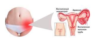
3. Pelvic level. MFS affecting the pelvic floor muscles occur almost exclusively in women [1]. This is primarily due to the anatomy of the female body and the structural changes that the female body experiences during reproductive life. During puberty after menarche, the girl's pelvis expands, the gluteal muscles increase in volume, and the hips rotate inward, leading to a lateral displacement of the patella. Constant internal rotation of the hips can negatively impact the pelvic diaphragm, which increases a woman's risk of developing pelvic floor spasms in the future. Pregnancy or weight gain increases this risk. Normally, the kneecap extends beyond the second toe, which helps maintain stable balance when standing. In many women, due to lateral deviation of the patella, the mobility of the joint decreases, which leads to a flattening of the arch of the foot. These structural changes in the lower extremities lead to disruption of the physiological maintenance of balance when standing and to excessive stress on the pelvic floor muscles. Ligaments in women are more extensible than in men, which is a necessary condition for maintaining joint stability and ensuring the process of physiological childbirth, but at the same time, this ability is a predisposing factor in the formation of fascial and ligament dysfunction in women. A fall on the buttocks can lead to limited mobility of the sacrum and the appearance of pelvic pain due to tension in the ligamentous apparatus of the pelvic floor muscles. In humans, the lower half of the body has more mass than the upper. Insufficiently developed muscles and muscle hypotonia can aggravate lumbar lordosis and increase the forward tilt of the pelvis. Increased lumbar lordosis is also observed during pregnancy. The reduction of estrogen during menopause is the main factor in the disruption of the physiological curves of the spine in old age. Changing the natural curves of the spine creates additional stress on the muscular frame, especially on the pelvic floor muscles. How to diagnose secondary muscle pain? Regardless of the primary source of pain and its pathogenetic characteristics, the muscles of the trunk are involved in the pathological process, becoming secondary sources of pain. Secondary pain occurs in the skeletal muscles outside the spinal motion segment due to a reflex increase in muscle tone. The physiological basis for muscle tension that follows any pain lies in the immobilization of the affected area of the body, the creation of a muscle corset. However, the muscle spasm itself leads to increased stimulation of the muscle's nociceptors. An increase in the flow of nociceptive impulses increases the activity of motor neurons in the anterior horns and contributes to increased muscle spasm. A reflex tonic muscle tension is formed. An additional factor in the development of painful muscle spasms is the antalgic posture. Transferring weight to one leg leads to curvature of the torso and asymmetrical position of the pelvis with the subsequent development of pain in the sacro-lumbar joints and the muscles that provide movement in these joints. The nature of secondary muscle pain is dull, aching, and pulling. Their intensity can vary widely. From a diagnostic point of view, it is important that pain is provoked by movements and is significantly intensified in positions in which the muscles surrounding the spinal column are stretched. Pain can also intensify when maintaining the same position for a long time (driving a car, sleeping in an uncomfortable position, long flight, etc.). There are no symptoms of loss.
With lumbar musculoskeletal pain, pseudo-Lasegue syndrome can be observed. If, when performing the Lasegue test, pain occurs only locally in the lower back, or hip, or under the knee, or in the lower leg, this is due to a stretch of spasmodic muscles (paravertebral or posterior thigh muscles) (“short” pain). On palpation, the paravertebral muscles are compacted, tense, and painful. Secondary muscle pain can become chronic and persist on its own even after the original cause has been eliminated. How to treat myofascial pain? Treatment of MFS requires multipronged approaches. Standard treatment includes: drug therapy with drugs such as nonsteroidal anti-inflammatory drugs (NSAIDs) and muscle relaxants; impact on TT, including physiotherapy; therapy aimed at restoring the normal functioning of muscle tissue: reducing muscle strain, strengthening the muscle frame, changing lifestyle. The main short-term task is to destroy trigger points, which leads to pain reduction. But the impact on TT should not be carried out in isolation. The long-term goal is to relax the muscles, restore the balance between postural and dynamic muscles, and neutralize predisposing factors, which reduces the risk of recurrence of pain. Warming the muscle can help it relax; for this, applications of “warming” ointments, gels, as well as hot wet wraps of the affected muscle, and wet warm compresses can be used. If you have certain skills, TT can be mechanically destroyed by injection of anesthetics (novocaine, lidocaine), which shortens the period of pain associated with the procedure. TT injections can provide excellent results. Less commonly used is “dry needling” without the use of an anesthetic. Specialized centers use muscle stretching exercises and gentle muscle relaxation techniques, such as post-isometric relaxation. In addition, traditional relaxation massage can be effective. The duration of therapy is significantly reduced with rapid and effective pain relief for the patient. Pain relief with NSAIDs is generally accepted for MFS - Nise, Diklak, Brufen SR, Movalis, etc. The prescription of NSAIDs is mandatory for any severity of pain - from mild (NSAID monotherapy) to severe (in combination with other drugs). Applications to painful areas of gels and ointments containing NSAIDs or their general dosage forms (tablets, suppositories, injection forms) can be used. The combination of NSAIDs and muscle relaxants in the treatment of MFS has become almost standard, making it possible to reduce treatment time. In addition, the simultaneous use of muscle relaxants and NSAIDs allows you to reduce the dose of the latter and, therefore, avoid the development of side effects of therapy. While taking muscle relaxants, post-isometric muscle relaxation, massage, and physical therapy are facilitated. It has been proven that the use of muscle relaxants makes it possible to rid the muscle not only of active, but also of latent TP, i.e., it improves the long-term prognosis, reducing the recurrence of MFS. Randomized controlled trials demonstrate the superiority of this class of drugs over placebo.
A study conducted by the Cochrane Physicians Society, which included over thirty controlled trials, also confirmed the usefulness of the use of benzodiazepine and antispastic muscle relaxants [2]. Domestic researchers prefer non-benzodiazepine central muscle relaxants. Usually tizanidine, tolperisone, and baclofen are used. These drugs have fewer side effects than benzodiazepines. Tizanidine (Sirdalud) is a prominent representative of central muscle relaxants. The drug is registered for the treatment of painful muscle spasm caused by musculoskeletal diseases and spasticity. The combination of tizanidine with NSAIDs demonstrates a more pronounced effect on pain reduction compared to NSAID monotherapy in patients with back pain [3, 4]. In addition to reducing pain, tizanidine reduces the need for NSAIDs and tranquilizers in patients, thereby reducing potential side effects from treatment. Placebo-controlled studies have shown the actual analgesic effect of tizanidine, as well as its effect on muscle tension and reduction of active trigger points [5]. Therapeutic tactics completely depend on the severity of the pain syndrome, its duration and the number of muscles affected by the MFS. In acute MFS, Sirdalud can be used in monotherapy. The recommended daily dose of Sirdalud is 6 mg per day in 2 or 3 divided doses. For severe MFS, combination treatment is used, combining pharmacological and non-pharmacological methods. Adding Sirdalud to a comprehensive treatment regimen allows you to reduce the duration of taking NSAIDs and avoid taking tranquilizers. Since the mild sedative effect of Sirdalud allows you to cope with mild anxiety without prescribing psychotropic therapy. Adjuvant treatments (antidepressants, anxiolytics, hypnotics): There are no high-quality randomized controlled trials on the use of these agents in patients with MFS. But numerous studies show the effectiveness of these drugs for treating chronic pain. It should be noted that chronic pain is often associated with depression, and effective treatment of depression can significantly reduce pain. The presence of comorbid syndromes requires mandatory targeted therapeutic efforts. A necessary component of treatment is the patient's physical activity. It is necessary to recommend that the patient return to normal daily activities. Physical therapy has a positive effect. Avoiding postural tension, daily physical therapy, mastering autogenic training with the ability to relax muscles is an effective defense against muscle pain. It is necessary to encourage the patient to make a positive change in life style (avoidance of anti-physiological poses, rational equipment of the workplace, stopping smoking, weight control, physical therapy, annual massage courses, mastering autogenic training with the ability to relax muscles).
Literature Vorobyova O.V. Painful spasm of the pelvic floor muscles as a cause of chronic pelvic pain in women // Farmateka. 2011, no. 5 (218): 51–55. Van Tulder MW, Touray T, Furlan AD, Solway S, Bouter LM Cochrane Back Review Group. Muscle relaxants for nonspecific low back pain: a systematic review within the framework of the cochrane collaboration // Spine. 2003, Sep 1; 28 (17): 1978–1992. Berry H., Hutchinson DR Tizanidine and ibuprofen in acute low-back pain: results of a double-blind multicentre study in general practice // J Int Med Res. 1988; 16:83–91. Pareek A., Chandurkar N., Chandanwale AS et al. Aceclofenac–tizanidine in the treatment of acute low back pain: a double-blind, double-dummy, randomized, multicentric, comparative study against aceclofenac alone // Eur Spine J. 2009; 18 (12): 1836–1842. Lepisto P. Muscle relaxants for nonspecific low back pain: a systematic review within the framework of the Cochrane Collaboration // J Int Med Res. 1981; 9 (6): 501–505.
The article was published in the journal The Attending Physician
Source: https://medblog.su/prochie-tematiki/miofastsialnye-bolevye-sindromy-lokalizovannye-v-oblasti-spiny.html © medical portal MedBlog.su
Link to publication: medblog.su
Symptoms and diagnostic methods
Symptoms of lordosis are the following::
- protruding belly , pelvis tilted back, knees turned in opposite directions;
- stretched muscles in the lumbar region , constant aching pain, which noticeably intensifies and becomes almost unbearable from standing for a long time;
- lowering of the abdominal organs , which provokes disturbances in their functioning;
- rapid fatigue , deterioration of night sleep, pain in any attempts to straighten the back, as a result of which the patient cannot take a position on his stomach while sleeping.
In case of pathological curvature of the spine, it is necessary to consult an orthopedist or surgeon. If there is a suspicion of lordosis, a person should first perform a self-diagnosis technique . To do this, you need to press your back against the wall so that your shoulder blades, buttocks and heels touch it at the same time.
Then you need to try to stick your hand into the gap between your lower back and the wall. Difficulty in this action will be a positive sign indicating the absence of this pathology.
If the hand passes freely, especially against the backdrop of pain in the spinal column, it is necessary to see a doctor as soon as possible with suspicion of developing lordosis.
A surgeon or traumatologist can most accurately make this diagnosis . As a rule, the doctor makes a conclusion based on a general examination, studying the medical history, and assessing the range of motion and muscle strength. Neurological status is also assessed. The most accurate diagnostic method in this case is radiography.
For existing somatic or neurological symptoms, methods are also used:
- magnetic resonance imaging (MRI);
- computed tomography;
- electroneuromyography.
If there is a suspicion of the development of inflammatory processes or tumors, the doctor may prescribe additional laboratory methods.
Main symptoms of the disease
Straightening the natural curve in the lower back does not go unnoticed. Already in the early stages, the patient notices changes in posture, difficulties with full alignment of the back and other negative signs. The main ones are:
Physiological curves of the spinal column
- Sharp pain in the lower back.
- The appearance of numbness in the morning in the damaged area.
- On palpation, a tightness is felt in the lower back.
- A forward tilt of the body that is difficult to correct by standing straight.
- Constant feeling of fatigue or decreased performance.
The lumbosacral region moves forward, and the gait changes greatly for the worse. The stomach protrudes slightly, the chest becomes sunken, the head is tilted forward. Some patients complain of severe pain in their legs. Degenerative processes that occur in the spine cause serious diseases, which in the future affect the functioning of the entire body.
Treatment
Did you know that...
Next fact
When treating lordosis, it is important to determine the root cause of the pathology. So, if the main cause is an infection that provoked a change, it is necessary to start treatment with it. In cases where the disease was caused by obesity, therapy begins with a special diet and weight-correcting procedures, including a physical training program.
Mandatory therapy for lordosis includes:
- massage course;
- therapeutic exercises;
- strength exercises to pump up the back muscles;
- correction of the daily diet;
- transition to the right lifestyle.
Manual therapy, swimming, wearing reclinators and other devices that correct posture may also be recommended.
Drugs
If the pathology of lordosis has progressed to more complex stages, the doctor may prescribe medication.
For this disease may be prescribed:
| Painkillers with anti-inflammatory effects: |
|
| Ointments and gels: |
|
| B vitamins: |
|
| Muscle relaxants: |
|
| Glucocorticoid hormones: |
|
| Therapeutic compresses: |
|
Surgery
Surgical treatment is used for congenital lordosis. Surgical intervention for the pathology of lumbar lordosis is used extremely rarely, in cases where non-surgical therapy methods do not bring results. Surgeries are also used for congenital lordosis.
This type of therapy includes spinal alignment using special metal structures that allow the operated area to be fixed.
Rehabilitation measures after surgery include wearing a bandage and corset, taking vitamins, special gymnastic exercises and massage.
Modern methods of surgical operations include the technique of implanting implants into intervertebral discs to ensure normal mobility of the spine .
Such operations are performed using endoscopic equipment. They involve removing the damaged disk and installing two metal plates on it, which secure a special “support” made of plastic.
After this, the implant performs the shock-absorbing and motor functions of the disc.
Exercises, physical therapy, massage
The most effective method of treating lordosis is a set of special exercises that combine strength training, relaxation exercises and stretching. It is important that therapeutic exercises are recommended by a doctor, and that the patient needs to perform them regularly and systematically.
Massage can also be of great benefit for lordosis. It will help normalize blood circulation, improve the outflow of venous blood, relieve pain, and relax muscles affected by spasms. Massage can also help prepare the spine for straightening.
Typically, the following types of massage are prescribed for lordosis::
- classical medicinal;
- lymphatic drainage;
- spot.
For this disease, the doctor may prescribe an intensive course of manual therapy , which will help improve the flexibility of the intervertebral discs, stretch the spinal segments, relax the muscles, and increase the tone of other back muscles. It is important to remember that traction with this therapy is only possible if the spinal segments are stable and there are no significant deformities.
Video: “How to correct lumbar lordosis with exercises?”
Treatment at home
Patients with severe curvature of the spine should have a special orthopedic mattress at home, designed to support a person’s weight .
At home, with lordosis, it is recommended to wear a bandage. Sleeping on your stomach , under which you need to place a cushion, will also be useful. The thickness of the roller must exactly correspond to the degree of curvature of the spinal column. The use of a roller during pregnancy, acute chronic diseases of internal organs and their exacerbation is strictly prohibited.
With lordosis, regular wearing of a bandage has a very effective effect on health . Most often it is prescribed to children who are obese, to patients whose severity of lordosis is low, to pregnant women, in cases where the pathology is associated with significant weakening of the back muscles. The bandage is selected strictly according to individual indicators.
It allows you to fix compromised areas of the spinal column and prevent further deformation. Pregnant women are most often prescribed a bandage after 16 weeks . It helps them when walking and serves as a prevention of disorders in the intervertebral joints after childbirth.
Prevention
In order to prevent the development of lordosis in the lumbar region, it is necessary to begin its prevention as early as possible. The best way to do this would be properly selected sports, such as swimming, yoga, Pilates, which will help develop and strengthen the muscle corset.
The following measures will also help for prevention::
- monitoring posture, even with severe fatigue;
- refusal of a soft bed;
- sleep on a hard orthopedic mattress.
Prevention
Preventive measures should include the necessary changes in lifestyle and the development of new habits:
- use of an orthopedic mattress;
- maintaining correct posture while working, walking, resting;
- it is necessary to sit on a chair, leaning on the back of the chair with your entire back, without loading the lumbar region;
- stand on two legs with an even distribution of body weight;
- you should lift the load without jerking, bending your legs at the knee joints and squatting slightly;
- normalization of the diet - it should be balanced;
- Regular swimming and gymnastics classes are required.
A timely visit to a doctor at the first signs of the disease and a course of treatment selected individually for each patient, taking into account the degree of pathological changes, will help restore a full quality of life and avoid the dire consequences of spinal pathology - limitation of motor activity.
Sign up for consultation and treatment at the “YES Medical Center!” You can fill out the form on the website or by calling 8(812)323-15-03.
Our licenses for medical activities
All rights reserved by copyright law. No part of the contents of the site may be used, reproduced, transmitted by any electronic, copying or other means without the prior written permission of the copyright owner.
Cervical hyperlordosis
It is characterized by an increase in the angle of deflection of the cervical spine to 45 degrees. The causes, as well as the types of this pathology, are similar to lumbar hyperlordosis, but its symptoms are specific:
- obsessive headaches;
- neck pain radiating to the shoulder girdle;
- progression of hearing and vision impairment, especially in adolescence;
- decreased concentration and memory;
- tinnitus and dizziness with preservation of normal peripheral pressure, but often a pronounced increase in intracranial pressure.
In addition to special gymnastics, in the treatment of cervical hyperlordosis, physiotherapeutic methods, kinesio taping, orthopedic styling and collars are used to relieve tension in the muscles.
Content:
- Causes of spinal curvature
- Classifications and types of curvature
- Differences in back curvature in adults and children
- Short description
The human spine is exposed to a lot of stress every day. Due to the influence of negative factors, posture changes. Spinal curvature can be congenital or acquired. The condition affects the patient’s well-being, the quality of the circulatory and nervous systems. The pathology is treated by an orthopedist who prescribes treatment individually for each patient.