Pancreatic enzymes play a critical role in the digestion of food from the stomach in the small intestine. Biocarbonate, which is the main component of pancreatic juice, creates an alkaline environment, neutralizes the acidity of the food mass with gastric juice in the duodenum, creating the pH range necessary for pancreatic enzymes. Participation in digestion and regulation of metabolism are the main functions of the pancreas, which is both an exocrine (secreting) and endocrine (increting) organ.
Exocrine function of the pancreas
The secretory function of the pancreas is divided into two types:
- • Ecbolic function - consists in the synthesis of more than twenty enzymes and proenzymes by its cells and their release into the duodenum. Digestive enzymes make up more than 90% of the proteins in pancreatic juice and are involved in the breakdown of food in the intestines.
- • Hydrokinetic function - consists of producing water, bicarbonates and other electrolytes. This function affects the neutralization of stomach contents, creating an alkaline environment in the intestines, favorable for the activity of pancreatic and intestinal enzymes.
Causes of pancreatitis
Factors that provoke the onset of the inflammatory process and persistent disruption of the enzymatic function of the pancreas:
- structural defects of the digestive system;
- dyskinesia of the biliary tract and gallbladder;
- severe stress;
- cystic fibrosis;
- hypothyroidism;
- infectious and inflammatory diseases of the gastrointestinal tract;
- intoxication with chemicals;
- parasitic infections;
- mechanical injuries of the abdominal organs as a result of compression, blows, falls;
- long-term use of medications: antibiotics, glucocorticosteroids.
Living in environmentally unfavorable regions and dietary errors can affect the health of the pancreas. The risk of pancreatitis increases consumption of alcohol, refined, fatty foods, fried foods, canned, smoked foods, and overeating.
Pancreatic enzymes
There are several types of pancreatic enzymes:
- • Amylolytic enzymes (amylases) break down complex carbohydrates into dextrins, maltose, maltooligosaccharides and glucose.
- • Lipolytic enzymes (lipases) break down fats into fatty acids and monoglycerides that pass through the enterocyte membrane.
- • Proteolytic enzymes (proteases) break down proteins by breaking internal bonds in the middle of amino acid chains and synthesizing peptides.
- • Nucleolytic enzymes (nucleases) break down nucleic acids. Phosphodiesterases present in pancreatic juice are divided into two groups: ribonuclease breaks down ribonucleic acid, and deoxyribonuclease hydrolyzes deoxyribonucleic acid.
Large glands of the digestive system
Liver
Hepar, liver
, is a voluminous glandular organ (weighing about 1500 g).
The functions of the liver
are diverse.
It is primarily a large digestive gland that produces bile, which enters the duodenum through the excretory duct. (This connection of the gland with the intestine is explained by its development from the epithelium of the midgut, from which the duodenum develops). It is characterized by a barrier function: toxic products of protein metabolism, delivered to the liver with the blood, are neutralized in the liver; in addition, the endothelium of the hepatic capillaries and Kupffer cells have phagocytic properties (reticuloendothelial system), which is important for the neutralization of substances absorbed in the intestine. The liver is involved in all types of metabolism; in particular, carbohydrates absorbed by the intestinal mucosa are converted in the liver into glycogen (“glycogen depot”). The liver is also credited with hormonal functions. In the embryonic period, it is characterized by the function of hematopoiesis, as it produces red blood cells. Thus, the liver is simultaneously an organ of digestion, blood circulation and metabolism of all types, including hormonal. The liver is located directly under the diaphragm in the upper part of the abdominal cavity on the right, so that only a relatively small part of the organ extends to the left of the midline in an adult; in a newborn it occupies most of the abdominal cavity, equal to 1/20 of the weight of the entire body, while in an adult the same ratio drops to approximately 1/50. The liver has two surfaces and two edges. The upper, or more precisely, the anterior superior, surface, fades diaphragmatica
, is convex according to the concavity of the diaphragm to which it is adjacent;
the lower surface, fades visceralis
, faces down and back and bears a number of impressions from the abdominal viscera to which it is adjacent.
The upper and lower surfaces are separated from each other by a sharp lower edge, margo inferior
. The other edge of the liver, the superoposterior, on the contrary, is so blunt that it can be considered as the posterior surface of the liver.
There are two lobes in the liver - the right lobe, lobus hepatis dexter
, and the smaller one - the left one,
lobus hepatis sinister
, which on the diaphragmatic surface are separated from each other by the falciform ligament of the liver, lig. falciforme hepatis. The free edge of this ligament contains a dense fibrous cord - the round ligament of the liver, lig. teres hepatis, which stretches from the navel, umbilicus, and is an overgrown umbilical vein, v. umbilicalis. The round ligament bends over the lower edge of the liver, forming a notch, incisura lig. teretis, and lies on the visceral surface of the liver in the left longitudinal groove, which on this surface is the boundary between the right and left lobes of the liver. The round ligament (overgrown with v. umbilicalis) occupies the anterior section of this groove - sulcus venae umbilicalis; the posterior section of the groove contains a continuation of the round ligament in the form of a thin fibrous cord - an overgrown venous duct, ductus venosus, which functioned in the embryonic period of life; this section of the groove is called fossa ductus venosi (Fig. 141).
Rice. 141. Gate of the liver. 1 - lig. venosum; 2 - v. hepatica sinistra; 3, 5 - v. cava inferior; 4 - lobus caudatus; 6 - v. portae; 7 - a. hepatica propria; 8 - ductus hepaticus communis; 9 - ductus choledochus; 10 - ductus cysticus; 11 - a. cystica; 12 - vesica fellea; 13 - fundus vesicae felleae; 14 - lobus quadratus; 15 - lig. teres hepatis; 16 - lig. falci forme hepatis; 17 - lobus sinister; 18 - r. sinister a. hepaticae propriae
The right lobe of the liver on the visceral surface is divided into secondary lobes by two grooves, or depressions. One of them runs parallel to the left longitudinal groove and in the anterior section, where the gallbladder is located, vesica fellea, is called fossa vesicae felleae; the posterior section of the groove, deeper, contains the inferior vena cava, v. cava inferior, and is called sulcus venae cavae. Fossa vesicae felleae and sulcus venae cavae are separated from each other by a relatively narrow isthmus of liver tissue called the caudate process, processus cauddtus
.
The deep transverse groove connecting the posterior ends of the sulcus venae umbilicalis and fossae vesicae felleae is called the portal of the liver, porta hepatis
. Through them enter a. hepatica and v. portae with the accompanying nerves and the lymphatic vessels and ductus hepaticus communis, which carries bile from the liver, emerge. The part of the right lobe of the liver, bounded at the back by the porta hepatis and at the sides by grooves (the gallbladder on the right and the umbilical vein on the left), is called the quadrate lobe, lobus quadrdtus. The area posterior to the gate of the liver between the fossa ductus venosi on the left and the sulcus venae cavae on the right constitutes the caudate lobe, lobus cauddtus.
Organs in contact with the surfaces of the liver form depressions on it, impressiones, which are called the organ in contact. The liver is covered for most of its length by peritoneum, with the exception of part of its posterior surface, where the liver is directly adjacent to the diaphragm.
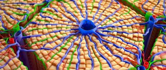
Structure
(Fig. 142). Beneath the serous membrane of the liver is a thin fibrous membrane, tunica fibrosa. In the area of the portal of the liver, together with the vessels, it enters the substance of the liver and continues into the thin layers of connective tissue surrounding the lobules of the liver, lobuli hepatis. In humans, the lobules are weakly separated from each other, but in some animals, such as pigs, the connective tissue layers between the lobules are more pronounced.
Rice. 142. Microstructure of the liver (according to R. D. Sinelnikov). 1 - hepar (lobus sinister); 2 - ductus hepaticus comm.; 3 - a. hepatica propria; 4 - ductus choledochus; 5 - v. portae; 6 - vesica fellea; 7 - vv. hepaticae; 8 - hepar (lobus dexter); 9 - v. cava inferior; 10 - interlobular blood vessels and bile ducts; 11 - network of primary bile canaliculi; 12 - v. centralis; 13 - aa. interlobulares; 14 - lobuli hepatis; 15 - vv. centrales
Liver cells in the lobule are grouped in the form of beams, which are located radially from the axial part of the lobule to the periphery. The lobules are surrounded by interlobular veins, venae interlobulares, which are branches of the portal vein, and interlobular arterial branches, arteriae interlobulares (from a. hepatica propria). Between the liver cells, which make up the liver lobules, located between the contacting surfaces of two liver cells, there are bile ducts, ductuli billferi (see Fig. 142). Leaving the lobule, the bile ducts flow into the interlobular ducts, ductuli interlobulares. An excretory duct emerges from each lobe of the liver. The ductus hepaticus communis is formed from the fusion of the right and left ducts
, removing bile from the liver, fel s. bills, and emerging from the portal of the liver. The common hepatic duct most often consists of two ducts, but sometimes of three, four and even five (G. A. Mikhailov, 1963). Inside the lobules, the endothelium of the hepatic capillaries consists of stellate cells (the so-called Kupffer cells), which have phagocytic properties.
Vesica
*
fellea
(sustis fellea)**,
the gall bladder
(see Fig. 141; Fig. 143) is pear-shaped.
Its wide end, extending slightly beyond the lower edge of the liver, is called the bottom, fundus vesicae felleae
.
The opposite narrow end of the gallbladder is called the neck, collum vesicae felleae
;
the middle part forms the body, corpus vesicae felleae
.
The cervix directly continues into the cystic duct, ductus cysticus
, about 3.5 cm long.
From the fusion of the ductus cysticus and the ductus hepaticus communis, the common bile duct is formed, ductus choledochus
, bile duct (from the Greek dechomai - accept).
The latter lies between two leaves of lig. hepatoduodenale, having the portal vein behind it, and the common hepatic artery to the left; then it descends down behind the upper part of the duodeni, pierces the medial wall of the pars descendens duodeni and opens together with the pancreatic duct with an opening into the expansion located inside the papilla duodeni major and called ampulla*** hepatopancredtica. At the confluence with the duodenum ductus choledochus, the circular layer of muscles of the duct wall is significantly strengthened and forms m. sphincter ductus choledochi, which regulates the flow of bile into the intestinal lumen; in the area of the ampulla there is another sphincter, m. sphincter ampullae hepatopancreaticae. The length of the ductus choledochus is about 7 cm. The gallbladder is covered with peritoneum only on the lower surface; its bottom is adjacent to the anterior abdominal wall in the corner between the right m. rectus abdominis and the lower edge of the ribs.
The muscular
lying under the serous
membrane , tunica muscularis, consists of smooth fibers with an admixture of fibrous tissue.
The mucosa
forms folds and contains many mucous glands. In the neck and in the ductus cysticus, there are a number of folds arranged spirally and forming a spiral flap, plica spiralis.
* (The correct pronunciation is vesica.
)
** (Greek - cholecystis, hence the inflammation - cholecystitis.
)
*** (The correct pronunciation is ampulla.
)
Rice. 143. Pathways for excretion of bile (top). Segmental structure of the liver (bottom). a - liver, gall bladder, pancreas and duodenum; b — the liver was removed, the intestine and pancreas were opened: 1 — lig. falciforme hepatis; 2 - lobus sinister hepatis; 3 - lobus dexter hepatis; 4, 5, 6 - lig. coronarium hepatis; 7 - vesica fellea; 8 - ductus cysticus; 9 - ductus hepaticus communis; 10 - ductus choledochus; 11 - corpus pancreatis; 12 - caput pancreatis; 13 - ductus pancreaticus (gland is opened); 14 - pars superior duodeni; 15 - pars descendens duodeni; 16 - pars horizontalis (inferior) duodeni; 17 - pars ascendens duodeni; 18 - beginning of the jejunum. A - diaphragmatic surface of the liver; B - visceral surface; B — segmental branches of the portal vein (projection on the visceral surface). Roman numerals indicate segment numbers (according to Couinaud)
Gallbladder of a living person
. An X-ray examination of the gallbladder (cholecystographia) reveals a shadow of the gallbladder, in which the neck, body and fundus can be distinguished. The latter is facing caudally. The contours of the bubble are clear, even and smooth. The shape of the bladder, depending on the degree of filling it with bile, can be pear-shaped, cylindrical and ovoid. The position of the bladder varies between the levels of the XII thoracic and V lumbar vertebrae, depending on the position of the liver, its excursions during breathing, etc.
Pathways for bile excretion
: since bile is produced in the liver around the clock and enters the intestines as needed, there is a need for a reservoir for storing bile. The gallbladder is such a reservoir. Its presence determines the structural features of the bile ducts (see Fig. 143).
Bile produced in the liver flows out of it through the hepatic duct, ductus hepaticus communis. If necessary, it goes directly to the duodenum through the ductus choledochus. If this need is not present, then the ductus choledochus and its sphincter are in a contracted state and do not allow bile into the intestine, as a result of which bile can only be directed to the ductus cysticus and further to the gallbladder, which is facilitated by the structure of the spiral fold, plicae spiralis.
When food enters the stomach and the corresponding reflex occurs, the muscular wall of the gallbladder contracts and the ductus choledochus muscles and sphincters relax simultaneously, resulting in bile entering the intestinal lumen.
Liver topography
. The liver is projected onto the anterior abdominal wall in the epigastric region. The borders of the liver, upper and lower, projected onto the anterolateral surface of the body, converge with one another at two points: on the right and on the left. The upper border of the liver begins in the tenth intercostal space on the right, along the middle axillary line. From here it rises steeply upward and to the left, corresponding to the projection of the diaphragm, to which the liver is adjacent, and along the right nipple line reaches the fourth intercostal space: from here the border gently descends to the left, crossing the sternum slightly above the base of the xiphoid process, and in the fifth intercostal space it reaches the middle of the distance between left sternal and left nipple lines. The lower border, starting in the same place in the tenth intercostal space as the upper border, goes from here obliquely and to the left, crosses the IX or X costal cartilage on the right, goes along the epigastric region obliquely to the left and up, crosses the costal arch at the level of the VII left costal cartilage and in the fifth intercostal space it connects with the upper border.
The liver is nourished by a. hepatica propria, but in a quarter of cases from the left gastric artery (G. A. Mikhailov, 1963).
Features of liver vessels
are that, in addition to arterial blood, it also receives venous blood. Through the gate, a. enters the substance of the liver. hepatica propria and v. portae. Entering the gate of the liver, v. portae, carrying blood from the unpaired organs of the abdominal cavity, branches into its thinnest branches located between the lobules - vv. interlobulares. The latter are accompanied by aa. interlobulares (branches of a. hepatica propia) and ductuli interlobulares. In the substance of the liver lobules themselves, capillary networks are formed from arteries and veins, from which all the blood collects in the central veins - vv. centrales. Vv. centrales, leaving the liver lobules, flow into the collecting veins, which, gradually connecting with each other, form vv. hepaticae. The hepatic veins have sphincters at the junction of the central veins (Gibson, 1959). Vv. hepaticae in the amount of 3-4 large and several small ones emerge from the liver on its posterior surface and flow into the v. cava inferior.
Thus, in the liver there are two venous systems: 1) portal, formed by the branches of v. portae, through which blood flows into the liver through its gates, and 2) caval, representing a set of vv hepaticae, carrying blood from the liver to v. cava inferior.
In the uterine period, a third, umbilical, vein system also functions; the latter are branches of v. umbilicalis, which becomes obliterated after birth. As for the lymphatic vessels, there are no true lymphatic capillaries inside the liver lobules: they exist only in the interlobular connective tissue and flow into the plexus of lymphatic vessels accompanying the branches of the portal vein, hepatic artery and bile ducts, on the one hand, and the roots of the hepatic veins on the other. The draining lymphatic vessels of the liver go to the lnn. hepatici. celiaci, gastrici dextri, pylorici and to the peri-aortic nodes in the abdominal cavity, as well as to the diaphragmatic and posterior mediastinal nodes (in the thoracic cavity). About half of the body's lymph is drained from the liver. The liver is innervated from the solar plexus via tr. sympathicus and n. vagus
Segmental structure of the liver
(see Fig. 143). In connection with the development of surgery and the development of hepatology, the doctrine of the segmental structure of the liver has now been created, which has changed the previous idea of \u200b\u200bdividing the liver only into lobes and segments.
As mentioned above, the liver has five tubular systems: 1) bile ducts, 2) arteries, 3) branches of the portal vein (portal system), 4) hepatic veins (caval system) and 5) lymphatic vessels (Fig. 144).
Rice. 144. Tubular systems of the liver (according to R. D. Sinelnikov). 1 - v. cava inf.; 2 - lobus hepatis dexter; 3 - ductus hepaticus comm.; 4 - v. portae; 5 - lymphatic vessels; 6 - a. hepatica; 7 - lobus hepaticus sinister; 8 - vv. hepaticae
The portal and caval vein systems do not coincide with each other, and the remaining tubular systems accompany the branches of the portal vein, run parallel to each other and form vascular-secretory bundles, to which nerves are attached.
All intraorgan tubular systems of the liver are located in 3 tiers (G. A. Mikhailov, 1963); the upper and lower tiers are occupied by systems passing through the gates of the liver (portal complex); the middle tier contains the hepatic veins, which carry blood from certain areas of the liver called zones. There are four such zones (Chen-Hao-de)*: in the right lobe - anterior and posterior and in the left - medial and lateral. According to the location of the vascular-secretory bundles of the portal complex, areas of the liver are distinguished that have, to some extent, separate blood supply, lymph circulation, bile outflow and invervation. These areas are called segments, and this structure of the organ is called segmental. A segment (not an embryological concept, but a spatial one) is a part of the liver corresponding to a large branch of the portal vein and the accompanying branches of the hepatic artery and bile ducts.
* (From the Department of Normal Anatomy of the 1st Leningrad Medical Institute named after. acad. I. P. Pavlova.
)
The number of segments varies according to the individual variability of the branches of the portal vein, as a result of which different researchers count different numbers of them.
The most widespread is the liver division scheme according to Couinaud (1957), according to which the liver has 2 lobes (right and left), 5 zones and 8 constantly occurring segments.
Recently, in connection with a more detailed study of the internal structure of the liver, the boundary between the right and left lobes is considered to be a plane drawn through the middle of the gallbladder bed, the left semicircle of the inferior vena cava and the middle hepatic vein (N. I. Odnoralov and B. P. Shmelev, 1963).
The following segments are distinguished in the liver, starting from the sulcus venae cavae to the left, clockwise (see Fig. 143);
I. Caudate segment of the left lobe, corresponding to the conominal lobe of the liver.
II. The posterior segment of the left lobe is localized in the posterior section of the lobe of the same name.
III. The anterior segment of the left lobe is located in its department of the same name.
IV. The square segment of the left lobe corresponds to the conominal lobe of the liver.
V. Middle upper anterior segment of the right lobe.
VI. Lateral inferoanterior segment of the right lobe.
VII. Lateral infero-posterior segment of the right lobe.
VIII. Middle superoposterior segment of the right lobe. (Segment names indicate areas of the right lobe.)
The segments, grouped along radii around the gate of the liver, are included in larger independent areas of the liver, called zones or sectors. There are five such sectors*:
* (Quote according to B.V. Petrovsky - “Surgical hepatology”. M, 1972.
)
1. The left lateral sector corresponds to segment II (monosegmental sector).
2. The left paramedian sector is formed by segments III and IV.
3. The right paramedian sector consists of segments V and VIII.
4. The right lateral sector includes segments VI and VII.
5. The dorsal sector corresponds to segment I (monosegmental sector).
Liver segments are formed already in uterine life and are clearly expressed at the time of birth (G. F. Vsevolodov, V. N. Verbitskaya and E. N. Dolgopolova, 1972). The doctrine of the segmental structure of the liver deepens the previous idea of dividing it only into lobes and segments. Thus, according to the latest data, lobes, zones or sectors, segments and lobules are distinguished in the liver.
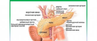
Pancreas
Pancreas, pancreas
, lies behind the stomach* on the posterior abdominal wall in the regio epigastrica, extending its left part also into the left hypochondrium. Posteriorly it is adjacent to the inferior vena cava, left renal vein and aorta.
* (When opening a corpse in a supine position, it actually lies under the stomach, hence its name. In newborns it is located higher than in adults, at the level of the XI-XII thoracic vertebrae (K. I. Kulchitsky).
)
The pancreas is divided into a head, caput pancreatis
, with a hooked process, processus uncindtus, on the body,
corpus pancreatis
, and tail,
cauda pancreatis
.
The head
of the gland is covered by the duodenum and is located at the level of the I and upper part of the II lumbar vertebra.
At its border with the body there is a deep notch, incisura pancreatis (the notch contains a. and v. mesentericae superiores), and sometimes a narrowed part - in the form of a neck. The body
is prismatic in shape and has three surfaces: front, back and bottom.
The anterior surface, facies anterior, is concave and adjacent to the stomach; near the junction of the head with the body, a bulge is usually noticeable on it towards the lesser omentum, called tuber omentdle. The posterior surface, fades posterior, faces the posterior abdominal wall. The lower surface, fdcies inferior, faces down and somewhat forward. The three surfaces are separated from each other by three edges: mdrgo superior, anterior and inferior. Along the upper edge on the right side there is a. hepatica communis, and the splenic artery stretches to the left along the edge, heading towards the spleen. The gland rises slightly from right to left, so that tail
lies higher than the head and approaches the lower part of the spleen. The pancreas does not have a capsule, due to which its lobular structure is strikingly obvious. The total length of the gland is 12-15 cm.
The peritoneum covers the anterior and lower surfaces of the pancreas, while the posterior surface is completely devoid of peritoneum. Pancreatic duct, ductus pancreaticus
, receives numerous branches that flow into it almost at right angles;
Having connected with the ductus choledochus, the duct opens with a common opening with the latter on the papilla duodeni major (see Fig. 143). This constructive connection of the ductus pancreaticus with the duodenum, in addition to its functional significance (processing of the duodeni contents by pancreatic juice), is also due to the development of the pancreas from that part of the primary intestine from which the duodenum is formed. In addition to the main duct, there is almost always an additional one, ductus pancreaticus accessorius
, which opens on the papilla duodeni minor (about 2 cm above the papilla duodeni major). Sometimes there are cases of accessory pancreas, pancreas accessorium. There is also a ring-shaped pancreas, causing compression of the duodenum (Hays, 1961; Hyden, 1962).
Structure
.
In its structure, the pancreas resembles a serous salivary gland of an alveolar or alveolar-tubular structure. There are two components in it: the main mass of the gland has an exocrine function, releasing its secretion through the excretory ducts into the duodenum; The smaller part of the gland in the form of the so-called islets of Langerhans
belongs to endocrine formations, releasing insulin into the blood (insula - islet), which regulates blood sugar.
Pancreas, as a mixed secretion gland, receives multiple sources of nutrition: aa. pancreaticoduodenales superiores et inferiores, aa. lienalis and gastroepiploica sin. etc. The sonominal veins flow into v. portae and its tributaries. Lymph flows to the nearest nodes: lnn. celiaci, pancreaticolienales, etc.
Innervation - from the solar plexus.
Pancreatic secretion
The quantity and quality composition of food consumed is directly proportional to the amount of pancreatic juice secreted and its enzymatic activity. Increased secretion of pancreatic juice causes a large volume of food, which stimulates an increase in acidity in the stomach. Ingestion of liquid food causes faster secretion from the pancreas than solid, fatty food slowly evacuated from the stomach. The production of pancreatic juice increases 2-3 minutes after food enters the stomach, its duration ranges from 6 to 14 hours. The greatest secretory response is caused by mixed food. Strong stimulants that affect the secretory enzymes of the pancreas are neutral fats, products of their digestion and proteins. Amino acids entering the intestinal tract (especially phenylalanine, choline, methionine) affect a sharp increase in the blood level of cholecystokinin, a hormone responsible for local stimulation of the activity of acinar cells that have the function of enzyme synthesis. The predominance of carbohydrates in the diet facilitates the activity of the pancreas, so this group of products is especially recommended in diets for patients suffering from exacerbation of chronic pancreatitis. Foods and drinks that stimulate the appetite (fruits, dairy products, products containing spices and seasonings, marinades, juices and alcohol) increase the production of gastric juice and hydrochloric acid, which, in turn, stimulates the onset of exocrine pancreas.
Forms of pathology
There are several forms of pancreatic inflammation. People of any age can suffer from them: children, adults and the elderly.
- Acute pancreatitis. Develops over several hours, usually after eating a large amount of food. Patients experience severe nausea with vomiting, pain in the upper abdomen, accompanied by diarrhea, bloating and belching. Severe fever is likely: heavy sweating, high body temperature, weakness. During the destructive process, abscesses and foci of necrosis arise, requiring immediate surgical intervention.
- Reactive interstitial pancreatitis is a type of acute form of inflammation, more typical for childhood. It is manifested by high anxiety temperature with sharp abdominal pain, accompanied by swelling of the gland and the release of the enzyme trypsin, which destroys its tissue. This process is reversible, unlike necrotizing, but also requires urgent medical intervention.
- Chronic pancreatitis. It may not make itself felt for decades, and the period of remission is asymptomatic. During exacerbations, it is manifested by severe dyspepsia, abdominal pain, loss of appetite, general intoxication, and fever. Often accompanied by chronic cholecystitis or gastritis, it requires regular maintenance therapy and lifestyle adjustments.
Pancreatitis causes disturbances in the functioning of the liver, gastrointestinal tract, vascular system, and kidneys. In most cases, the inflammatory process does not affect the endocrine functions of the gland and is not associated with the development of diabetes mellitus.
Disorders of the pancreas
Disorders of the digestive process in the small intestine are directly related to changes in enzyme synthesis caused by pancreatic dysfunction. In the presence of pathological processes in it, the action of lipase, which hydrolyzes fats, is inhibited, which leads to their insufficient digestion and excretion with feces up to 80% of the mass entering the body. The breakdown of proteins is also impaired, as may be evidenced by creatorrhea, a characteristic feature of which is the presence of an increased volume of muscle fibers in the stool. Digestive insufficiency can lead to such negative consequences as dyspeptic syndrome, dehydration, acid-base imbalance and intestinal autointoxication. External secretion of the pancreas is caused by the presence in the body of a number of inflammatory diseases and some other pathological processes:
- pancreatitis in acute and chronic stages;
- cholelithiasis;
- the presence of parasites in the intestines;
- duodenitis, duodenal ulcer, which results in a decrease in the production of pancreatic juice;
- tumors;
- vaterites;
- excess body weight, hormonal imbalance, resulting from metabolic disorders, causing dystrophic lesions of the pancreas;
- allergic restructuring of the body;
- Vagal dystrophy, long-term atropinization;
- tumors that destroy the pancreas.
The development of obstructive processes in the pancreas that impede or completely block the outflow of pancreatic juice into the duodenum, and the resulting hypertension that results from this pathology can cause acute pain in this area, as well as internal ruptures and destruction of the parenchyma of the organ. As a result of the destruction of the pancreas, its enzymes and destruction products are absorbed into the blood and surrounding organs, which leads to the development of necrosis and the formation of a body intoxication syndrome.
Methods for diagnosing pancreatitis
For most patients, the reason for going to the clinic is regular abdominal pain, loss of appetite, heaviness in the stomach and other obvious signs of pathology. Chronic inflammation can be discovered accidentally during medical examination. Methods for diagnosing pancreatitis:
- Blood chemistry. Classic signs of health problems: high ESR, leukocytosis. With pancreatitis, the levels of trypsin, amylase and lipase increase.
- Examination of urine and feces samples. They show an increase in amylase and changes in the levels of other enzymes. The coprogram shows the presence of undigested food residues.
- Ultrasound of the abdominal organs. An informative method that determines changes in the size and structure of the pancreas, concomitant inflammation of nearby bile ducts and liver.
- MRI. Prescribed for the differential diagnosis of pancreatitis in complex or controversial cases, with a severe course of the process.
Autophagy
- Cellular damage and apoptosis are associated with autophagy, the process of removing damaged organelles from the cell through lysosomal degradation and the formation of new ones in their place.
- Some diseases, including diabetes, are believed to have multiple etiologies. In particular, autophagy is called among the reasons.
- Studies examining the relationship between diabetes and autophagy in humans have found that when β cells and islet cells of Langerhans are exposed to hyperglycemia or hyperlipidemia, autophagy stops.
- This leads to the accumulation of oxidized, damaged or incorrectly folded proteins in cells.
- β death is also observed as a result of oxidative stress.
- The process of autophagy is regulated by various signals.
- The mTOR signaling complexes 1 and 2 proteins (mTORC1 and mTORC2) are of particular importance in the mechanism.
- These proteins are kinases involved in several cellular processes, for example: the formation of insulin resistance (IR);
- adipogenesis;
- angiogenesis;
- autophagy.
- improper protein folding;
Treatment of pancreatitis
The treatment strategy for pancreatitis depends on the severity of the disease. This may include taking various medications to relieve stress on the pancreas, parenteral nutrition, and surgery.
Treatment of acute pancreatitis
Patients diagnosed with acute pancreatitis are sent to the hospital. The doctor examines the clinical picture and refers for tests and examination. In severe cases of pancreatitis, the patient is sent to the intensive care unit; in non-severe cases, the patient is sent to the surgical department. The main task of doctors will be to ensure that the patient rests and the pancreas is completely hungry.
Treatment of non-severe forms includes probing the stomach contents, prescribing analgesics and antispasmodics, and administering infusion solutions. In severe cases, the patient is referred for surgery if endoscopic methods are not able to eliminate the complications of pancreatitis (bleeding, acute intestinal obstruction, etc.). An important component of treatment is antibiotic therapy.
Treatment of chronic pancreatitis
For chronic pancreatitis, doctors choose conservative or surgical treatment. Before making a decision, the condition of the entire digestive system is assessed. Lack of surgery can lead to loss of pancreatic function, while early surgery carries the risk of severe postoperative consequences. Doctors resort to surgery when there are complications of pancreatitis and there is a possibility of damage to other organs of the digestive system. Cysts, fistulas and obstruction of the gastrointestinal tract are other indications for surgery.
With conservative treatment, rest, cold on the stomach, antibiotics and analgesics, as well as vitamins (A, D, E and K) are prescribed. To restore the intestinal microflora, bifidobacteria and lactobacilli are prescribed. To replenish enzymes (amylase, trypsin and lipase), which the pancreas is not able to produce in the required quantities, enzyme preparations are prescribed. The doctor selects the appropriate drug and determines its dosage and duration of treatment.
Important! The daily dose of the enzyme preparation should be consumed throughout the day with meals. It is necessary to strictly follow the doctor's instructions about diet, avoid alcohol and junk food. Fatty foods are especially contraindicated.
Diet, nutrition for pancreatitis
Diet is extremely important for the treatment of pancreatitis. Disruption of the pancreas affects the functioning of other organs: it becomes more difficult for them to digest food. Therefore, the lighter the food, the better the peristalsis. This is not only about the absence of fatty foods and alcohol in the diet. It is also important to exclude foods that cause bile secretion and gas formation. It is better to eat fractionally, cut food into small pieces. You should choose foods that are low in fat and protein.
You will need to follow the diet throughout your life. Doctors advise sticking to table No. 5.
Diet No. 5 exists in a basic and extended version. The basic option is indicated for relapses of chronic pancreatitis and for the acute nature of the disease. In the acute phase, the diet is more strict with many restrictions. It is aimed at unloading the pancreas and relieving the symptoms of acute inflammation. In the first 3 days of the acute stage, the patient is recommended to fast. Then, for 3-7 days, carbohydrate foods are allowed in small portions at short intervals. After the symptoms of acute pancreatitis are relieved, the diet is expanded and other foods are added to it, the amount of protein in the diet and the total caloric content of the diet increases.
Taken from the article: “Diet for pancreatitis”
What can you eat
Among the permitted products for pancreatitis:
- Stale bread, dry biscuits and biscuits, crackers,
- Boiled or steamed vegetables (potatoes, beets, zucchini, carrots, cauliflower, pumpkin, broccoli, Brussels sprouts),
- Vegetable puree soups,
- Chicken, turkey, beef, veal, rabbit (boiled or steamed),
- Lean fish (pike perch, cod, pike, perch),
- Porridge with water (oatmeal, buckwheat, semolina, rice),
- Low-fat dairy products: milk (preferably in a dish), yogurt, cottage cheese, sour cream, kefir, cheese,
- Unsalted and low-fat butter in limited quantities,
- Sweets in limited quantities: honey, marshmallows, jam, preserves,
- Fruits: non-acidic apples, strawberries, bananas (not during an exacerbation of the disease and preferably in the form of puree or baked), non-acidic blueberries and cherries,
- Drinks: dried fruit compotes, jelly, weak unsweetened tea, non-acidic juices, still water,
- Omelette or eggs in the oven.
What not to eat
The diet for pancreatitis is designed to spare the pancreas.
The National Pancreatitis Foundation recommends that patients limit their fat intake to 30 to 50 grams per day, with no meal containing more than 10 grams of fat.
Digestive and fatty foods are prohibited (video 1):
- Fresh bread, pizza, dumplings and dumplings,
- Fresh vegetables: beans, peas, white cabbage, cucumbers, tomatoes, mushrooms, turnips, radishes, sorrel,
- Soups with broths (fish or meat), okroshka, beetroot soup, spicy soups,
- Lamb, pork, duck, goose, sausages, sausages, smoked meat, bacon, canned food, lard,
- Fish: fatty varieties, canned food, smoked fish, caviar, salted fish, seafood,
- Porridge: millet, pearl barley, corn,
- Fatty dairy products: full-fat milk, cheese, sour cream, cream,
- Margarine, mayonnaise, ketchup, mustard,
- Cakes, pastries, buns, ice cream, chocolate, jam,
- Bananas, citrus fruits, grapes, dates, sour fruits,
- Alcohol, coffee, cocoa, hot chocolate, carbonated drinks, packaged juices, energy cocktails, sparkling water, strong tea,
- Eggs: fried or hard-boiled.
Important! The digestive system of some patients diagnosed with pancreatitis does not digest fresh vegetables, particularly tomatoes. In this case, doctors advise being careful with the consumption of tomatoes, cucumbers and some other fresh vegetables and steaming or stewing them.
Video 1. Diet for pancreatitis.
Traditional methods
Pancreatitis is a serious disease that can lead to damage to all organs of the digestive system. Self-medication with folk remedies without seeking help from a doctor can lead to serious consequences. Thus, the use of any choleretic drugs is absolutely contraindicated: artichoke leaves, milk thistle, turmeric, celandine, etc.
The National Pancreatitis Foundation advises adults to do yoga three times a week to reduce pain, meditate to relieve stress, and move more. The digestive system generally welcomes physical activity. Doctors talk about the need for early movement even after abdominal surgery. This improves peristalsis and helps the organs quickly establish the processes of absorption and digestion of food.
Naturally, the use of folk recipes will neither lead to relief from pancreatitis nor to its aggravation. But the lack of timely qualified treatment can have disastrous results. It is important to remember that untreated pancreatitis can be fatal.
Features of the pancreas in children and adults
The pancreas is a rather sensitive organ. At the slightest disruption in its work, a person’s well-being immediately worsens. In adults, the size of this organ reaches 22 cm, weight up to 80 g. The formation of the organ occurs 4-5 weeks after conception. The pancreas is formed from endoderm and mesenchyme and is located near the liver. During development, the organ acquires parts: tail, body, head. The pancreas begins to perform its main functions already at the end of the 1st trimester of pregnancy. For some time the organ is mobile, which is explained by the lack of reliable fixation. Closer to 6 years of age, he occupies a permanent position.