Embryology
The parotid gland, like other major salivary glands, develops from the epithelium of the oral cavity. The gland bud appears in the embryo at the 6th week of development in the depths of the groove separating the cheek from the gum, in the form of an epithelial cord, which grows towards the ear. At the 8th week. embryonic development, the distal end of this cord begins to branch and gives rise to excretory ducts and terminal secretory sections of the liver. At the beginning of the 3rd month, gaps appear in the anlage of the excretory ducts, their epithelial lining becomes double-rowed, and in large excretory ducts multilayered. Differentiation of glandular epithelium in the terminal secretory sections of the liver. occurs somewhat later than in other salivary glands.
Operations
When removing the submandibular gland, a 7-8 cm long incision is made parallel to the lower edge of the body of the lower jaw, 2 cm away from it. The skin, subcutaneous tissue, subcutaneous muscle of the neck, and the superficial plate of the fascia of the neck are dissected, the gland capsule is opened, and the facial vein is ligated. . P.J. easily separates from the tissues of the bed. When separating the inner surface of the pancreas. The facial artery is isolated and ligated. In the region of the upper pole of the pancreas. the excretory duct is isolated and ligated and the facial artery is re-ligated. If the gland is removed due to an inflammatory process, then one should not go beyond its capsule so as not to damage the facial nerve (marginal branch of the lower jaw). The wound is sutured in layers, leaving the wound closed for 24 hours.
Bibliography:
Zedgenidze G. A. X-ray diagnosis of diseases of the salivary glands, L., 1953, bibliogr.; Kasatkin S.N. Anatomy of the salivary glands, Stalingrad, 1948; Sazama L. Diseases of the salivary glands, trans. from Czech., Prague, 1971, bibliogr.; Solntsev A. M. and Kolesov V. S. Surgery of the salivary glands, Kyiv, 1979, bibliogr.; Burch RJ a. Woodward HW Differential diagnosis and surgery of the submaxillary gland, J. oral Surg., v. 18, p. 470, 1960; Rauch S. Die Speicheldriisen des Menschen, Stuttgart, 1959; Schulz HG Das Rontgenbild der Kopfspeicheldriisen, Lpz., 1969, Bibliogr.
I. F. Romacheva; V. S. Speransky (an., hist.).
Anatomy
Rice.
1. Variants of the parotid gland and parotid duct (but B. G. Ali-Zade and M. K. Artemova, according to S. N. Kasatkin): a - trapezoidal parotid gland and straight parotid duct; b - semilunar-shaped parotid gland and arcuate parotid duct; c - triangular-shaped parotid gland and geniculate parotid duct; d — oval-shaped parotid gland and the ascending direction of the parotid duct; 1 - parotid gland, 2 - parotid duct; 3 - chewing muscle. In the Parotid Gland, there is a superficial part (pars superficialis), adjacent to the masticatory muscle, and a deep part (pars profunda), extending into the mandibular fossa (fossa retromandibularis). Sometimes a pharyngeal process extends from the inner edge of the gland. O.J. most often it has an irregular pyramidal or trapezoidal shape, sometimes crescentic, triangular or oval (Fig. 1).
In a newborn O. has a mass of 1.8 g, contains a lot of loose connective tissue and blood vessels, its secretory function in the first 6 weeks. insignificant. The gland grows most intensively up to 2 years, increasing 5-6 times. At the end of the 2nd year of life, gistol ends. differentiation of the skin, its growth slows down.
In an adult O. weighs 20-30 g; its vertical size is 4-6.5 cm, sagittal 3-5 cm, horizontal 2-3.8 cm. In old age, the size and weight of the body. are decreasing.
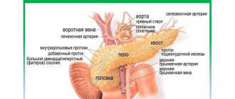
Rice. 2. Scheme of the parotid gland bed (horizontal section): 1 - skin; 2 - subcutaneous tissue; 3 - superficial layer of the fascia of the parotid gland; 4 - chewing muscle; 5 - lower jaw; 6 - medial pterygoid muscle; 7 - wall of the pharynx; 8 - deep layer of fascia of the parotid gland; 9 - styloid process; 10 - internal carotid artery; 11 - internal jugular vein; 12 - digastric muscle; 13 - sternocleidomastoid muscle. Rice. 3. Bed of the parotid gland: 1 - temporomandibular joint; 2 - external carotid artery; 3 - chewing muscle; 4 - septum between the parotid gland and the submandibular gland; 5 - submandibular gland; 6 - digastric muscle; 7 - sternocleidomastoid muscle; 8 - styloid process; 9 — wall of the external auditory canal; 10 - external auditory opening.
In front, the parotid gland is adjacent to the masticatory muscle (m. masseter), the branch of the lower jaw (r. mandibulae) and the medial pterygoid muscle (m. pterygoideus med.); at the back it borders with the sternocleidomastoid muscle (m. sternocleidomastoideus), the posterior belly of the digastric muscle (venter post m. digastrici) and the mastoid process (processus mastoideus); medially adjacent to the styloid process (processus styloideus) and the stylohyoid (m. stylohyoideus) and styloglossus (m. styloglossus) muscles, the internal carotid artery (a. carotis int.) and the internal jugular vein (v. jugularis) extending from it int.), hypoglossal nerve (n. hypoglossus) and peripharyngeal tissue; from above it adjoins the zygomatic arch (areus zygomaticus) and the external auditory canal (porus acusticus ext.). These formations limit the bed of the liver. (Fig. 2), which is lined by the fascia of the liver. (fascia parotidea). Fascia O. g. fused with the fascia of the surrounding muscles and attached to the edge of the lower jaw, zygomatic arch, mastoid and styloid processes. Between the angle of the lower jaw and the sternocleidomastoid muscle, the fascia forms a dense septum (Fig. 3), separating the O. from the submandibular gland (submandibular gland, T.; gl. submandibularis).
Rice. 4. Topography of the parotid gland: 1 - auriculotemporal nerve; 2 - superficial temporal arteries and vein; 3 - zygomatic arch; 4 - temporal branch of the facial nerve; 5 - zygomatic branch of the facial nerve; 6 - parotid duct; 7 - buccal branches of the facial nerve; 8 - facial arteries and vein; 9 - chewing muscle; 10 - marginal branch of the facial nerve; 11 - submandibular gland; 12 - cervical branch of the facial nerve; 13 - external carotid artery; 14 - subcutaneous muscle of the neck; 15 - internal jugular vein; 16 - sternocleidomastoid muscle; 17 - external jugular vein; 18 - v. retromandibularis; 19 - parotid gland; 20 - maxillary artery; 21— parotid plexus of the facial nerve; 22 - facial nerve; 23 - auricle; 24 - accessory parotid gland.
Through the thickness of O. zh. large vessels and nerves pass through; external carotid artery (a. carotis ext.) with the maxillary (a. maxillaris) and superficial temporal arteries (a. temporalis superficialis) branching off from it, v. retromandibularis, nerves auriculotemporal (n. auriculotemporalis) and facial (n. facialis). The facial nerve (see) forms the parotid plexus (plexus parotideus) in the thickness of the gland, the branches of which, leaving the gland, fan out to the facial muscles (Fig. 4). This determines the radial direction of the incisions of the gland during operations.
The system of excretory ducts of the gland is represented by intralobular, interlobular and interlobar ducts, which merge into the common parotid duct (ductus parotideus), or Stenon duct, which was first described by the Danish scientist N. Stenon in 1661. Length of the parotid duct 40-70 mm, its dia. 3-5 mm. The parotid duct usually originates from the upper third of the gland, bends around the edge of the masseter muscle and the fatty body of the cheek (corpus adiposum buccae) and opens into the vestibule of the mouth at the level of the upper second molar. In this place on the mucous membrane of the cheek there is a papilla of the liver. (papilla parotidea). According to S. N. Kasatkin (1948), in 44% of cases the parotid duct is ascending, in 23% - descending, less common are straight, cranked, arcuate (Fig. 1), S-shaped and bifurcated parotid duct. In half of the cases, the duct of the accessory parotid gland (glandula parotis accessoria) flows into it. Sometimes a blind canaliculus, the so-called, departs from the parotid duct near its mouth. Shievich's organ is a rudimentary salivary duct. The parotid duct contains valves and terminal siphons that regulate the excretion of saliva.
Blood supply is provided by the branches of the external carotid artery, superficial temporal artery, transverse artery of the face (a. transversa faciei), posterior and deep auricular arteries (aa. auriculares post, et profunda). Intraorgan arteries and veins pass through the interlobular septa. Venous drainage occurs into the pterygoid plexus (plexus pterygoideus) and the mandibular vein.
Lymphatic vessels of the lake. drain into the superficial and deep parotid lymph nodes (nodi lymphatici parotidei superficiales et profundi); their efferent vessels go to the superficial and deep cervical lymph nodes (nodi lymphatici cervicales superficiales et profundi).
Innervation is carried out by sympathetic and parasympathetic nerves. Preganglionic sympathetic fibers originate in the gray matter of the upper thoracic segments of the spinal cord and are interrupted in the superior cervical ganglion (gangl, cervicale sup.). Postganglionic sympathetic fibers go to the lake. as part of the external carotid plexus (plexus caroticus ext.). Sympathetic nerves constrict blood vessels and inhibit the secretion of saliva. The gland receives parasympathetic innervation from the lower salivary nucleus (nucleus salivatorius inf.) of the glossopharyngeus nerve. Preganglionic fibers go as part of this nerve and its branches (n. tympanicus, n. petrosus minor) to the ear ganglion (gangl, oticum). Postganglionic fibers reach the gland along the branches of the auriculotemporal nerve. Parasympathetic fibers stimulate secretion and dilate blood vessels.
X-ray anatomy
Sialography of the Parotid gland (see Sialography) in the anterior direct projection allows you to detect the shadow of the gland. outward from the branch of the lower jaw. In the lateral projection of the lake. projected onto the ramus of the mandible and the fossa retromandibularis area. The parotid duct on the sialogram in the lateral projection runs in an oblique direction from back to front and upward. Its intraglandular part crosses the posterior edge of the ramus of the lower jaw at the border of its middle and lower thirds or lies on the angle of the lower jaw. At the level of the anterior edge of the mandibular ramus, the duct emerges from the gland, and its extraglandular part is located at the base of the coronoid process of the mandible. Interlobar ducts of the lake. They merge into the parotid duct at different angles and have different numbers of branches. Depending on this, S. N. Kasatkin (1948) divides the glands into many branched, moderately branched and few branched.
Theory and practical experience in ultrasound diagnosis of salivary gland pathology
Ultrasound scanner WS80
An ideal tool for prenatal research.
Unique image quality and a full range of diagnostic programs for an expert assessment of a woman’s health.
In domestic and foreign literature there are many works devoted to sialogy (from the Greek Sialon - saliva and logos - study) - the science of diseases and injuries of the salivary glands, methods of their diagnosis and treatment. According to various authors, diseases of the salivary glands account for up to 24% of all dental pathologies. Currently, in clinical practice, the most common are dystrophic, inflammatory diseases of the salivary glands (sialoadenoses, sialadenitis), as well as tumors and congenital malformations of the salivary glands. In addition, pathological changes in the salivary glands often accompany other diseases (diabetes mellitus, bronchiectasis, sarcoidosis, liver cirrhosis, hypertriglyceridemia, lymphogranulomatosis, etc.).
Various instrumental methods are used to diagnose diseases of the salivary glands [1]:
- radiography (if the formation of stones in the ducts of the salivary glands is suspected, but in 20% of the stones of the submandibular salivary glands and 80% of the parotid salivary glands are non-radiographically opaque);
- sialography (examination of the ducts of the salivary glands with a radiopaque substance, is rarely useful in differentiating tumors from inflammatory processes, but it can help differentiate the mass formation of the salivary glands from formations in neighboring tissues. In patients with suspected autoimmune disease of the salivary glands, a characteristic pattern of saccular expansion may be detected ductal system. In case of acute infection of the salivary glands, sialography should not be performed [2]);
- computed tomography together with sialography;
- ultrasound method (is the most accessible, safe and informative in the process of differential diagnosis of the pathological condition of the salivary glands).
Anatomy of the salivary glands [3]
There are three pairs of major salivary glands (SG) and many small ones. The large ones include paired parotid, submandibular and sublingual SGs. The parotid salivary gland (PSG) is located on the outer surface of the branch of the lower jaw at the anterior edge of the sternocleidomastoid muscle, as well as in the retromandibular fossa. Dimensions vary widely: length 48-86 mm, width 42-74 mm, thickness 22-45 mm. The OSJ is covered by the parotid fascia, which is its capsule and is tightly fused with it. Sometimes, at the anterior edge of the parotid duct, there is an additional lobule measuring 10-20 mm, which has its own duct flowing into the parotid. The parotid duct emerges from the gland at the border of its upper and middle thirds, then it passes along the outer surface of the masticatory muscle parallel to the zygomatic arch and turns 90° inward, penetrating the fatty tissue and buccal muscle. The projection of the parotid duct onto the skin of the cheek is determined on the line connecting the tragus of the auricle and the corner of the mouth. The parotid duct opens in the vestibule of the oral cavity at the level of 1-2 large molars. The diameter of the duct is on average 1.5-3.0 mm, its length is 15-40 mm. The thickness of the gland contains the branches of the external carotid artery, the facial nerve and its branches, and the auriculotemporal nerve. There are many lymph nodes around the OUSG and in its parenchyma (Fig. 1), which can serve as a primary or secondary collector for draining lymph from teeth and oral tissues.
Rice. 1.
Lymph nodes in the thickness of the parotid salivary gland.
The submandibular salivary gland (MSG) is located in the submandibular triangle between the body of the mandible and the anterior and posterior bellies of the digastric muscle. The dimensions of the gland are: anteroposterior 20-40 mm, lateral 8-23 mm, superior-inferior 13-37 mm. Posteriorly, the PNJ is separated from the OSJ by a process of the fascia propria of the neck. The medial surface of the gland in the anterior section lies on the mylohyoid muscle. The submandibular duct, bending over the posterior edge of this muscle, is located on the lateral surface of the hyoglossus muscle. Then it goes between the medial surface of the hyoid gland and the genioglossus muscle to the point of its exit in the area of the hyoid papilla. The facial artery and its branches, the lingual artery and the veins of the same name pass through the gland.
The sublingual salivary gland (SSG) is located on the floor of the mouth in the sublingual region parallel to the body of the lower jaw. The dimensions of the gland are: longitudinal 15-30 mm, transverse 4-10 mm and vertical 8-12 mm. The duct of the parathyroid gland passes along its inner surface and opens in the region of the anterior section of the sublingual ridge independently or together with the submandibular duct. Sometimes the PJS duct flows into the middle section of the PJS duct.
The minor salivary glands - labial, buccal, lingual, palatine, incisive - are located in the corresponding areas of the mucous membrane. They can be a source of development of adenocarcinomas of the oral cavity.
Pathology of the salivary glands
SG malformations are rare. The most common are anomalies in the size of the glands (agenesis and aplasia, congenital hyperplasia (Fig. 2) and hypoplasia), their location (heterotopia, accessory glands), and anomalies of the excretory ducts (atresia, stenosis, ectasia, cystic transformation, ductal dystopia).
Rice. 2.
Hyperplasia of the left sublingual salivary gland.
Sialadenitis is a large group of polyetiological inflammatory diseases of the gastrointestinal tract (Fig. 3). Primary sialadenitis - sialadenitis considered as an independent disease (for example, mumps). Secondary sialadenitis is sialadenitis that is a complication or manifestation of other diseases (for example, sialadenitis with influenza). The echographic picture for different etiologies is not very specific. Etiology has clinical significance in determining treatment tactics.
Rice. 3.
Sialadenitis of the right submandibular salivary gland.
According to the etiological factor, sialadenitis is classified [4] into:
- sialadenitis developing under the influence of physical factors (traumatic sialadenitis, radiation sialadenitis (Fig. 4) occurs during radiation therapy of malignant tumors of the head and neck);
- sialadenitis developing under the influence of chemical factors (toxic sialadenitis);
- infectious sialadenitis (routes of infection penetration into the fluid: stomatogenic (through ducts), contact, hematogenous and lymphogenous);
- allergic and autoimmune sialadenitis (recurrent allergic, Sjogren's disease and syndrome, etc.);
- myoepithelial sialadenitis caused by a pathological process, previously designated as a benign lymphoepithelial lesion. The term benign lymphoepithelial lesion was first used by JT Godwin in 1952, replacing the concept of Mikulicz disease;
- obstructive sialadenitis, which develops when there is difficulty in the outflow of saliva due to obstruction of the excretory duct with a stone (Fig. 5-7) or thickened secretion, as well as due to cicatricial stenosis of the duct. According to the prevalence of the process, they distinguish between focal, diffuse sialadenitis and sialodochitis - inflammation of the excretory duct. The course of the process can be acute or chronic;
- pneumosialadenitis, which develops when there is air in the gastric tissue in the absence of a bacterial gas-forming infection. Air enters the gland from the oral cavity when the pressure there increases through the duct. Pneumosialadenitis is typical for a number of professions, primarily for glassblowers and musicians playing wind instruments.
Rice. 4.
Post-radiation sialadenitis.
Rice. 5.
Stone of the duct of the submandibular salivary gland.
Rice. 6.
Stone in the parenchyma of the submandibular salivary gland.
Rice. 7.
Stone in the duct of the submandibular salivary gland.
Tumors of the salivary glands
Tumors of the salivary glands are divided into two groups: epithelial and non-epithelial. Epithelial tumors predominate in adults (95%). In children with SG, epithelial and non-epithelial tumors are equally common. In addition to true tumors, processes resembling tumors (tumor-like lesions) develop in the GS.
Among epithelial tumors of the gastrointestinal tract, benign neoplasms are distinguished, as well as malignant ones - carcinomas.
Benign epithelial neoplasms of the stomach include ductal papillomas, adenomas and benign sialoblastoma. SG adenomas are divided into two groups: polymorphic (the most common SG adenoma) and monomorphic (all other) adenomas. Tumors of different structure, origin and prognosis were artificially included in the group of monomorphic adenomas.
Pleomorphic (polymorphic) adenoma (mixed tumor of the gland) is a adenoma of the gland, built from two types of cells: ductal epithelium and myoepithelial cells. Macromorphological picture. The tumor is usually an elastic or firm nodule of lobulated grayish-white tissue, usually partially encapsulated. Typical of a pleomorphic adenoma is the so-called chondroid stroma, resembling hyaline cartilage. Variants of the echographic image of pleomorphic adenomas are presented in Figure 8.
Rice. 8.
Pleomorphic adenoma of the gastrointestinal tract.
Warthin's tumor is an adenolymphoma in which multiple cystic cavities are formed, covered with double-layered epithelium. The papillae protrude into the lumen of the cysts. A pronounced proliferation of lymphoid tissue occurs in the tumor stroma. This tumor almost exclusively develops in the parotid gland.
Other types of benign tumors are less common. These are benign oncocytoma (oxyphilic adenoma), basal cell adenoma, tubular adenoma, benign cystadenoma sialoblastoma.
Among benign primary non-epithelial tumors, the most common are hemangioma, lymphangioma, neurofibroma and lipoma.
Among malignant non-epithelial tumors, malignant lymphomas are more often found (they arise, as a rule, against the background of myoepithelial sialadenitis, Sjögren's disease and syndrome).
Tumor-like lesions of the salivary glands
Rice. 9.
Salivary gland cysts.
- Salivary gland cysts (mucoceles). There are two types of mucocele of the gland: the retention type (retention cyst of the small gland, formed when saliva is retained in the excretory duct) and the type of interstitial secretion, when, when the wall of the duct is injured, saliva enters directly into the fibrous tissue surrounding the gland. Mucoceles in the floor of the mouth are also called ranulae.
- Cysts of the excretory ducts of large SGs are pronounced dilatation of the excretory duct due to retention of secretions in it. Blockage of salivary outflow can be caused by various reasons: tumor, stone, thickened mucus, post-inflammatory stenosis, even cicatricial obliteration of the lumen.
- Sialoadenosis (sialosis) is a non-tumor and non-inflammatory symmetrical increase in SF due to hyperplasia and hypertrophy of secretory cells. The outcome of sialosis is often SG lipomatosis. The process has a chronic relapsing course. Sialosis occurs in a number of diseases and conditions: diabetes mellitus, hypothyroidism, malnutrition, alcoholism, liver cirrhosis, hormonal disorders (hypoestrogenemia), reactions to medications (most often antihypertensive), neurological disorders.
Adenomatoid hyperplasia of small SGs leads to their increase to 0.5-3.0 cm in diameter. The causes of adenomatoid hyperplasia are trauma and prolonged exposure to ionizing radiation.
Oncocytosis is age-related changes in secretory cells and epithelium of the ducts of the gastrointestinal tract. In this case, the SF may slightly increase, but usually their value does not change.
To summarize, I would like to note that ultrasound using Doppler sonography in many of our observations helped to accurately determine the nature of the pathological process in the gastrointestinal tract. However, this diagnostic method does not allow one to unambiguously confirm or refute the malignant nature of the formation of the salivary glands.
Literature
- Benign and malignant tumors of soft tissues and bones of the face. A.G. Shargorodsky, N.F. Rutsky. M.: GOU VUNMTs, 1999.
- Topographic anatomy and operative surgery. I.I. Kagan, S.V. Chemezov. M.: GEOTAR-Media, 2011.
- Salivary glands. Diseases and injuries. V.V. Afanasiev. M.: GEOTAR-Media, 2012.
- Inflammatory diseases of the tissues of the maxillofacial area and neck. A.G. Shargorodsky. M.: GOU VUNMTs, 2001.
Ultrasound scanner WS80
An ideal tool for prenatal research.
Unique image quality and a full range of diagnostic programs for an expert assessment of a woman’s health.
Histology
The parotid gland has a lobular structure. Connective tissue septa extending from the fascia of the stomach divide its parenchyma into lobules, which in turn form 5-7 larger lobes.
Rice. 5. Schematic representation of the histological structure of the parotid gland. A - at a magnification of 200 times: 1 - intercalary duct; 2 - striated duct; 3 - terminal secretory department; 4—fat cell; 5 - interlobular duct; 6 - interlobular septum; 7 - blood vessel; hematoxylin-eosin staining; B - with a magnification of 600 times: 1 - fat cell; 2 - serocytes of the terminal secretory section; 3 - striated duct; 4 — myoepitheliocytes; 5 - intercalary duct; hematoxylin-eosin staining.
O.J. is a complex alveolar gland (see Glands). Its lobules are formed from terminal secretory sections tightly adjacent to each other - acini and excretory intralobular ducts: intercalated and striated (Fig. 5).
The terminal secretory sections are formed by glandular epithelial cells - serocytes, located on the basement membrane, consisting of a dense network of reticular fibers. The serocyte has a pyramidal shape, its cytoplasm contains a basally located rounded nucleus, a well-developed endoplasmic reticulum with numerous cisterns, a lamellar complex and secretory granules of various types.
In the terminal secretory sections of the lake. secretory tubules are distinguished, which are located between serocytes and have the appearance of thin tubes without their own walls.
Intercellular secretory tubules are characteristic of glands with protein secretion. The apical pole of serocytes, as well as their surface facing the intercellular secretory tubules, are covered with microvilli.
The terminal secretory sections pass further into narrow intercalary ducts lined with cuboidal epithelium, and then into striated ducts, or salivary tubes, lined with single-layer columnar epithelium. The characteristic basal striation of the epithelium of striated ducts depends on the accumulation of mitochondria lying between the folds of the plasmalemma of the basal part of the cell. It is believed that the striated ducts play a role in changing the concentration of primary saliva.
Between the cells and the basement membrane of the terminal secretory sections, intercalary and striated ducts there are myoepithelial cells, or basket cells, which are contractile elements that contribute to the secretion of secretions and maintaining the tone of the gland ducts.
Intralobular ducts pass into interlobular ducts, which run in layers of connective tissue between the lobules of the gland, merge with each other, forming interlobular ducts and, finally, the parotid duct. Interlobular ducts of the lake. lined with double-row prismatic epithelium, which, as the excretory ducts thicken, becomes multilayered. Along the parotid duct the epithelium is multilayered cubic, and at the mouth of the duct it is multilayered squamous.
Physiology
As an organ of the digestive system, the parotid gland, together with other salivary glands, secretes saliva into the oral cavity; its edges have a complex composition and perform various functions (see Saliva), exocrine (salivary) function of the o. performs starting from the 4th month of embryonic development. In this period, according to E. Sh. Gerlovin (1963), the secretion of O. zh. is mucous in nature, at the end of the 2nd year of life it becomes serous. The amount of saliva secreted is not constant and depends on the state of the internal environment of the body, the type and smell of food, the nature of irritation of specific receptors of the oral mucosa, etc. (see Salivation).
Cells of O. perform an excretory function, accumulating and removing various medicinal substances, poisons, toxins, and, in patients with diabetes, sugar from the body with saliva.
There is evidence indicating the endocrine function of the lake. Thus, biologically active substances (parotin, nerve growth factor, epithelial growth factor) were extracted from gland cells. Ito (I. Ito, I960) found that parotin has the properties of a hormone and affects protein and mineral metabolism. From O. zh. insulin-like protein is isolated. A histofunctional connection was revealed. with the reproductive, parathyroid, thyroid, pancreas, pituitary and adrenal glands.
Research methods
When identifying pathology of the Parotid gland, questioning and examining the patient, palpation of the liver are of great importance, which allow us to make an assumption about a particular disease of the bladder. (inflammation, tumor, damage, etc.).
Laboratory, instrumental, and X-ray radiological methods play a significant role in clarifying the diagnosis. research.
Probing of the parotid duct allows one to determine its patency and the presence of dense foreign bodies in it.
Cytological examination of the secretion of the gland, as well as puncture biopsy with histol, examination of organ tissues help to identify morphological changes in the gland, in particular in the presence of a tumor.
Secretory function of the lake. studied using sialometry (measurement of the amount of saliva released per unit of time), as well as radioisotope methods—radiosialography and radiosialometry, based on the ability of the parenchyma of the liver. concentrate and release radioactive isotopes 131I, 99Tc with saliva.
To determine foreign bodies and morphol, changes in the structure of the ducts and parenchyma of the lake. (chron, inflammation, tumor) X-rays of the gland are performed without contrast and with contrast of the ducts (see Sialography).
Layer-by-layer images of the organ are obtained using tomography (see), and the use of panoramic tomography (see Pantomography) makes it possible to simultaneously examine and compare the right and left O.
Ultrasonic dowsing (see Ultrasound diagnostics) is a method for diagnosing tumor processes in the liver. and, in addition, allows one to judge the size of the gland and the degree of sclerosis of its parenchyma.
Scanning of the body. using 99Tc (see Scanning) allows you to visualize the parenchyma of the gland, identify the localization of its non-functioning areas, which is also an indirect sign of a violation of its function.
Thermal imaging (see Thermography) is carried out to measure the temperature in the tissues of the body. An increase in the cut is a sign of acute inflammation, a malignant tumor, Sjögren's syndrome, and a decrease is a sign of a benign tumor, cystic formations, certain forms of hron, mumps and etc.
Pathology
Rice.
2. A patient with chronic sialadenitis of the left submandibular gland: the contours of an enlarged submandibular gland are visible in the submandibular region. The main symptoms indicating a pathological process in the submandibular gland are dysfunction (decrease, increase, delay, as well as a qualitative change in the secretion released from the duct), an increase in the size of the gland (Fig. 2). With a decrease in secretion, patients feel dryness in the oral cavity (see Xerostomia); the delay in secretion is manifested by the so-called salivary colic (tingling, bursting pain in the area of the gland that appears during eating), with increased secretion, salivation occurs (see Salivation).
Damage
Submandibular glands are rarely observed. According to E. E. Babitskaya, during the Great Patriotic War, damage to the pancreas. with the formation of external salivary fistulas (see) was observed in 2% of cases of injuries to all salivary glands. There are cases of damage to the submandibular duct by a disc during the preparation of teeth for crowns. Wound of P. and its duct can lead to the formation of a parenchymal defect, stenosis or complete obliteration of the submandibular duct. A sign of impaired patency of the submandibular duct is swelling of the pancreas, especially during meals. The localization and degree of damage to the gland or its excretory duct is established using sialography (see). To eliminate narrowing of the duct, bougienage with a special probe is used. Obstruction of the duct in the anterior sections is eliminated surgically. In this case, the central end of the duct is prepared, split longitudinally over 1-1.5 cm from the site of fusion and sutured to the mucous membrane of the sublingual region.
Reactive-dystrophic changes
P.J. occur with certain diseases: Mikulicz's disease (see Mikulicz syndrome), Sjögren's syndrome (see Sjögren's syndrome), damage to the endocrine glands, collagen diseases (see), etc.
Acute inflammatory diseases of the pancreas. (see Sialadenitis) occur when they penetrate into the duct of the pancreas. a foreign body, for example, fruit peels, blades of grass, with phlegmon of the submandibular and sublingual areas (due to the spread of the process from surrounding tissues), with an atypical course of mumps (see Epidemic mumps). In these cases, the disease is acute, with a deterioration in general health, an increase in body temperature, discharge of pus from the duct, pain and swelling of the pancreas; purulent melting of the pancreas is not excluded. In the complex to treat. procedures for acute sialadenitis include the introduction of drugs (antibiotics, bacteriophage, proteolytic enzymes) into the ducts of the gland, novocaine blockade of the gland area. In case of viral infection, it is recommended to irrigate the oral mucosa with interferon.
Rice. 3. Sialogram of the left submandibular gland with chronic sialodochitis: the ducts of the gland are unevenly dilated, their contours are clear (indicated by arrows).
Chronic inflammation of the submandibular gland mainly occurs as calculous sialadenitis (see Sialolithiasis). Less common is non-calculous hron, sialadenitis: interstitial (the so-called inflammatory tumor of Küttner or damage to the pancreas in Mikulicz's disease) and parenchymal (in Sjögren's syndrome). Sialodohit P. zh. It is less common than the parotid gland and can be bilateral. Patients complain of periodically appearing swelling in the submandibular region, tingling, bursting pain in the gland area during eating, and the release of salty saliva into the oral cavity. The disease lasts for years with periodic exacerbations, during which severe pain and purulent discharge from the duct are observed. For the diagnosis of chronic sialodochitis of the pancreas. sialography (Fig. 3) or pantomosialography is used. For calculous sialadenitis, treatment is surgical (see Sialolithiasis). For non-calculous sialadenitis and sialodochitis, treatment is aimed at increasing the body's resistance, as well as eliminating concomitant diseases.
Specific inflammatory diseases of the pancreas. (tuberculosis, syphilis and actinomycosis) occur relatively rarely (see Syphilis, Tuberculosis, Actinomycosis).
Relatively rare in P. There are retention cysts that arise as a result of atresia of the salivary ducts. Salivary duct atresia can be congenital or occur as a result of injury or inflammation. Surgical treatment: the cyst is removed along with the pancreas.
Damage to the submandibular gland by the tumor process is observed much less frequently than the parotid gland (see Salivary glands).