Osgood-Schlatter disease can present as a painful lump in the area below the kneecap during childhood and adolescence as puberty begins. Osgood-Schlatter disease occurs most often in children who participate in sports, especially sports such as running, jumping, or sports that require rapid changes in movement trajectories, such as football, basketball, figure skating and gymnastics.
And although Osgood-Schlatter disease is more common in boys, the gender gap narrows as girls become more involved in sports. Osgood-Schlatter disease affects more adolescents who play sports (by a ratio of one to five). The age range of incidence has a gender factor, as girls experience puberty earlier than boys. Osgood-Schlatter disease usually occurs in boys between 13 and 14 years of age and in girls between 11 and 12 years of age. The disease usually goes away on its own as bone growth stops.
General information
Osgood-Schlatter disease is a specific disease of the musculoskeletal system, namely the knee joints, characterized by dystrophic damage to the tibia in the area of its tuberosity.
Such aseptic destruction of bone tissue occurs against the background of permanent or acute trauma and usually affects only young people at the stage of intensive skeletal development. Clinically, the disease is manifested by swelling of the knee joint, the formation of a kind of growth (bump) under it and pain in its lower part, which occurs during normal physical activity (running, squats, etc.) or even without it.
This pathology was first described in 1878 by the French surgeon O. M. Lannelong under the name “Apophysitis of the tibia,” and in 1903, thanks to the work of the American orthopedist R. B. Osgood and similar works of the Swiss surgeon K. Schlatter (Schlatter), it appeared its more detailed nosography. Wikipedia defines this painful condition with the term “Osteochondropathy of the tibial tuberosity,” and the international classification assigned it the ICD-10 code – M92.5 “Juvenile osteochondrosis of the tibia and fibula.” Despite this, in medical practice this disease is still most often referred to as “Osgood-Schlatter disease” or simply “Schlatter disease”.
Children
Due to insufficient development of the large, medially located shin bone in a child under eighteen years of age. Most of the motor impact is transmitted to the ligamentous apparatus of the knee. This distribution of pressure on the joint provokes a failure of the nutritional function of the tibia under the knee.
Schlatter's disease in children involves changes in the undulation of the tibia. The indicated piece of bone under the knee is localized. A characteristic anatomical formation is responsible for attachment to the patella. The roughness of the bone is located near the apophysis (the place that ensures the development of the musculoskeletal system). This factor influences the development of pathology.
Active growth in children
The bone process near the epiphysis has its own capillaries. They are responsible for providing oxygen and nutrients to the growth zone. Active growth in children causes the blood vessels to not keep pace with the growth of bone mass. This is manifested by a lack of nutrients and hypoxia. The consequence of this process is considered to be an increased susceptibility of the bone area to damage. He becomes very fragile.
The body fights the disease
The body tries to cope with the disease on its own. It seems to be patching up a defect, which manifests itself in the formation of a lump under the knee. Usually only one limb is affected. But there are situations when bumps appear under the knees of both limbs.
A disorder of the blood supply to the tibia roughness zone is accompanied by the following disorders:
- Necrosis of the roughness of the tibia;
- inflammation of an aseptic nature in the area of the bags;
- minor accumulations of blood;
- separation of the ligaments of the knee joint.
Pathology was first described by Osgood-Schlatter. This happened at the beginning of the twentieth century. The disease described was named after him. Doctors can also use another term for the disease in question - osteochondropathy of the tibial tuberosity. It can be used by employees of traumatology and orthopedics departments. Therefore, we can conclude that the pathology belongs to the group of osteochondropathy. The disease has a non-inflammatory genesis. At the same time, necrosis of bone tissue is observed.
Pathogenesis
The mechanism of occurrence and further development of Osgood-Schlatter syndrome is directly related to the patient’s age and physical activity. According to statistics, in the vast majority of cases, doctors diagnose Schlatter's disease in children and adolescents in the age group from 10 to 18 years, while young people involved in sports suffer from it 5 times more often than their peers leading a passive lifestyle . The same reason for more intense physical activity explains the fact that this osteochondropathy mainly affects boys.
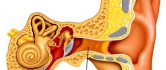
As is known, two large bones are involved in the formation of the human knee joint - the femur (above the knee) and the tibia (below the knee). In the upper part of the last of them there is a special area (tuberosity), to which the quadriceps femoris muscle is attached by means of a tendon. It is this part of the bone that is responsible for its growth in childhood and adolescence and is therefore particularly susceptible to various injuries and damage. During active physical activity, in some cases, the knee joint is subject to a large load and the quadriceps muscle is overstrained, which leads to stretching or tearing of the tendon and a lack of blood supply in this area. As a result of such a traumatic effect and a decrease in nutrition in the area of the tibial tuberosity, gradual necrotic changes develop in it, up to the death of individual parts of its core.
In addition, any injury to the knee joint or constant impact on its musculoskeletal structure (for example, jumping) can cause cracks and microfractures of the tibial tuberosity, which the growing body tries to quickly compensate for by the growth of new connective tissue. As a result of this, a person develops a bone growth (bump), typical of Osgood-Schlatter osteochondropathy, that forms just below the knee. Such a pathological process usually involves one leg, but bilateral involvement of the lower extremities is also possible.
Military service with Schlatter's disease
As you know, men in our country are drafted into the army at the age of 18. At this age, symptoms of this disease are not observed. Consequently, this pathology cannot become a reason for the release of young people from military duties.
In some cases, it is possible to delay conscription for medical reasons - due to the need for a medical course. A young man can be released from military service only in a situation where the pathological process has provoked a significant deterioration in the motor activity of the joint.
Classification
In the orthopedic environment, this pathology is usually classified according to the degree of its severity and the severity of the observed external and internal symptoms. Regarding this, there are three degrees of Schlatter’s disease, namely:
- initial – visual manifestations in the form of a lump-like growth under the knee are absent or minimal, pain in the area of the knee joint is episodic, mild and occurs mainly at the time of physical activity on the leg;
- an increase in symptoms - swelling of the soft tissues around the affected knee appears, a lump becomes visually visible directly below it, pain syndrome manifests itself during the period of loads on the leg and for a certain period of time after them;
- chronic - a lump-like formation is clearly visible under the knee, which is most often surrounded by swelling, discomfort and pain in the joint is persistent and is observed even at rest.
Nature of pain
The duration of pain in the initial stages of the disease is short or may not be noticeable at all. Over time, the duration increases.
It always appears after exercise. In the beginning, full training time is enough for pain to appear. In the last stages, pain appears already during warm-up.
The location is always limited to the tuberosity zone. Pain does not spread to the hip or ankle joint.
The intensity of pain depends on the extent and severity of the process. Mild pain is observed in the early stages and does not create any particular inconvenience. Severe pain is noted at a late stage. With severe pain, it is sometimes difficult for a person to even step on the sore leg.
Causes
There are two main physical activity-related underlying causes of Osgood-Schlatter disease in adolescents and children:
- direct injuries to the tissues of the knee joint (subluxations and dislocations, sprains, bruises, fractures);
- systematic microtraumas (external and internal) of the knee joint that occur as a result of intense sports or other activities associated with excessive physical stress on the lower extremities.
The greatest risk factors for Schlatter's disease in adolescents and children are:
- football, basketball, handball, hockey, volleyball, tennis;
- track and field athletics, acrobatics, gymnastics;
- judo, kickboxing, sambo;
- skiing, sports tourism, figure skating, cycling;
- ballet, sports and ballroom dancing.
Surgery
Often, the disease does not require treatment at the initial stage, since it regresses on its own over a certain period of time. To do this, you just need to adhere to certain conditions. The main task is to prevent stress and damage to the limb. But if the disease reduces the quality of life, impairs the functioning of the legs and causes pain, then the patient is prescribed complex therapy.
In most cases, as indicated by information from the forums, the disease can be dealt with through conservative treatment at home, which includes taking medications and physical therapy. You need to get rid of the disease by taking certain medications and following the doctor's instructions. Do not forget about the regime, diet, taping, use of orthopedic insoles and exclusion from sports. All these methods are aimed at preventing the development of the disease and its complete elimination.
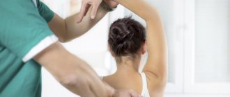
Conservative treatment includes massage and other physical therapy methods
To eliminate symptoms, therapeutic massage, gymnastics and exercise therapy, electrophoresis, ointments and even essential oils are used. Developing the limb will speed up recovery.
Unfortunately, there are cases when conservative treatment does not give the desired result. In this case, doctors recommend an operation aimed at eliminating symptoms and restoring normal function of the limb. Indications for the procedure are as follows:
- the disease develops over 2 years or more;
- conservative treatment, which was carried out for 9 months, did not give the desired result;
- the degree of damage is very high - the presence of bone fragmentation and ligament separation;
- At the time of diagnosis of the disease, the patient was already 18 years old.
When performing an operation, specialists adhere to the basic principle: to achieve high results with minimal damage. In this way, the surgeon eliminates the necrotic focus and sutures the fixing tuberosity of the bone graft. After this, the traumatologist applies a pressure bandage on the left or right leg. The patient must wear such a knee brace for one month. However, it is not recommended to exercise.
After the operation, the patient undergoes a rehabilitation period, during which special exercises and medications are prescribed. This allows for faster pain relief and recovery of the limb after the intervention.
https://youtube.com/watch?v=6p3xX4DyC5s
Symptoms of Osgood-Schlatter disease
The severity of the negative manifestations of this pathology in different patients may differ depending on the nature of the injuries received, the degree of physical activity and the personal characteristics of the body.
At the beginning of the development of the disease, the patient begins to experience vague pain in the knee area, which usually appears after or during physical activity on the affected limb. As a rule, such pain is not yet associated with an internal pathological process and therefore there are quite few visits to the doctor during this period.
Over time, pain symptoms begin to increase, are localized in one place and can appear not only during physical activity, but also at rest. At the same time, swelling caused by edema appears around the affected knee, and a lump-like growth appears just below it. During this period of illness, it becomes increasingly difficult for the patient (especially the athlete) to perform his usual exercises, and sometimes even natural leg movements. The greatest intensity of pain is observed in the body position - kneeling.
Photo of a “bump” in Osgood-Schlatter disease
In addition, the patient may experience other negative symptoms:
- tension in the leg muscles (mainly the thigh muscles);
- limited mobility of the knee joint;
- outbreaks of sharp “shooting” pain in the knee area, arising when it is overstrained;
- severe morning swelling in the upper or lower part of the knee, which forms the day after physical activity.
When you independently palpate the affected knee, points of pain are felt, as well as smoothness of the contours of the tibia. The texture of the knee joint is felt as densely elastic, and a hard lump-like formation is felt under the swollen soft tissues. The general well-being of the patient, despite the accompanying pain and pathological processes in the knee, does not change significantly. The skin over the affected joint does not turn red, temperature indicators remain normal.
In most clinical cases, this disease occurs in a measured chronic form, but sometimes its wave-like course can be observed with periods of sudden exacerbation and relative calm. Without medical intervention and with continued physical activity, negative symptoms can persist for many months and worsen against the background of further mechanical damage to the knee joint. However, the manifestations of the disease gradually disappear on their own over 1-2 years, and by the time the period of bone tissue growth ends (approximately 17-19 years) they usually eliminate themselves. Before treating Osgood-Schlatter, the need for such therapy should be comprehensively and individually assessed, since in some cases it may be inappropriate.
Clinic
The leading symptom of this disease is pain, which appears and intensifies during physical activity - running, jumping, cycling, kneeling, going up and down stairs, and hitting a ball (in the phase when the knee extends). Most often, pain intensifies when playing sports such as basketball, volleyball, football and tennis. Clinically, the disease manifests itself as pain localized in the LBD area. In some cases, there may be swelling and hypertrophy, as well as excessive tension in the quadriceps femoris muscle. Characteristics such as local fever and intra-articular edema are not important in this disease. There is rarely hyperthermia in this area, but swelling, tenderness and pain often occur.
Clinical symptoms include the following:
- pain on palpation of the LBD;
- pain in the tuberosity area, which intensifies after physical activity or sports;
- increasing pain when squatting, walking up stairs or jumping.
Tests and diagnostics
In general, the doctor can suspect the development of Schlatter’s disease due to the complexity of the patient’s clinical manifestations and the localization of the pathological process typical for this disease. The gender and age of the patient also play an important role in correct diagnosis, since adults, as a rule, are not exposed to this type of damage. Even through a simple visual examination and the usual collection of anamnesis regarding previous injuries or overloads of the knee joint, an experienced orthopedic traumatologist is able to make the correct diagnosis, but it would be useful to confirm it using some hardware diagnostic methods.
The decisive factor in making an unambiguous diagnosis of Osgood-Schlatter disease in children and adolescents was and remains radiography , which, in order to increase the information content of the pathology, is best carried out over time. To exclude other orthopedic diseases, such an examination of the affected knee joint must be carried out in two projections, namely lateral and direct.
In the initial phase of the development of the disease, X-ray images show a flattening of the tibial tuberosity in its soft part and a rise in the lower edge of the clearing, corresponding to the adipose tissue located in the anterior lobe of the knee joint. The last discrepancy with the norm is caused by an increase in the size of the infrapatellar bursa, which occurs as a result of its aseptic inflammation. There are most often no visible changes in the ossification nucleus itself at this stage of Schlatter’s disease.
X-ray of the knee joint in Osgood-Schlatter disease
As the pathology progresses, the x-ray picture changes for the worse. The photographs show a shift of the ossification nucleus by 2-5 mm upward and forward relative to the standard location of the tuberosity or its fragmentation. In some cases, there may be unevenness of the natural contours and unclear structure of the ossification nucleus, as well as signs of gradual resorption of its parts, but most often it fuses with the main body of the bone with the formation of a bone conglomerate in the form of a spiky protrusion. This “bump”, characteristic of Schlatter’s disease, in the later stages of the disease is especially clearly visible on a lateral radiograph and is clearly palpable during palpation in the area of the tuberosity.
In some atypical cases, it may be necessary to prescribe an MRI , CT and/or ultrasound of the problem knee and adjacent tissues to clarify the expected diagnosis. It is also possible to use a technique such as densitometry , which will provide comprehensive data on the structural state of the bones being studied. Other laboratory diagnostic methods, including PCR studies and blood tests for rheumatoid factor and C-reactive protein, are carried out in order to exclude the possible infectious nature of problems with the knee joint (mainly nonspecific and specific arthritis ).
Differential diagnosis of Osgood-Schlatter syndrome must be carried out with any fractures in the knee joint, bone tuberculosis , patellar tendinitis osteomyelitis , infrapatellar bursitis , Sinding-Larsen-Johanson disease and tumor neoplasms.
Treatment of Schlatter's disease (osteochondropathies)
To cure Schlatter’s disease in a minimum period of time, it is important to provide an integrated approach, which involves the implementation of the following measures:
- Use of various bandages and orthoses;
- Application of special elastic bands in the knee area (kinesiological taping);
- Use of medications;
- The use of therapeutic massage and physiotherapy;
- Regularly performing a special set of exercises to maintain tone in the joint;
- Surgery;
- Methods of traditional medicine.
Fixative for Schlatter's disease
Taking into account the stage of development of the pathological process, the doctor prescribes special fixatives. They have different design features and different stiffness levels:
- Knee braces for the treatment of Schlatter's disease. They are made mainly from wool or other natural fabrics. In addition to the fixing effect, they have a warming effect on the affected area;
- Neoprene fixing device. Has a long service life, securely fixes the knee joint;
- Cut. Unlike the orthopedic devices described above, the orthosis, which is worn on the knee joint with Schlatter's disease, promotes the most effective fixation. This is achieved through the use of side plates that fit securely to the surface of the body;
- Tutor. An orthopedic product that guarantees extremely rigid fixation, comparable only to a plaster splint. The key advantage of such a device is the ability to maintain the number of movements.
Kinesiology taping
Very little time has passed since the advent of this technique. It uses a special tape – kinesio tape. It is glued to the surface of the skin, which allows you to gradually increase the load on the knee joint.
Most often, the tape patch is used by the patient for 2 weeks. Moreover, its use does not have a significant impact on the daily routine and allows you to visit the bathhouse.
Make an appointment Online booking
- Clinic on Krasnopresnenskaya +7 (499) 252-41-35 Volkov lane, 21
- Clinic on Varshavskaya +7 (499) 610-02-09 Varshavskoe highway, 75, building 1
- Clinic in Annino +7 (495) 388-08-08 Varshavskoe highway, 154, building 1
Medications
In case of osteochondropathy, the patient is provided with clinical recommendations, within the framework of which the use of medications in various forms is allowed. In the early stages of the disease, special injections can be used to restore blood circulation and relieve pain. Subsequently, the patient is prescribed medications, as well as creams, gels and ointments.
When forming a treatment plan for the younger generation, in order to determine the one-time and daily dosage, the following are taken into account: the child’s body weight, the intensity of symptoms.
| Group of drugs | Name | Therapeutic effect |
| Nonsteroidal anti-inflammatory drugs | Nimesulide, Diclofenac, Ketoprofen, Ibuprofen, Meloxicam | Reduces pain, relieves swelling in the knee joint |
| Drugs that improve the condition of the cardiovascular system | Eufillin, Trental, Xanthinol nicotinate | Improve blood flow and vascular permeability, have a vasodilating and tonic effect |
| Medicines used to treat osteoarthritis and tendinopathies | Chondroxide, Teraflex, Dona, Structum, Artra | Improves metabolic processes in bone tissue, restores the elasticity of cartilage |
| Vitamin B complexes included | Combilipen, Pentovit, Neuromultivit, | Have a beneficial effect on the peripheral nervous system and normalize metabolic processes |
Gymnastics for knee joints
To achieve the desired healing effect, a set of gymnastic exercises must be performed on a daily basis at strictly prescribed times.
In this case, special attention should be paid to relaxing the thigh muscles and knee ligaments.
Thus, to improve the condition of a patient with Schlatter’s disease, physical therapy should include the following exercises:
- While in an upright position, the patient should lift with bent legs (alternately), pulling the knees to the chest as high as possible. When lowering the limbs, the socks are pulled toward themselves, which provides the necessary tension in the lower leg muscles;
- Perform emphasis on one leg. Next, you need to try to maintain your balance with your eyes closed. Moreover, the second limb should be in a bent position;
- The patient needs to take a “sitting” position and stretch his legs in front of him. Subsequently, alternating movements are performed with the fingertips - away from you, towards you;
- While in the “lying” position, the patient performs movements identical to riding a bicycle.
Each exercise must be repeated at least 10 times.
Massage for Schlatter's disease
It is impossible to cure Schlatter's disease without therapeutic massage. However, such procedures must be performed by specialists with impressive professional experience and capable of correctly deciphering research results. Only in this case can you count on obtaining an effective and prolonged result.
This involves rubbing, kneading and vibration, which are characteristic of traditional massage procedures. This allows you to strengthen muscle tissue and restore blood circulation to the affected area. Acupuncture and vacuum massage are also allowed.
Physiotherapy for Schlatter's disease
Physiotherapeutic techniques that allow you to obtain the best treatment results include: ozokerite-paraffin applications, exposure to therapeutic mud, UHF, MRI and shock wave therapy. In cases of significant pain, electrophoresis with anesthetic drugs is used. And during the recovery period, exposure to electric current should be carried out in combination with solutions of calcium salts.
Shock wave therapy for Schlatter's disease
Shock wave therapy for Schlatter pathology gives a noticeable positive effect in the subacute stage of the disease. At the same time, under the influence of shock waves, it is possible to crush the existing salt formations and normalize blood circulation in the affected area, as well as improve the nutrition process in this area.
At the Health Plus clinic, shock wave therapy is performed using the latest Swiss equipment, which allows you to get the best effect for Schlatter’s disease. At the same time, specialists who know all the intricacies of this destructive process and are able to correctly decipher the diagnostic results will work with you. This guarantees a quick and complete recovery for each patient.
Make an appointment Online booking
- Clinic on Krasnopresnenskaya +7 (499) 252-41-35 Volkov lane, 21
- Clinic on Varshavskaya +7 (499) 610-02-09 Varshavskoe highway, 75, building 1
- Clinic in Annino +7 (495) 388-08-08 Varshavskoe highway, 154, building 1
In what cases is surgery performed for Schlatter's disease?
The need for surgical intervention for this disease arises in exceptional cases. Most often, in case of Schlatter's disease, surgery is prescribed for pronounced destructive changes in the bone structure.
During the procedure, the specialist removes areas with necrotic changes and transplants a graft, which allows the tibial tuberosity to be fixed.
Traditional methods
The use of traditional methods to relieve anxiety symptoms in children is prohibited. The possibility of using herbal infusions and ointments in the presence of Schlatter's disease in adolescents is also excluded.
However, you can still resort to the help of some recipes - after consultation with an experienced specialist. For example, with Schlatter's disease, a compress with an infusion of black root and comfrey rhizomes can be applied to the knee. To obtain a decoction, 10 tablespoons of the herbal mixture are poured with boiling water and left to infuse for 12 hours.
Fir or olive oil also helps relieve pain.
Treatment with folk remedies
With the permission of the attending physician and in addition to traditional methods of treating Schlatter's disease, the use of folk remedies is allowed, which mainly boil down to the use of various compresses and rubbing that relieve pain and inflammation. The following recipes have proven themselves well in this direction.
Honey compress
To make such a product, natural fresh honey should be mixed in equal proportions with medical alcohol and heated in a water bath until the honey is completely liquefied. Immediately after this, you need to moisten a clean piece of gauze in this mixture, apply it to the problem joint and wrap it first with cellophane and then with a warm cloth (preferably wool). Such procedures can be carried out twice a day for a month, keeping the compress on the knee for approximately 2 hours.
St. John's wort and yarrow
A kind of ointment is prepared from a crushed mixture of these herbs (in equal proportions), for which they are mixed with rendered pork fat, and then heated over low heat for 15 minutes. After cooling, the ointment is considered ready for use and can be rubbed into the skin around the injured knee 2-3 times a day.
Garlic
Two medium heads of garlic are peeled, passed through a garlic press and mixed with 400 ml of regular apple cider vinegar. Before use, this drug should be infused for a week in a dark glass container, where it can then be stored for six months. The method of application is to rub a small volume of this tincture into the damaged knee area 2-3 times a day.
Burdock
Finely chop a few fresh burdock leaves, place them on clean gauze and wrap it around the painful part of the leg for 3 hours. This dry compress is placed at night and applied once every 24 hours for one month (instead of burdock, you can take cabbage or plantain leaves).
Onion
Grate two small peeled onions on a fine grater and mix them with 1 tsp. granulated sugar. The resulting mixture is used for night compresses for about a month.
Healing oils
Camphor, clove, eucalyptus, menthol oil and aloe juice should be carefully mixed in equal proportions. This mixture should be rubbed into the skin over the damaged area several times a day, and then wrapped with a warm cloth.
Traditional healing recipes
As an additional treatment at home, after consultation with the doctor, you can use alternative medicine methods:
- An infusion of dry comfrey and black root rhizomes is very suitable for a compress. To prepare the infusion, take 5 spoons of each ingredient, after which they are poured with boiling water and infused for 10-12 hours. The bandage with the compress should remain on the knee for no more than 8 hours.
- Fir oil will help relieve pain if used morning and evening.
- The use of sunflower seed oil or olive oil for a compress is also allowed.
Prevention
Prevention of the first occurrence or re-development of Schlatter's disease in general consists of controlling the intensity of physical activity performed by a child or adolescent on the lower extremities, especially if he is actively involved in sports, dancing, etc. This largely depends on the parents, since young people are rarely aware of the adequacy of their own training and can constantly overexert themselves. Also, an important role in the preservation of joints and the entire skeletal system during the period of its growth is played by nutritious nutrition, which should include the entire complex of minerals and vitamins . In addition, it is imperative to undergo full professional treatment for any injuries sustained by children, even if at first glance they seem insignificant.
Proper nutrition for osteochondropathy
For Schlatter's disease, effectiveness is demonstrated not only by a knee brace, physiotherapeutic procedures, physical therapy or shock wave therapy, but also by changing the diet.
First of all, you need to include foods containing fiber, calcium and vitamins in your daily menu. These include:
- Vegetables;
- Citrus fruits: oranges, lemons, apricots;
- Dairy products;
- Lean meat;
- Seafood.
Osgood-Schlatter disease in adults
The age group at increased risk of developing Schlatter's disease includes only children and adolescents, whose tibia in the area of their tuberosity are in the process of intensive growth. As it stops and the body naturally matures, the tuberosity zone becomes stronger and eventually completely ossifies, which in itself excludes the development of this disease in adults. The only thing that can connect adults with this osteochondropathy is its residual changes in the form of small tubercles under the knees.
Symptoms of knee arthralgia
It is quite difficult for doctors to make an accurate diagnosis based only on the patient’s complaints, since the symptoms of arthralgia are quite vague, while there are no signs of joint damage at all. Painful sensations in a patient with arthralgia can occur either in only one or in several joints at once. Pain may also vary from patient to patient. One of them experiences pain more vaguely, while the other, on the contrary, complains of acute pain. In addition, the pain may be throbbing or quite vague.
But since in this article we are talking about arthralgia of the knee joint, we should talk about it in more detail. A disease that affects only one knee joint by its presence is called “monoarthralgia.” If both joints are affected at once, then a diagnosis of “oligoarthralgia” is made.
Depending on how the disease progresses, arthralgia is divided into dull and acute. And depending on the intensity - weak and moderate. In addition, arthralgia can also be permanent or transient.
If we are talking about an infectious form of arthralgia of the knee joint in children and adolescents, then its main symptoms are:
- a feeling of aching in the affected parts of the body;
- pain;
- myalgia.
It is very noteworthy that the functions of the joint are completely preserved during the course of this disease, and the pain disappears soon after the infection leaves the body.
Complications and consequences of Osgood-Schlatter
Most often, Osgood-Schlatter disease does not lead to any serious complications in the damaged knee joint and goes away over time with virtually no consequences. Sometimes, at first after treatment, local swelling or minor pain persists in the knee area, which usually occurs after excessive physical exertion.
Also, quite often, in the area of the previously affected lower leg, a formed bone growth remains noticeable, which, as a rule, does not affect the mobility of the knee joint and does not cause a feeling of discomfort both in everyday life and during sports. In rare cases, with severe cases and/or improper treatment of Schlatter's disease, such a bone growth can provoke deformation and displacement of the patella. osteoarthritis as adults and may experience pain when kneeling, as well as aching pain when weather conditions change.
Possible complications
If the disease is diagnosed in time and treatment is started, complications usually do not arise.
But no doctor can confidently predict the outcome of events, so prevention is necessary and important
If prolonged stress is placed on the tibia tuberosity, the patella will begin to dislodge. This will disrupt the functioning of the knee joint, which will lead to complete immobility of the lower leg. Any movement will be accompanied by pain.
There have been cases in medicine when the joint developed incorrectly, which led to its deformation and degenerative processes. Subsequently, arthrosis may develop, and with this disease, pain will always be present, even with minimal physical exertion. In addition, there is stiffness and inflexibility in the knee joint.
List of sources
- Abalmasova E.A. Osteochondropathies // Orthopedics and traumatology of childhood. - M., 1983. - P. 385-393.
- Gorodnik A.G., Lantsov V.P. The problem of Osgood Schlatter's disease // Vestn. X-ray Radiol. — 1963.- No. 38.-С14-17.
- Pozharsky V.F., Osteochondropathy of the tibial tuberosity (Osgood Schlatter disease) // Medical assistant Obstetrics.- 1982.- No. 47(9).- P.53.
- Pudovnikov S.P., Tarabykin A.N. “Method of surgical intervention for Osgood-Schlatter disease” // Military Medical Journal 1987. - No. 7. - P. 62.
- Esedov E.M. “Osgood-Schlatter syndrome” in the practice of a therapist // “Clinical Medicine”. - 1990, - No. 1. - P. 109-111.
How to recognize?
Pronounced symptoms of damage to the heel bone are observed in girls during the formation of hormonal levels. The main symptom of the disease is pain, which leads to changes in gait and rapid fatigue of muscle tissue.
Pain occurs acutely during physical activity and even prolonged stay in a static position. If there is bilateral damage to the legs, the child stops leaning on his heels when walking and places emphasis on his toes.
The disease provokes severe degenerative damage to bone tissue.
Changes in gait have a pathological effect not only on the feet, but also on other parts of the lower extremities. The disease can spread to the area of the talus of the ankle, hip and spinal column.
The risk of developing pathology of the sesamoid bone of the first metatarsophalangeal joint increases. If Schinz's disease or osteochondrosis of the calcaneus occurs, the symptoms are complemented by an increase in local temperature, swelling and hyperemia of the skin, as well as an increase in the intensity of pain and a significant impairment of mobility in the affected areas.
Possible complications
Only sometimes doctors observe complications of the pathology in question. They are represented by the following points:
- Constant pain;
- Presence of local edema.
To eliminate these symptoms, you can use ice packs to cool the joint. When the signs of pathology are eliminated, the possibility of a bone lump in the knee area remains. The formed tubercle is present throughout life. It does not have any specific negative effects on the knee. The healthy functioning of the joint is not impaired.
If the disease starts
If the disease is neglected, the functioning of the joint may be impaired. In case of complications, the patella moves slightly upward and becomes deformed.
Osteoarthritis of the knee joint may also occur and develop. With this pathology, the patient will experience pain when he leans on a bent knee.
After a course of therapy, some patients complain about the presence of unpleasant sensations. They are bothered by aching aches. A similar symptom appears when the weather changes.
Progression
The progression of the disease contributes to the development of severe pain in the lower part of the knee joint. Severe pain occurs during active flexion of the joint. It usually subsides after rest.
Sharp and stabbing pains
There are sharp and stabbing pains. They are palpable in the knee area (this is the area where the knee joint ligament attaches to the tibial tuberosity). During an attack of the disease, a swelling of the knee appears. The general condition of the patient usually does not change. There are also no local signs of inflammation (redness of the epidermis, fever).
Examination of the knee provides the following symptoms of pathology:
- Swelling of the knee. Because of it, the contours of the tuberosity are smoothed out. The swelling is tightly elastic;
- Local pain that does not extend beyond the joint. It is felt using a physical method of medical diagnosis, carried out by palpating the patient’s body for the area of inflammation;
- the presence of a solid protrusion. It can be felt by palpating. Hardness is felt through the swelling;
- strong tension in the muscle fibers of the body, especially in the thigh muscle area;
- severe pain with varying degrees of intensity that occurs after active movements in the knee.
The course of the pathology in question is usually chronic. The process lasts 1 – 2 years. The patient's recovery is observed after the completion of bone development (17 - 19 years).